Comparative Analysis of Artifact Expression in Zirconia and Graphene Crowns in CBCT Images From Different Systems
Sâmia Mouzinho-Machado, Rocharles Cavalcante Fontenele, Fernanda Bulhões Fagundes, Bahaaeldeen M. Elgarba, Reinhilde Jacobs, Sergio Lins de-Azevedo-Vaz

TL;DR
This study compares how zirconia and graphene crowns create imaging artifacts in CBCT scans, finding that graphene produces fewer artifacts and that scan settings can also reduce them.
Contribution
The study introduces a comparative analysis of zirconia and graphene crowns in CBCT imaging, emphasizing artifact reduction through material choice and protocol optimization.
Findings
Zirconia crowns showed significantly more volumetric and surface artifacts than graphene crowns across all CBCT systems.
The Veraview X800 system demonstrated higher artifact expression with medium FOV compared to small FOV protocols.
Optimizing CBCT protocols can reduce artifacts independently of material density.
Abstract
Comparing artifact expression in emerging materials, like zirconia, known for high artifact generation, to lower‐density materials like graphene is essential to find ways to minimize it. Evaluating these materials across cone‐beam computed tomography (CBCT) systems and acquisition protocols provides a comprehensive performance assessment. To compare zirconia and graphene‐reinforced crowns’ volumetric alteration artifact, surface area distortion, and general artifact expression on CBCT images using three systems and three protocols. An anthropomorphic phantom covered with Mix‐D soft tissue‐simulator material was scanned using 3 CBCT systems: 3D Accuitomo 170, NewTom VGi evo, and Veraview X800. A single zirconia or graphene crown was placed on the right mandibular second premolar. Three acquisition protocols were used: medium field of view (FOV) with standard resolution (SR), small FOV…
Genes, proteins, chemicals, diseases, species, mutations and cell lines named across the full text — each resolved to its canonical identifier and authoritative record.
Click any figure to enlarge with its caption.
 Figure 1
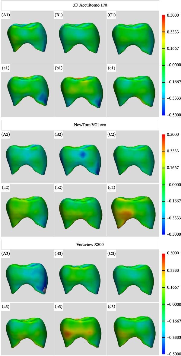 Figure 2
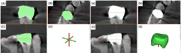 Figure 3
 Figure 4
 Figure 5
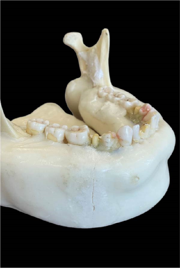 Figure 6
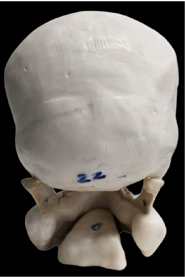 Figure 7
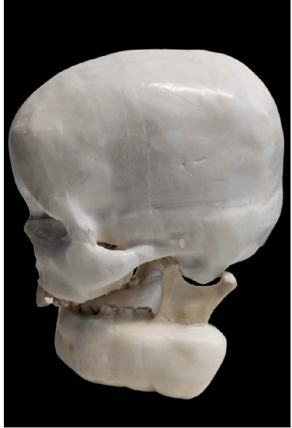 Figure 8
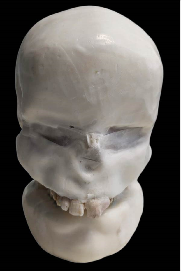 Figure 9
Peer Reviews
No public reviews on file for this paper yet. If you reviewed it on a platform where reviews are public (OpenReview, ICLR, NeurIPS, ICML), you can paste yours below so the community can read it here.
Videos
No videos yet. Explain this paper in a talk, walkthrough, or lecture? Add one.
Taxonomy
TopicsDental Radiography and Imaging · Advanced X-ray and CT Imaging · Medical Imaging Techniques and Applications