Greater medial arterial supply revealed by 7‐Tesla quantitative magnetic resonance imaging, histology and high‐resolution computed tomography of the patellar tendon
Maximilian M. Mueller, Craig E. Klinger, Sebastian Conner‐Rilk, Jerry Wang, Kevin G. Shea, Gregory S. DiFelice, Ryan Brown, Maneeza Bilal, Peter K. Sculco, Scott A. Rodeo, Daniel W. Green

TL;DR
This study shows that the medial side of the patellar tendon has a greater blood supply, using advanced imaging and histology techniques.
Contribution
The study introduces a novel combination of 7T-qMRI, histology, and micro-CT to quantitatively map the vascular supply of the patellar tendon.
Findings
The medial region of the patellar tendon has significantly higher arterial contributions compared to the lateral region.
A peripatellar circular vascular network was identified, extending from the medial margin into the posterior tendon layers.
Histology confirmed the greater medial arterial supply, with 8.3% more supply than the lateral side.
Abstract
To quantitatively assess relative arterial contributions to the patellar tendon (PT) across predefined anatomic regions with 7‐Tesla quantitative magnetic resonance imaging (7T‐qMRI), algorithm‐based histological analysis and high‐resolution computed tomography (micro‐CT) in a cadaveric model. Seven fresh‐frozen human cadaveric knee pairs (mean age 41.9 ± 15.5 years) underwent limited vascular dissection and arterial cannulation. Pre‐ and post‐contrast 7T‐qMRI, with a volumetric interpolated breath‐hold examination (VIBE) three‐dimensional T1‐weighted gradient echo pulse sequence, quantified tendonous vascularity by measuring contrast enhancement. Subsequent quantitative algorithm‐based histologic analysis with hematoxylin and eosin (H&E) staining followed, and two additional specimens underwent high‐resolution (98 μm) micro‐CT for qualitative vascular assessment. In the transverse…
Genes, proteins, chemicals, diseases, species, mutations and cell lines named across the full text — each resolved to its canonical identifier and authoritative record.
Click any figure to enlarge with its caption.
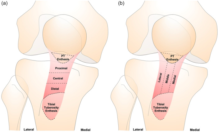 Figure 1
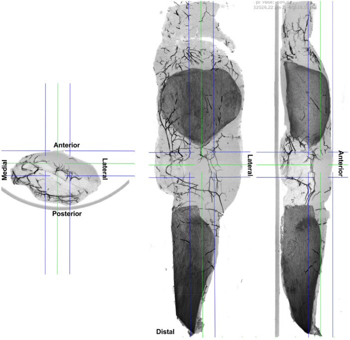 Figure 2
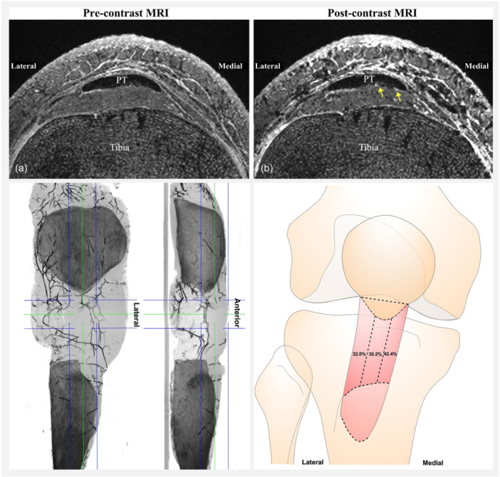 Figure 3
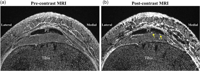 Figure 4
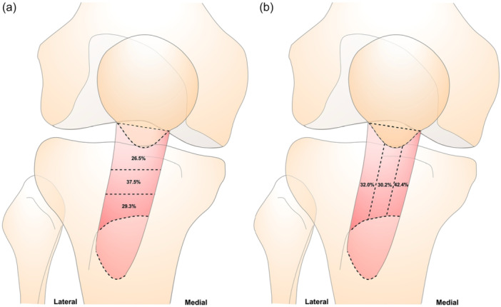 Figure 5
Peer Reviews
No public reviews on file for this paper yet. If you reviewed it on a platform where reviews are public (OpenReview, ICLR, NeurIPS, ICML), you can paste yours below so the community can read it here.
Videos
No videos yet. Explain this paper in a talk, walkthrough, or lecture? Add one.
Taxonomy
TopicsLower Extremity Biomechanics and Pathologies · Knee injuries and reconstruction techniques · Tendon Structure and Treatment