Trimodal cytological integration of micronuclei assay, argyrophilic nucleolar organizer region staining, and cytomorphometry enhances diagnostic discrimination of canine gingival masses
Poppapak Hoonpo, Tawewan Issarankura Na Ayudhaya, Kridsada Chaichoun, Panpanga Sangsuriya, Thanongsak Mamom, Parin Suwannaprapha

TL;DR
A new three-part cytological method improves the accuracy of diagnosing canine oral lesions by combining shape analysis, cell proliferation markers, and DNA damage indicators.
Contribution
A trimodal cytological framework integrating cytomorphometry, AgNOR staining, and micronuclei assay is developed and validated for canine gingival mass diagnosis.
Findings
Malignant neoplasms showed significantly higher AgNOR counts and micronuclei frequency compared to benign and hyperplastic lesions.
The nuclear-to-cytoplasmic ratio was highest in benign neoplasms and correlated with AgNOR and micronuclei counts.
The trimodal approach provides a quantitative framework that reduces subjective interpretation in diagnosing canine oral lesions.
Abstract
Canine gingival masses are common oral lesions with variable biological behavior, ranging from reactive hyperplasia to malignant neoplasia. Although routine cytology is widely used for initial evaluation, diagnostic overlap between benign and malignant lesions may limit accuracy when relying solely on morphology. This study aimed to develop and validate a trimodal cytological framework that integrates cytomorphometric analysis, argyrophilic nucleolar organizer region (AgNOR) staining, and micronuclei assay to enhance cytological differentiation and objectively characterize proliferative and genotoxic alterations in canine gingival masses. Cytological specimens were obtained through fine-needle aspiration from gingival masses of 46 dogs and classified as epithelial hyperplasia (n = 11), benign neoplasms (n = 14), and malignant neoplasms (n = 21), with histopathology serving as the…
Genes, proteins, chemicals, diseases, species, mutations and cell lines named across the full text — each resolved to its canonical identifier and authoritative record.
Click any figure to enlarge with its caption.
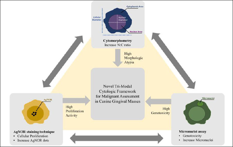 Figure 1
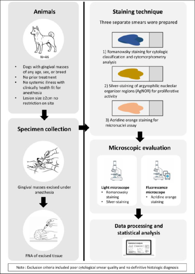 Figure 2
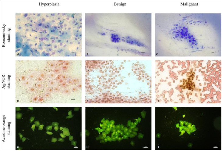 Figure 3
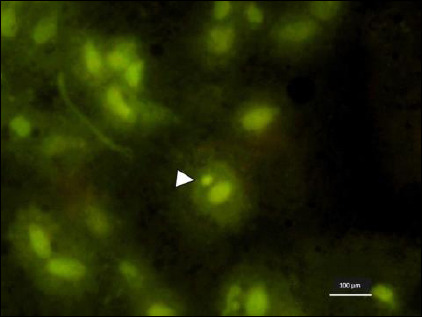 Figure 4
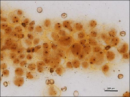 Figure 5
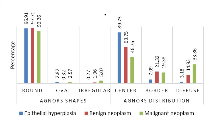 Figure 6
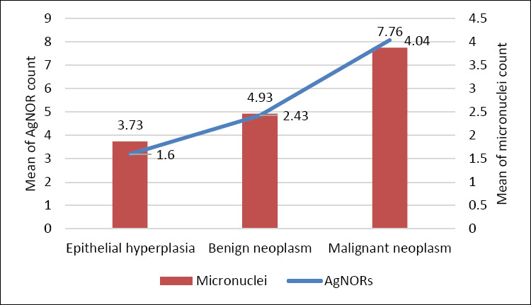 Figure 7
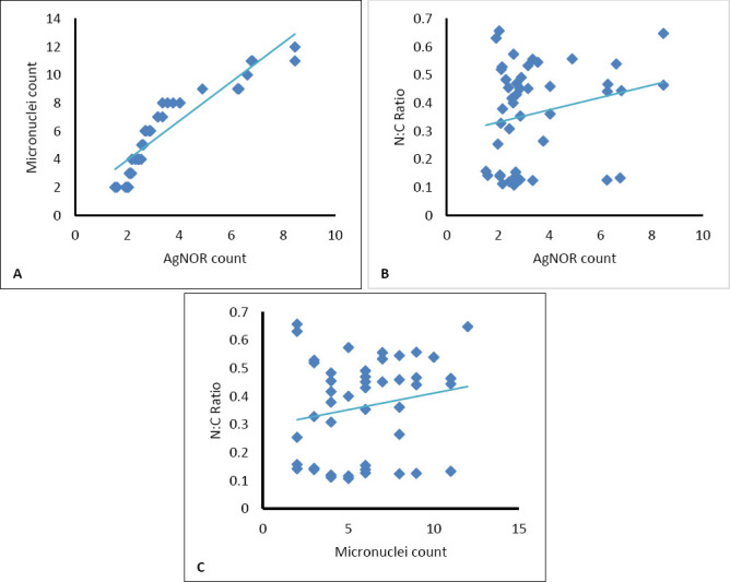 Figure 8
Peer Reviews
No public reviews on file for this paper yet. If you reviewed it on a platform where reviews are public (OpenReview, ICLR, NeurIPS, ICML), you can paste yours below so the community can read it here.
Videos
No videos yet. Explain this paper in a talk, walkthrough, or lecture? Add one.
Taxonomy
TopicsVeterinary Oncology Research · Oral and Maxillofacial Pathology · Salivary Gland Tumors Diagnosis and Treatment