Diffusion MRI sampling schemes bias diffusion metrics and tractography
Ivanei Bramati, Diego Szczupak, Marina Carneiro Monteiro, Fernanda Meireles, Daniel Menezes Guimarães, Ryan J. Dean, Lynn K. Paul, Fernanda Tovar-Moll

TL;DR
Different MRI scanning methods can lead to biased results in brain imaging studies, affecting how we understand white matter structures.
Contribution
The study reveals systematic differences in diffusion metrics and tractography outcomes across four common diffusion MRI sampling schemes.
Findings
Fractional anisotropy and mean diffusivity showed moderate cross-scheme correlations but rare matched means.
Tract volumes varied significantly between schemes, with DSI and HCPms yielding similar results in most regions.
Harmonization methods could reduce residual bias and enable pooled analyses across diverse protocols.
Abstract
Diffusion MRI is increasingly used to study white-matter architecture, but tractography and diffusion metrics can be biased by different sampling schemes. We assessed systematic differences across four common protocols—single-shell high-angular resolution diffusion imaging (HARDI), Siemens clinical multi-shell (Sms), diffusion spectrum imaging (DSI), and Human Connectome Project multi-shell (HCPms)—in healthy adults and individuals with corpus callosum dysgenesis (CCD). All data were acquired on a single 3 T scanner and processed uniformly to extract fractional anisotropy (FA), mean diffusivity (MD), effective contrast-to-noise ratio (eCNR), and orientation dispersion within the corpus callosum (CC), corona radiata (CR), and centrum semiovale (CSO). In controls, we measured tract volumes for CC, bilateral CR, anterior commissure (AC) and posterior commissure (PC), and streamline counts…
Genes, proteins, chemicals, diseases, species, mutations and cell lines named across the full text — each resolved to its canonical identifier and authoritative record.
Click any figure to enlarge with its caption.
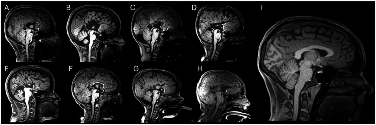 Figure 1
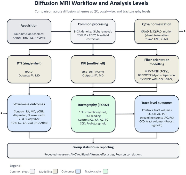 Figure 2
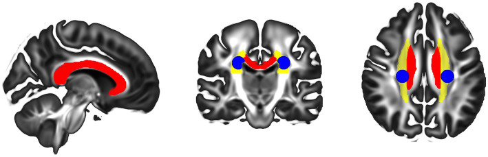 Figure 3
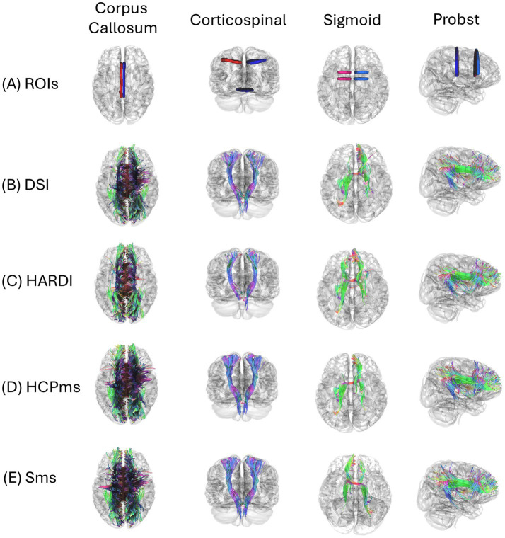 Figure 4
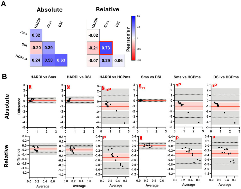 Figure 5
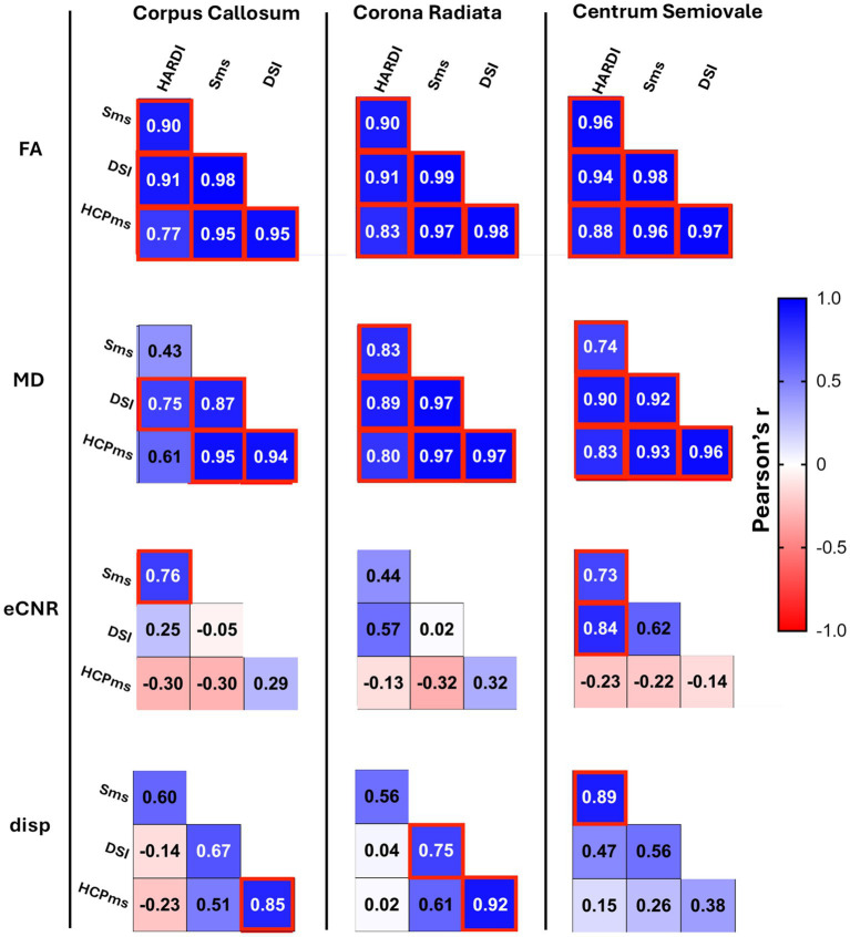 Figure 6
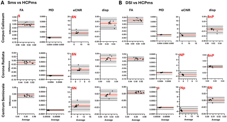 Figure 7
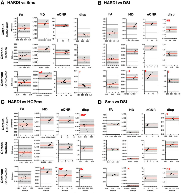 Figure 8
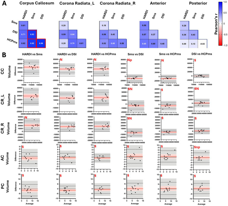 Figure 9
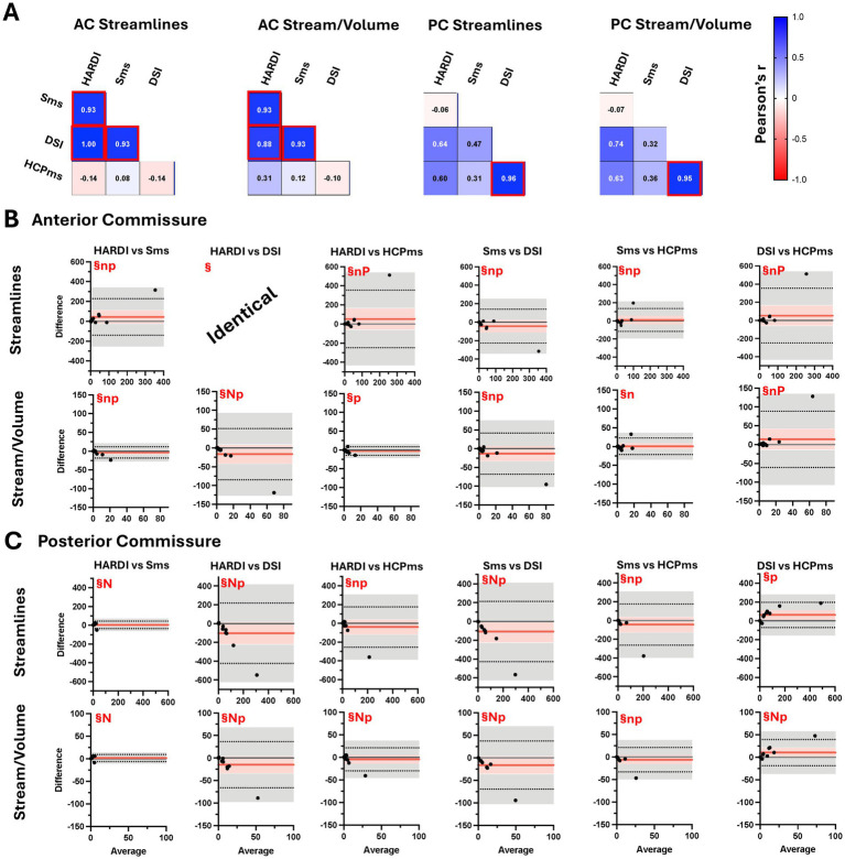 Figure 10
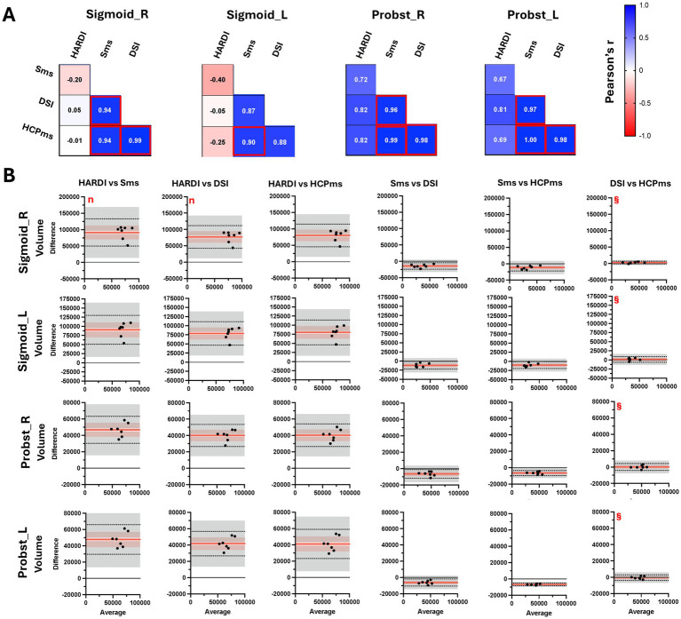 Figure 11
Peer Reviews
No public reviews on file for this paper yet. If you reviewed it on a platform where reviews are public (OpenReview, ICLR, NeurIPS, ICML), you can paste yours below so the community can read it here.
Videos
No videos yet. Explain this paper in a talk, walkthrough, or lecture? Add one.
Taxonomy
TopicsAdvanced Neuroimaging Techniques and Applications · Fetal and Pediatric Neurological Disorders · Epilepsy research and treatment