Fluorescence-guided surgery combined with intraoperative photodynamic therapy for recurrent atypical and anaplastic intracranial meningiomas: a prospective feasibility study
Anastasiia Nechaeva, Konstantin Kukanov, Alexey Ulitin, Victor Olyushin, Daria Sitovskaya, Danila Bobkov, Vseslav Ushanov, Stephanie E. Combs, Konstantin Samochernykh, Maxim Shevtsov

TL;DR
This study shows that combining fluorescence-guided surgery with photodynamic therapy is a safe and effective new approach for treating hard-to-manage brain tumors called meningiomas.
Contribution
The novel integration of fluorescence-guided surgery and intraoperative photodynamic therapy for recurrent meningiomas is introduced and evaluated for feasibility and efficacy.
Findings
The FGS+PDT protocol achieved 95.6% gross-total resection in treated patients, significantly higher than the 77.1% in the control group.
PDT induced profound biological effects, including increased apoptosis and mitochondrial dysfunction in tumor and peritumoral tissues.
No recurrences were observed in the experimental group over a median 16-month follow-up period.
Abstract
Recurrent intracranial meningiomas are a significant therapeutic challenge due to their invasive growth and high recurrence risk after surgery and radiotherapy. This study investigates the feasibility of a novel integrated approach combining 5-aminolevulinic acid (5-ALA) fluorescence-guided surgery (FGS) with intraoperative photodynamic therapy (PDT) for recurrent atypical and anaplastic meningiomas. In a single-center, prospective cohort study, 23 patients with recurrent atypical and anaplastic meningiomas received the experimental treatment protocol (FGS+PDT). A retrospective control group (n=35) underwent conventional microsurgery. The intervention included preoperative 5-ALA administration, FGS with visual (Fluorescence Intensity Score, FIS) and quantitative biospectroscopy (Fluorescence Index, FI) guidance, tumor resection, and subsequent PDT (635 nm laser) applied to the…
Genes, proteins, chemicals, diseases, species, mutations and cell lines named across the full text — each resolved to its canonical identifier and authoritative record.
Click any figure to enlarge with its caption.
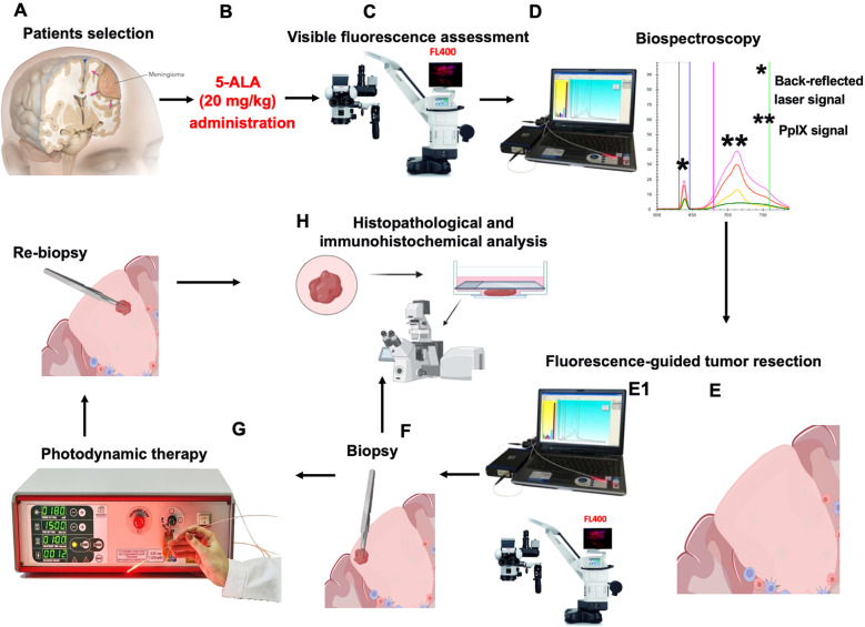 Figure 1
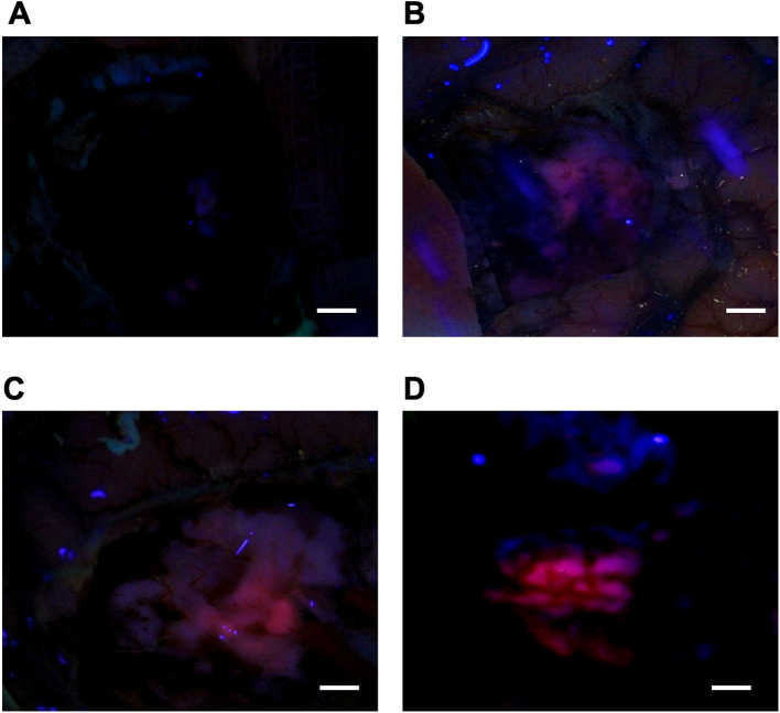 Figure 2
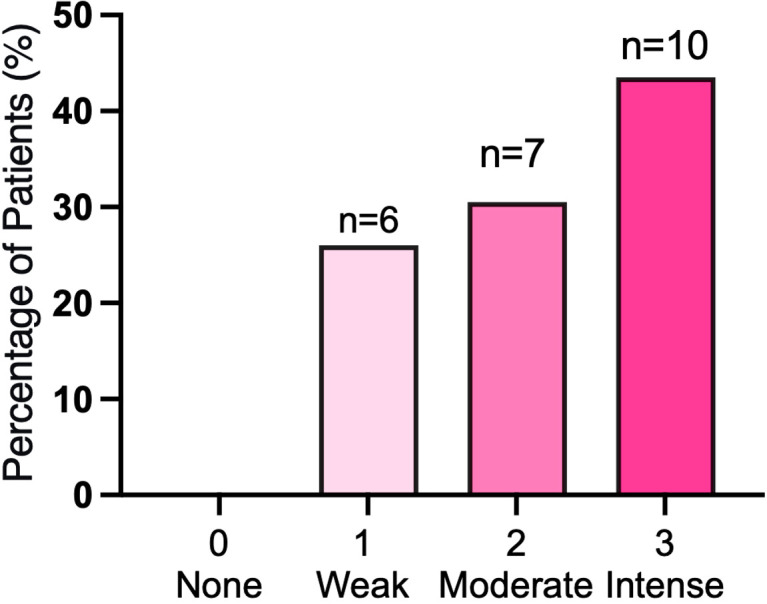 Figure 3
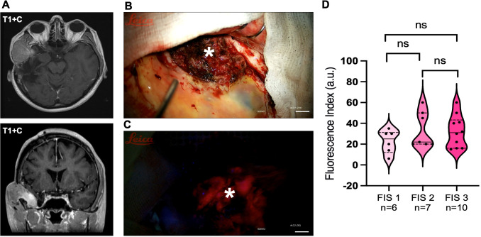 Figure 4
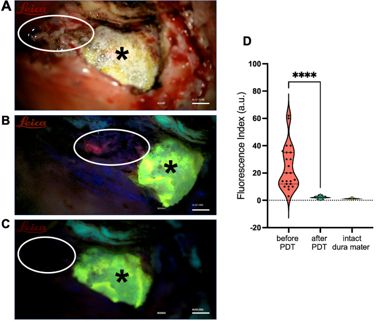 Figure 5
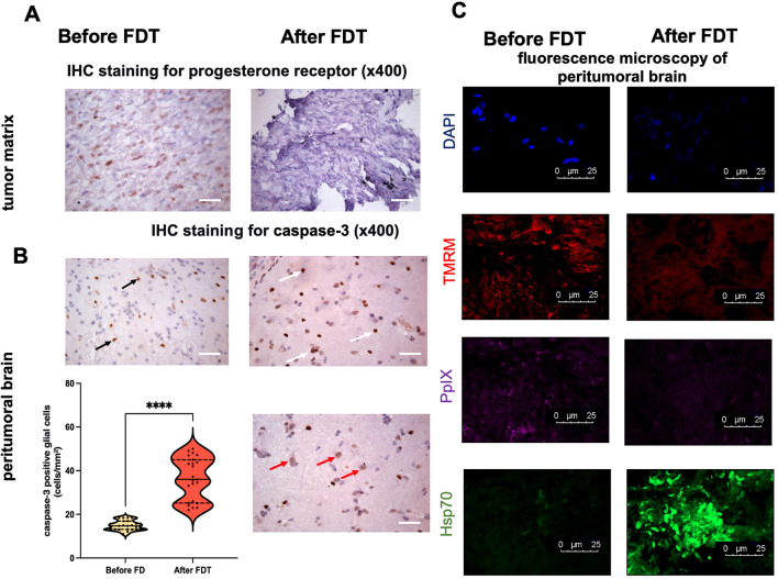 Figure 6
Peer Reviews
No public reviews on file for this paper yet. If you reviewed it on a platform where reviews are public (OpenReview, ICLR, NeurIPS, ICML), you can paste yours below so the community can read it here.
Videos
No videos yet. Explain this paper in a talk, walkthrough, or lecture? Add one.
Taxonomy
TopicsMeningioma and schwannoma management · Nanoplatforms for cancer theranostics · Optical Imaging and Spectroscopy Techniques