The exosomal miR-26b-3p derived from Crohn’s disease-associated mesenteric adipose tissue induces M1 macrophage polarization and exacerbates ileocolonic anastomosis inflammation via the p38-MAPK signaling pathway
Enhao Wu, Wenwei Qian, Xi Zhang, Lili Gu, Zhen Guo, Zeqian Yu, Yi Li, Weiming Zhu

TL;DR
Exosomal miR-26b-3p from Crohn’s disease mesenteric fat worsens intestinal inflammation by promoting harmful macrophage activity.
Contribution
Identifies miR-26b-3p in exosomes from CD mesenteric tissue as a driver of M1 macrophage polarization and inflammation via the p38-MAPK pathway.
Findings
Exosomal miR-26b-3p from hypertrophic mesenteric tissue promotes M1 macrophage polarization and inflammation in ileocolonic anastomosis.
miR-26b-3p activates the p38-MAPK pathway by targeting TRIM33, exacerbating inflammation in a mouse model of Crohn’s disease.
miR-26b-3p levels correlate with the severity of ileocolonic anastomosis inflammation in Crohn’s disease patients.
Abstract
Crohn’s Disease (CD) is a chronic inflammatory condition characterized by intestinal inflammation, especially in the progression of postoperative anastomotic recurrence. Recent evidence implicates mesenteric adipose tissue (MAT) in CD pathogenesis, particularly through its exosome secretion, which may influence inflammation pathways. The molecular mechanisms driving this inflammation remain inadequately understood. Exosomes were isolated from MAT of the diseased bowel and macroscopically normal MAT from the surgical margins of patients with CD. We induced chronic intestinal inflammation in mice using dinitrobenzene sulfonic acid (DNBS), simulating CD-like MAT. Using a surgical model of IL10-knockout mice, we performed a series of experiments in vitro and in vivo to assess the effects of exosomes on ileocolonic anastomosis inflammation and macrophage M1 polarization. We performed…
Genes, proteins, chemicals, diseases, species, mutations and cell lines named across the full text — each resolved to its canonical identifier and authoritative record.
Click any figure to enlarge with its caption.
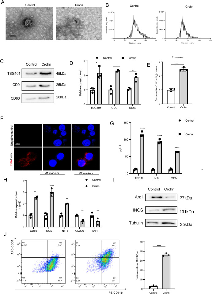 Figure 1
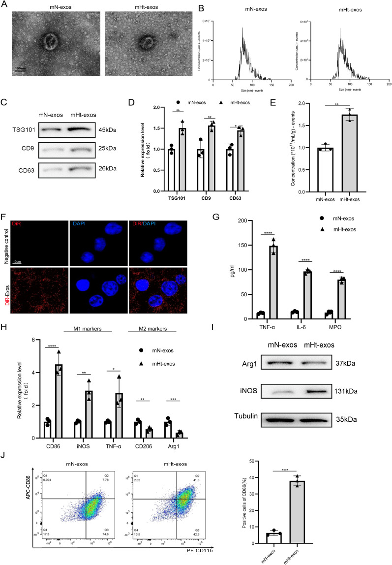 Figure 2
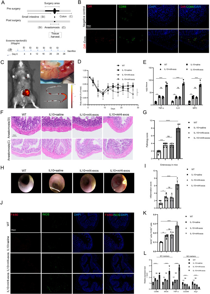 Figure 3
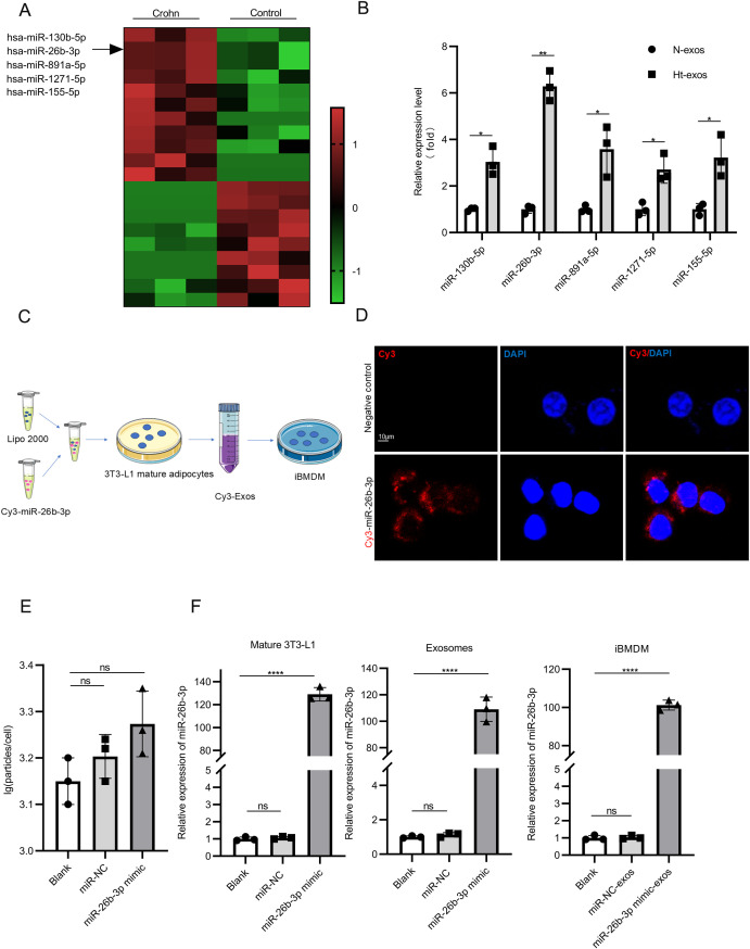 Figure 4
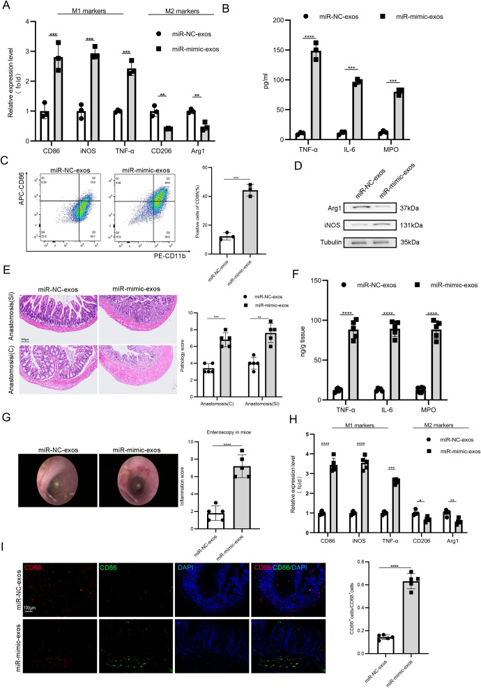 Figure 5
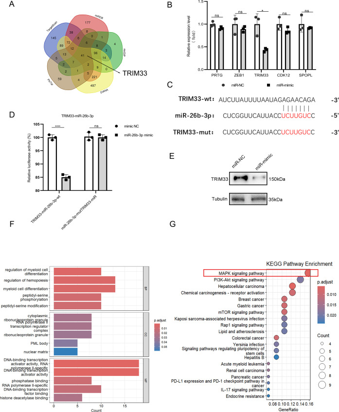 Figure 6
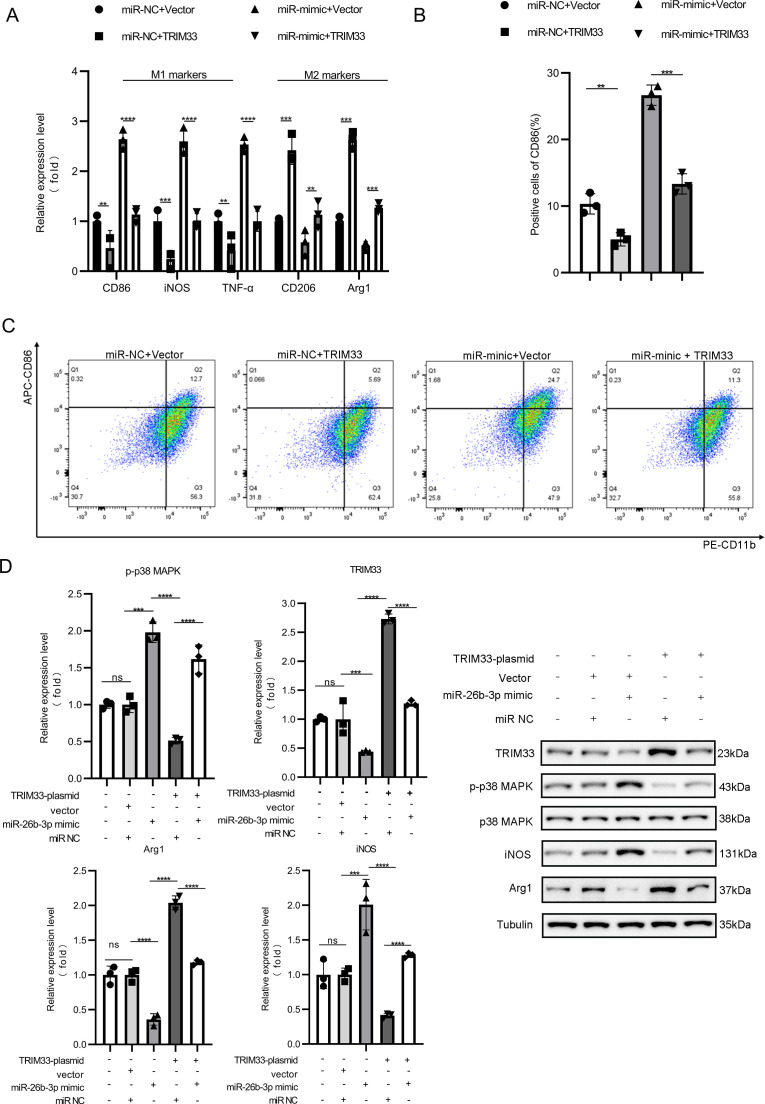 Figure 7
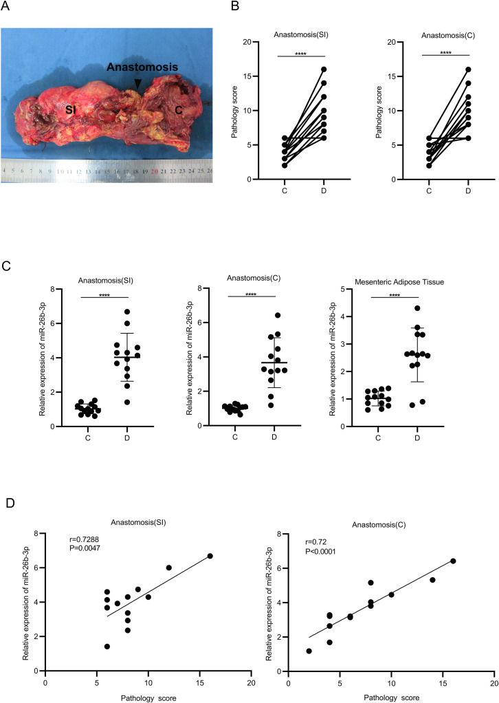 Figure 8
Peer Reviews
No public reviews on file for this paper yet. If you reviewed it on a platform where reviews are public (OpenReview, ICLR, NeurIPS, ICML), you can paste yours below so the community can read it here.
Videos
No videos yet. Explain this paper in a talk, walkthrough, or lecture? Add one.
Taxonomy
TopicsInflammatory Bowel Disease · Extracellular vesicles in disease · Immune cells in cancer