Artificial intelligence-based characterization of multi-organ ultrasound congestion across the heart failure Spectrum
Lavinia Del Punta, Giacomo Aru, Alina Sirbu, Nicolò De Biase, Stefano Taddei, Giuseppe Prencipe, Stefano Masi, Nicola Riccardo Pugliese

TL;DR
This study uses artificial intelligence to analyze ultrasound signs of congestion in heart failure patients, revealing a multidimensional congestion phenotype across different stages of the disease.
Contribution
The novel use of AI to integrate multi-organ ultrasound findings with clinical and echocardiographic data to characterize congestion in heart failure.
Findings
AI models identified key predictors of multi-organ congestion, including pulmonary artery pressures and medication use.
Multi-organ congestion was observed in 274 patients with ≥2 ultrasound signs of congestion.
Congestion features clustered into four domains: medical history, biohumoral variables, left heart function, and right heart/pulmonary circulation.
Abstract
To investigate, using artificial intelligence (AI), the relationships between ultrasound (US)-defined systemic congestion and demographic, echocardiographic, and biohumoral parameters across the heart failure (HF) spectrum. A total of 1588 subjects (651 Stage A–B, 376 HF with reduced left ventricular ejection fraction [HFrEF, <50%], and 561 HF with preserved ejection fraction [HFpEF, ≥50%]) underwent comprehensive clinical evaluation, laboratory testing, echocardiography, and US assessment of congestion, including inferior vena cava (IVC), lung ultrasound (LUS), renal venous flow (RVF), portal venous flow (PVF), and hepatic venous flow (HVF). Assessment of IVC, LUS, and RVF was available in the entire cohort, whereas HVF and PVF were performed in 359 and 289 patients, respectively. Overall, 856 patients had no US signs of congestion, 458 had one US sign, and 274 had ≥2 US signs…
Genes, proteins, chemicals, diseases, species, mutations and cell lines named across the full text — each resolved to its canonical identifier and authoritative record.
Click any figure to enlarge with its caption.
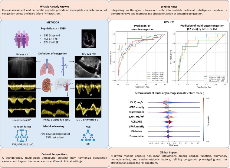 Figure 1
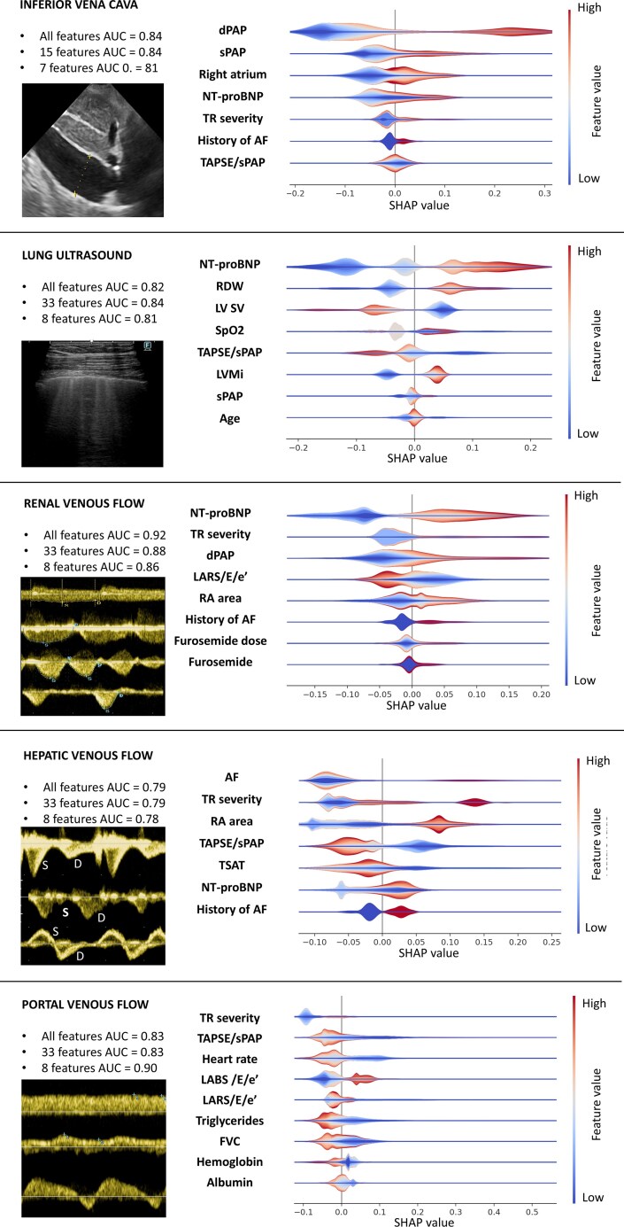 Figure 2
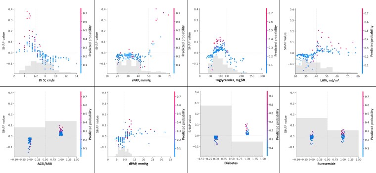 Figure 3
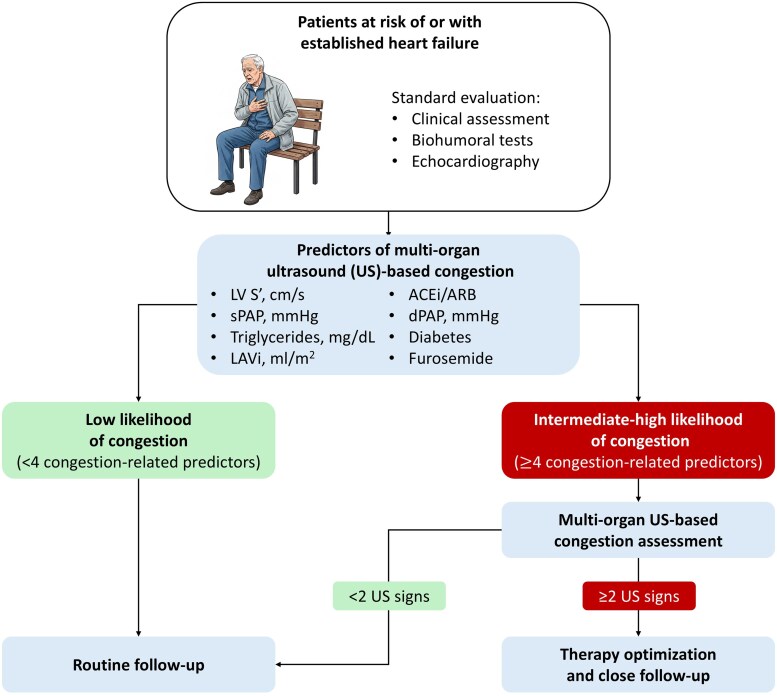 Figure 4
 Figure 5
Peer Reviews
No public reviews on file for this paper yet. If you reviewed it on a platform where reviews are public (OpenReview, ICLR, NeurIPS, ICML), you can paste yours below so the community can read it here.
Videos
No videos yet. Explain this paper in a talk, walkthrough, or lecture? Add one.
Taxonomy
TopicsUltrasound in Clinical Applications · Hemodynamic Monitoring and Therapy · Venous Thromboembolism Diagnosis and Management