Entomo-virological investigation during the epizootic outbreak of sylvatic yellow fever in Rio Grande do Sul, Brazil, between 2021 and 2022
Nícolas Felipe Drumm Müller, Marcelo de Moura Lima, Edmilson dos Santos, Aline Alves Scarpellini Campos, Thomas Rosa Menegazzi, Alanis Silva Melgarejo, Bruna Paredes-Galarza, Lina Violet-Lozano, Martha Trindade Oliveira, Cirilo Henrique Oliveira, Paulo Michel Roehe

TL;DR
This study investigated mosquitoes in Brazil during a yellow fever outbreak but found no evidence of the virus in the collected samples.
Contribution
The study provides insights into the low detection of yellow fever virus in mosquitoes despite active circulation among primates.
Findings
A total of 1,210 mosquitoes from 26 species were collected across 17 municipalities.
RT-qPCR analysis failed to detect YFV or other Orthoflaviviruses in any mosquito samples.
Haemagogus leucocelaenus, a key YFV vector, was among the most frequently captured species.
Abstract
Yellow fever virus (YFV) re-emerged among non-human primates (NHPs) in Rio Grande do Sul in early 2021, more than a decade after its last detection in the state. The spread of the virus was accompanied by increased mortality among NHPs. To conduct entomological surveillance and molecular detection of YFV and other Orthoflavivirus species in mosquito samples collected from affected and potentially receptive areas. Mosquitoes were collected during epizootics using human landing catches, BG-Pro traps, and ovitraps. Virus detection was performed using reverse transcription real-time polymerase chain reaction (RT-qPCR) assays targeting YFV and pan-Orthoflavivirus sequences. A total of 1,210 mosquitoes, representing 26 taxa, were collected across 17 municipalities. Psorophora ferox was the most abundant species, followed by Culex (Culex) spp., accounting for 27% and 12% of the specimens,…
Genes, proteins, chemicals, diseases, species, mutations and cell lines named across the full text — each resolved to its canonical identifier and authoritative record.
Click any figure to enlarge with its caption.
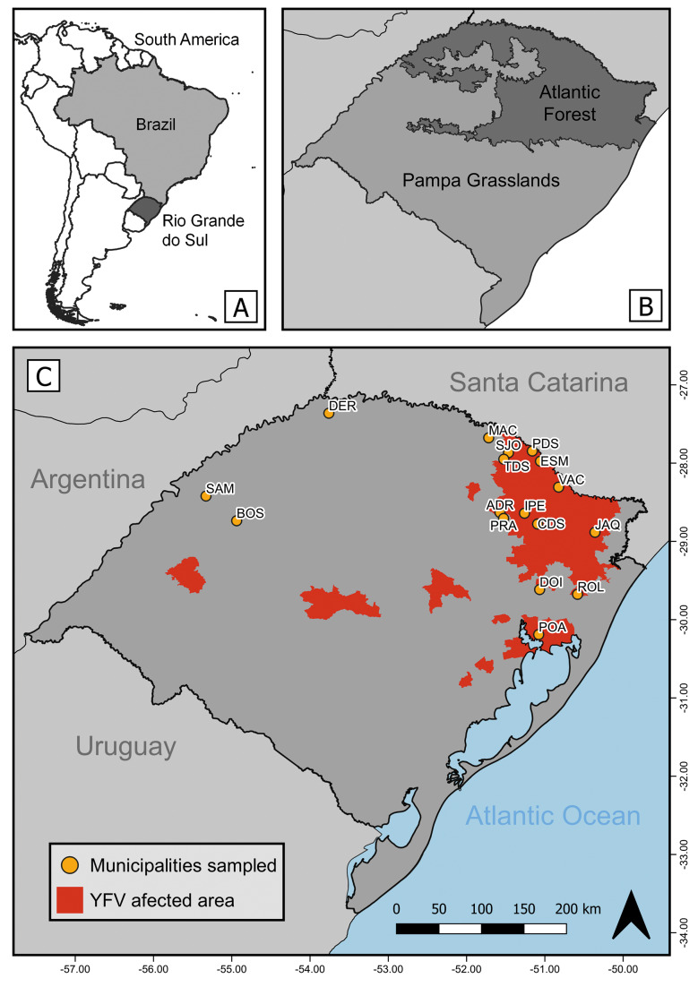 Fig. 1
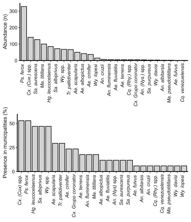 Fig. 2
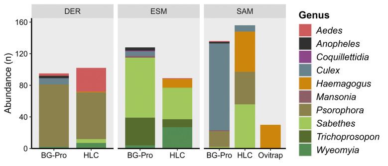 Fig. 3
| qPCR assay | Primer & Probe | Sequence (5'- 3') |
|---|---|---|
| YFV | YFallF | GCTAATTGAGGTGYATTGGTCTGC |
| YFallR | CTGCTAATCGCTCAAMGAACG | |
| YFallP | FAM-ATCGAGTTGCTAGGCAATAAACAC-TAMRA | |
| Pan-Orthoflavivirus | Flavi all S2 | TACAACATGATGGGMAAACGYGARAA |
| Flavi all AS4 | GTGTCCCAGCCNGCKGTRTCRTC | |
| Flavi all probe 1 | FAM-TGGTWYATGTGGYTNGGRGC-TAMRA | |
| Flavi all probe 2 | FAM-CCGTGCCATATGGTATATGTGGCTGGGAGC-TAMRA | |
| Flavi all probe 3 | FAM-TTTCTGGAATTTGAAGCCCTGGGTTT-TAMRA |
| Municipality | |||||||||||||||||
|---|---|---|---|---|---|---|---|---|---|---|---|---|---|---|---|---|---|
| Taxa | ADR* | BOS | CDS* | DER | DOI* | ESM* | IPE* | JAQ* | MAC | PDS* | POA* | PRA* | ROL* | SAM | SJO* | TDS* | VAC* |
| Anophelinae | |||||||||||||||||
|
| 2 | 2 | 2 | ||||||||||||||
|
| 7 | ||||||||||||||||
|
| 2 | ||||||||||||||||
|
| 1 | 3 | |||||||||||||||
| Culicinae | |||||||||||||||||
| Aedini | |||||||||||||||||
|
| 2 | 3 | |||||||||||||||
|
| 2 | 1 | 7 | 18 | |||||||||||||
|
| 1 | ||||||||||||||||
|
| 32 | 14 | 10 | 2 | 1 | ||||||||||||
|
| 2 | 2 | 1 | ||||||||||||||
|
| 1 | 41 | |||||||||||||||
|
| 13 ** | 1 | 15 ** | 1 ** | 2 ** | 1 ** | 51 | 1 | |||||||||
|
| 2 | 1 | 138 | 1 | 92 | 1 | 31 | 61 | 1 | ||||||||
| Culicini | |||||||||||||||||
|
| 2 | 1 | 1 | 1 | |||||||||||||
|
| 4 | 8 | 1 | 4 | 1 | 1 | 118 | 2 | 2 | ||||||||
| Mansoniini | |||||||||||||||||
|
| 5 | ||||||||||||||||
|
| 1 | ||||||||||||||||
|
| 2 | ||||||||||||||||
|
| 97 | 2 | 1 | ||||||||||||||
| Sabethini | |||||||||||||||||
|
| 126 | 1 | |||||||||||||||
|
| 7 | 4 | 1 | 1 | 1 | 1 | 56 | 1 | |||||||||
|
| 1 | 2 | |||||||||||||||
|
| 4 | 3 | 9 | 43 | 2 | 2 | |||||||||||
|
| 2 | 3 | |||||||||||||||
|
| 2 | 59 | 2 | 2 | 1 | ||||||||||||
|
| 3 | ||||||||||||||||
|
| 15 | ||||||||||||||||
| Abundance | 6 | 29 | 7 | 197 | 7 | 258 | 7 | 13 | 4 | 128 | 180 | 2 | 38 | 292 | 1 | 6 | 5 |
| Richness | 3 | 5 | 3 | 10 | 5 | 12 | 5 | 5 | 2 | 6 | 9 | 2 | 4 | 8 | 1 | 3 | 4 |
- —CNPq/Decit/SCTIE/MS
Peer Reviews
No public reviews on file for this paper yet. If you reviewed it on a platform where reviews are public (OpenReview, ICLR, NeurIPS, ICML), you can paste yours below so the community can read it here.
Videos
No videos yet. Explain this paper in a talk, walkthrough, or lecture? Add one.
Taxonomy
TopicsMosquito-borne diseases and control · Indigenous Health and Education · Zoonotic diseases and public health
Orthoflavivirus flavi, widely known as yellow fever virus (YFV), is the prototype virus of the genus Orthoflavivirus, family Flaviviridae. It is an endemic arbovirus in tropical and subtropical regions.1 In South America, YFV is maintained through two distinct transmission cycles: urban and sylvatic. The urban cycle involves the mosquito Aedes aegypti and humans.2 Due to the temporary eradication of Ae. aegypti between the 1940s and 1960s, along with human vaccination efforts, the urban transmission cycle was interrupted, and the virus remained confined to forested areas, where the sylvatic cycle persists. In these areas, native mosquitoes of the genera Haemagogus and Sabethes serve as vectors, non-human primates (NHPs) act as amplifier hosts.3 In Brazil, NHPs are used as sentinels for YFV surveillance due to their high susceptibility to infection.4 Humans, within the sylvatic cycle, are considered incidental hosts, with infections occurring sporadically, developing disease and viraemia, although they generally do not sustain transmission.5
In Brazil, YFV is endemic in the Amazon rainforest, but epidemic activity has extended to the southern limits of the Atlantic Forest. YFV circulation in extra-Amazonian regions has been reported since the early 2000s.3 From 2014 onwards, the virus expanded from the Cerrado biome in northern Brazil to other regions, causing high mortality in NHPs.3 Between 2016 and 2019, YFV reached the Atlantic Forest in the southeast, the most densely populated region of the country, triggering the largest sylvatic YFV outbreak ever recorded in Brazil.4 ,6 During this period, more than 2,000 human cases and over 700 fatalities were reported, this significant outbreak was primarily attributed to the lack of vaccination among the local population.7 Since 2019, YFV has also been detected among NHPs in southern Brazil8 and, in early 2021, the virus reached Rio Grande do Sul (RS), Brazil's southernmost state, after more than a decade without recorded activity.9
In the 21st century, the first YFV epizootic outbreak in RS occurred between 2001 and 2002, and was restricted to the northwestern region of the state. During this event, NHP deaths were recorded, but no human cases were reported.10 The largest sylvatic YFV outbreak in the RS took place between 2008 and 2009. Again, NHP deaths began in the northwest and subsequently spread to central and northern regions.11 This outbreak resulted in 21 confirmed human cases, including nine fatalities. Additionally, 204 NHP deaths were confirmed by laboratory diagnosis, although the number of NHPs affected is estimated to exceed 2,000.12 In the 2021 epizootic outbreak, the YFV dispersion wave reached the northeastern region of RS via neighbouring states, a marked shift from previous outbreaks, which typically entered through the northwest, bordering Argentina.9 The virus then progressed southward, and also affected areas in the central and northwestern regions of the state.13 In total, 420 NHP deaths were reported in 2021, of which 126 were laboratory-confirmed as YFV-positive. Notably, the majority of NHP cases occurred during the first semester of the year, with a marked decline in reports coinciding with the onset of the colder season, which is unfavourable for mosquito activity.13 As a result of prior vaccination efforts and heightened public awareness, no human infections were reported throughout 2021.13
In addition to the circulation of YFV, there is evidence supporting the presence of other orthoflaviviruses in RS. In urban settings, dengue virus, transmitted by Ae. aegypti, has been circulating autochthonously in humans since 2007, with a marked increase in human incidence observed from 2021 onward.13 ,14 Zika virus has also been generating human cases since 2017, although with a lower incidence compared to dengue.13 ,15 Although no human cases have been officially reported in RS for other orthoflaviviruses, indirect historical evidence suggests their circulation among wild and livestock animals, indicating potential zoonotic risk. Antibodies against Orthoflavivirus nilense (West Nile virus, WNV) have been detected in birds and horses,16 ,17 and antibodies for Orthoflavivirus louisense (St. Louis encephalitis virus, SLEV) and Orthoflavivirus ilheusense (Ilheus virus, ILHV) have been found in NHPs.18 ,19 Considering the evidence of arbovirus circulation in RS, this study aimed to analyse sylvatic mosquitoes collected during the YFV outbreak for the presence of YFV and other orthoflaviviruses.
MATERIALS AND METHODS
Study area - Rio Grande do Sul is the southernmost state in Brazil, bordering Uruguay and Argentina. The state covers an area of 281,707 km² and comprises 497 municipalities, with an estimated population of over 10 million people. Two major biomes occur in RS: the Pampa, a grassland biome where forests are limited to riparian zones and isolated patches, and the Atlantic Forest, a forest biome influenced by its proximity to the ocean.20
Mosquito sampling and taxonomic identification - The study was conducted with support from the Environmental Health Surveillance Division (DVAS) of the Rio Grande do Sul State Health Surveillance Centre (CEVS). Mosquito collections were carried out from February 2021 to February 2022 during the summer and spring seasons in forested areas of several municipalities in RS. Due to the emergency context of an active YFV outbreak, sampling procedures were not standardized across locations. Field strategies were dynamically adjusted based on real-time notifications of sick or dead NHPs recorded by DVAS, prioritizing newly affected areas (Fig. 1). Mosquito sampling was carried out concurrently with epizootic investigations and up to 25 days thereafter. Additionally, collections were extended to ecologically connected municipalities, areas with YFV circulation in previous years and two municipalities [Pinhal da Serra (PDS) and Esmeralda (ESM)] revisited approximately one year after the initial epizootic events [Supplementary data (Table)].
study area in Rio Grande do Sul (RS) State. (A) Map highlighting the spacial position of Brazil and RS. (B) Biome division of RS (Atlantic Forest and Pampa grasslands). (C) RS map highlighting the affected area by the yellow fever virus (YFV) (in red) according with the criteria established by the Environmental Health Surveillance Division (DVAS) of the Rio Grande do Sul State Health Surveillance Centre (CEVS) and the 17 municipalities where mosquitos are collected (in yellow): André da Rocha (ADR), Bossoroca (BOS), Campestre da Serra (CDS), Derrubadas (DER), Dois Irmãos (DOI), Esmeralda (ESM), Ipê (IPE), Jaquirana (JAQ), Machadinho (MAC), Pinhal da Serra (PDS), Porto Alegre (POA), Protásio Alves (PRA), Rolante (ROL), Santo Antônio das Missões (SAM), São José do Ouro (SJO), Tupanci do Sul (TDS), Vacaria (VAC).
In all areas, adult mosquitoes were collected using the human landing catch (HLC) method, which relies on the natural production of carbon dioxide (CO₂), heat, and human odour to attract mosquitoes seeking hosts. Between two and five collectors performed the HLC method using entomological nets and manual aspirators, operating during the day (9:00 a.m.-4:00 p.m.), for variable periods depending on field conditions.
At municipalities Santo Antônio das Missões (SAM), Derrubadas (DER), and ESM, additional collections were conducted using BG-Pro traps (Biogents AG, Regensburg, Germany). Each trap was baited with 2 kg of dry ice (as a CO₂ source) and an artificial attractant (BG-Lure unit; Biogents AG). Five traps were installed 1.5 metres above ground level, in the same locations where the HLC sampling was performed, and operated for 24 hours.
In SAM, 10 ovitraps baited with an infusion of dried forest floor leaves were installed 1.5 metres above ground level, each containing two wooden paddles. The traps remained in place for 15 days. Wooden paddles bearing mosquito eggs were immersed in water to induce hatching, and the resulting larvae were reared to adult stage in the laboratory for identification.
Mosquito handling and identification - After collection, the insects were quickly frozen, transferred to cryogenic tubes, and stored in containers filled with dry ice (-70 ºC). The specimens remained frozen during transport to the laboratory, where they were stored at -80ºC until identification. Mosquitoes were identified under a stereomicroscope on a cold table set at -20ºC, using dichotomous keys based on female morphological characteristics.21 ,22 ,23 Identified adult specimens were grouped into pools (≤ 10 individuals) according to species, sampling site, and collection date.
Statistical analysis - Species richness and abundance indices were calculated for the entire mosquito community based on the total number of captured specimens. In areas where multiple collection methods were applied, the differences in species richness and abundance among methods were evaluated. The average number of specimens captured by each method was assessed for normality using the Shapiro-Wilk test and compared using analysis of variance (ANOVA), followed by Tukey's post-hoc test. Statistical significance was set at p < 0.05. Analyses were performed using R software.
Virus detection - Mosquito pools were homogenised in 300 μL of L-15 culture medium containing 1% penicillin-streptomycin, and 10% foetal bovine serum (FBS) using the Precellys® 24 tissue homogeniser (Bertin Technologies, France) with glass beads. The homogenates were clarified by centrifugation at 12,500 g for 5 min at 4ºC. Supernatants were collected and subjected to RNA extraction using the Extracta® Kit Fast (Loccus, Cotia, Brazil) in the EXTRACTA® 96 automated extractor (Loccus), following the manufacturer's instructions.
Reverse transcription was performed using the High-Capacity cDNA Reverse Transcription Kit (Applied Biosystems™, Foster City, USA) with 1.5-2 μg of total RNA in a 20 μL reaction, according to the manufacturer's protocol. Quantitative polymerase chain reaction (qPCR) specific for YFV was carried out using primers and probes targeting a 97 bp region of the YFV NS1 gene.24 To detect other members of the Orthoflavivirus genus, an additional qPCR was performed using a pan-Orthoflavivirus assay.25 The list of primers used for amplification is provided in Table I.
To determine the detection limit of the pan-Orthoflavivirus assay, serial dilutions of a plasmid (10-10⁷ copies/μL) were prepared in triplicate. The plasmid was generated by cloning a 260 bp fragment of the NS5 gene (obtained from the YFV vaccine strain) into the TOPO TA vector (Invitrogen), following the manufacturer's instructions. Plasmid concentration was measured by spectrophotometry, and the same plasmid was used as a positive control in all PCR assays. Considering that the first dilution of the control plasmid contained 14 x 10⁸ genomic copies per microliter of the target fragment, the detection limit obtained was between 1 and 10 copies/reaction. For both assays' amplifications were performed under the following conditions: incubation at 55ºC for 2 min, polymerase activation and initial denaturation at 95ºC for 20 s and 45 cycles of denaturation at 95ºC for 1 s and extension/annealing at 60ºC for 25 s.
RESULTS
Between February 2021 and February 2022, 17 municipalities in RS were sampled, 14 of which were considered YFV affected areas, based on the detection of the virus in NHPs. In total, 1,210 individuals from the Culicidae family were collected. It was possible to categorize the specimens into 26 taxa; of these, 20 species were identified (Table II).
The most abundant mosquito species was Psorophora ferox, followed by Cx. (Cux.) spp., comprising 27% and 12% of the mosquitoes collected respectively. These taxa were also the most widespread, being present in more than half of the sampled municipalities (52.9%). The yellow fever vector Hg. leucocelaenus was the fifth most abundant (7%) and was present in almost half of the municipalities sampled (47%). Species of the genus Sabethes and Aedes also appear among the most collected mosquitoes in this study (Fig. 2).
abundance (A) and presence (B) of 26 mosquito taxa collected between February 2021 and February 2022 in Rio Grande do Sul.
When comparing the HLC and BG-Pro collection methods, there were no statistical differences between the total mosquito capture averages (p = 0.82). However, when comparing the capture of specific genera, it was observed that the BG-pro traps captured more Culex, but these traps did not capture any mosquitoes of the genus Haemagogus. When ovitraps were used, 30 individuals were collected, all belonging to the species Hg. leucocelaenus (Fig. 3).
abundance of different genera collected in the municipalities of Derrubadas (DER), Esmeralda (ESM) and Santo Antônio das Missões (SAM), where collections were carried out with human landing catch (HLC), BG-Pro traps and ovitraps. The bar colours represent the different genera of mosquitoes sampled.
In total, 203 pools of mosquitoes, separated by species, location and date of collection, were processed and submitted to a specific RT-qPCR for YFV and to a Pan-Orthoflavivirus RT-qPCR. All 203 pools tested negative in both RT-qPCR assays.
DISCUSSION
At the beginning of 2021, YFV reached RS, the southernmost state in Brazil. Virus spread in different municipalities was accompanied by deaths of native NHPs.9 Seeking to identify YFV and other arboviruses circulating in sylvatic areas in RS, mosquitoes were obtained from the same sites where circulation of YFV was identified when samples from NHPs were examined.
Regarding the abundance and distribution of the mosquito species found, Ps. ferox was the most abundant in the sampled areas. This mosquito is notably aggressive and exhibits a generalist feeding behaviour, obtaining blood from multiple mammalian groups, including NHPs, and to a lesser extent, from birds.26 Due to this behaviour, this species has been found naturally infected with several viruses, such as YFV, although in vector competence tests it has not been able to transmit this virus.6 The high proportion of Ps. ferox described here corroborates with previous findings in RS.27 Specimens classified as Cx. (Cux.) spp. were the second most abundant and constant in the sampled areas. These were identified to the subgenus level due to the high similarity between species. Members of this group are probable vectors of SLEV and WNV in South America.28 ,29 Corroborating this study, a review shows that this genus is widely distributed throughout RS.30 Hg. leucocelaneous was also frequently found in the sampled areas. This species has a wider distribution, occurring from the Amazon to the southern limit of the Atlantic Forest.31 It is noteworthy to mention that in Brazil and in other countries the genus Haemagogus has a major importance as the main vectors in the YFV sylvatic transmission cycle. The epidemiological importance of Hg. leucocelaneous increases in south of Brazil, where other species of this genus are absent.32 Although not the most abundant, this species has been consistently recorded in studies conducted in the Atlantic Forest of Rio Grande do Sul.27 ,32 Our findings reveal the presence of this species in municipalities where it had not been previously recorded, highlighting the potential susceptibility of these areas to YFV circulation. Here, the most abundant and widespread species of genus Sabethes, which can also be related to YFV spread, was Sa. albiprivus, that has already been found naturally infected by YFV in Argentina, close to the west border of RS.33 This species was also found infected during the dry period in the Brazilian Cerrado.34 Despite limited evidence of natural infections of this species, its role as secondary vector of YFV cannot be neglected. Sa. purpureus and Sa. aurescens were also found, however, to this date, they have not been found naturally infected by YFV. Members of the genus Aedes were also found here, including Ae. scapularis and Ae. albopictus, both previously found infected with YFV in Brazil.35 ,36 The latter is very opportunistic and strongly anthropophilic, so it's frequently captured in urbanised areas and in the transition between forest and city.37 ,38 The likelihood of spillover events is closely linked to the ecological and behavioural traits of mosquito species, particularly their host-feeding preferences, population density, and adaptability to anthropogenic environments. For this reason, Ae. albopictus is often highlighted as a possible bridge between the urban and sylvatic cycle of YFV.39 In addition to reports of natural infection, vector competence assays have confirmed its ability to acquire and transmit YFV, although at low rates.40 As in previous studies carried out in forested areas of Porto Alegre (POA), the Ae. albopictus has been captured in higher abundance than Hg. leucocelaenus.37 ,38
Although there were no significant differences between the total average of captures between the BG-Pro traps and HLC, in this study these traps did not capture mosquitoes of the genus Haemagogus; however, they captured more Culex, which are nocturnal vectors and were captured in smaller quantities manually. These findings are consistent with another study carried out in forest areas in the metropolitan region of RS, where these traps captured a large number of nocturnal vectors, mainly of the Culex genus and only one individual of the Haemagogus genus.38 Others studies conducted on the Amazon rainforest also demonstrate low efficiency in capturing Haemagogus mosquitoes from BG-Pro traps compared with HCL.41 ,42 A study comparing different collection methods carried out in the Atlantic Forest of the State of São Paulo also showed low efficiency in capturing YFV vectors using light-traps baited with the artificial attractant BG-Lure.43 Even though these traps are highly effective in capturing urban vectors, the few studies using these traps to collect wild mosquitoes in Brazil indicate low efficiency in capturing mosquitoes of the genus Haemagogus. The ovitraps installed at SAM captured only Hg. leucocelanus eggs, demonstrating the strong potential of this method in monitoring the main vector of YFV in RS. This methodology has already proven effective in capturing Haemagogus mosquitoes in the Atlantic Forest.44 It also enables molecular detection of YFV in mosquito samples after development into adult stage.45
In the present study, viral RNA was not detected following application of RT-qPCRs (either when specific primers for YFV or when primers for a wider group of orthoflaviviruses were used). In the zoonotic outbreak of YFV recorded in the RS between 2001 and 2002, the YFV was isolated from the vector Hg. leucocelaenus, the infection rate found in that occasion was 8.7%.10 The largest YFV outbreak recorded in Rio Grande do Sul occurred between 2008 and 2009. During this period, YFV was again isolated from the vector Hg. leucocelaenus, with an infection rate of 3.7%. On that occasion, Ae. serratus was also found infected and was considered a potential secondary vector.32 In the present study, however, this species was not sampled. Notably, these previous studies differed from the current approach by collecting mosquito specimens from a limited number of municipalities and employing intracerebral inoculation in mice as the primary method for viral detection. The low YFV infection rates reported in those studies may explain the absence of YFV-positive samples in the present survey.
Although no positive sample for orthoflaviviruses were detected in the present study, this result agrees with other studies carried out in Brazil. These methods have already been used on mosquito samples collected in urban parks in São Paulo, mainly with Ae. aegypti and Cx. spp. and, similar to this study, no positive samples were found.46 These primers and probes were applied to historical samples, the Iguape virus was detected in samples of Anopheles cruzii, and Ilheus virus in samples of Culex sp., Coquillettidia juxtamansonia and An. triannulatus.47 This same RT-qPCR was also used for viral identification after inoculation of mosquito samples from Brazil in cell culture. After inoculation in C6/36 cells it was detected Zika virus in pools of An. cruzii, Limatus durhamii and Weomyia confusa, dengue virus serotype 2 in Cx. spp. and Cx. vaxus and the insect specific virus (ISV), virus Guapiaçu, from samples of Ae. scapularis and Ae. Terrens.48 ,49
It is possible that strategies that aim to increase the initial viral load, such as previous inoculation in cell culture or in mice brain, could improve the detection of Orthoflavivirus in samples with low viral loads. Furthermore, although orthoflaviviruses were not detected, the possibility of circulation of ISV and arboviruses belonging to other genera, such as Alphavirus or Orthobunyavirus, cannot be ruled out. In addition, it is important to highlight that the presence of potential YFV vectors in the study areas indicate the possibility of virus circulation as soon as it reaches enough hosts to establish an outbreak. This fact, by itself, shows how imperative is to keep the surveillance of mosquitoes in areas where previous outbreaks of YFV were recognised.
SUPPLEMENTARY MATERIALS
Supplementary material
The reference list from the paper itself. Each links out to its DOI / PubMed record.
- 1Postler TS Beer M Blitvich BJ Bukh J Lamballerie X Drexler JF Renaming of the genus Flavivirus to Orthoflavivirus and extension of binomial species names within the family Flaviviridae Arch Virol 202316892243756116810.1007/s 00705-023-05835-1 · doi ↗ · pubmed ↗
- 2Gabiane G Yen P Failloux A Aedes mosquitoes in the emerging threat of urban yellow fever transmission Rev Med Virol 2022324 e 23333512485910.1002/rmv.2333 PMC 9541788 · doi ↗ · pubmed ↗
- 3Silva NIO Sacchetto L De Rezende IM Trindade GS La Beaud AD Thoisy B Recent sylvatic yellow fever virus transmission in Brazil: the news from an old disease Virol J 202017193197372710.1186/s 12985-019-1277-7PMC 6979359 · doi ↗ · pubmed ↗
- 4Cunha MS Da Costa AC Fernandes NCCA Guerra JM Santos FCP Nogueira JS Epizootics due to yellow fever virus in São Paulo State, Brazil: viral dissemination to new areas (2016-2017)Sci Rep 20199154743094086710.1038/s 41598-019-41950-3PMC 6445104 · doi ↗ · pubmed ↗
- 5Hamlet A Ramos DG Gaythorpe KAM Romano APM Garske T Ferguson NM Seasonality of agricultural exposure as an important predictor of seasonal yellow fever spillover in Brazil Nat Commun 202112136473413112810.1038/s 41467-021-23926-y PMC 8206143 · doi ↗ · pubmed ↗
- 6Abreu FVS Ribeiro IP Ferreira-de-Brito A Santos AAC Miranda RM Bonelly IS Haemagogus leucocelaenus and Haemagogus janthinomys are the primary vectors in the major yellow fever outbreak in Brazil, 2016-2018 Emerg Microbes Infect 2019812182313086677510.1080/22221751.2019.1568180 PMC 6455131 · doi ↗ · pubmed ↗
- 7Giovanetti M De Mendonça MCL Fonseca V Mares-Guia MA Fabri A Xavier A Yellow fever virus reemergence and spread in southeast Brazil, 2016-2019 J Virol 2019941 e 01623-193159777310.1128/JVI.01623-19PMC 6912119 · doi ↗ · pubmed ↗
- 8Giovanetti M Pinotti F Zanluca C Fonseca V Nakase T Koishi AC Genomic epidemiology unveils the dynamics and spatial corridor behind the yellow fever virus outbreak in Southern Brazil Sci Adv 2023935 eadg 92043765678210.1126/sciadv.adg 9204 PMC 10854437 · doi ↗ · pubmed ↗