Miracidia as main source for autofluorescence of Schistosoma mansoni eggs
Danielle Segóvia Chrysóstomo de Almeida Pereira, Laila Oliveira Vaz Oliveira Oliveira, Felipe Tonon Firmino, Thomas Hanscheid, Rock Pulak, Malcolm Jones, Silvio Dolabella, Deborah Negrão-Corrêa, Carlos Graeff-Teixeira

TL;DR
This study explores the use of autofluorescence in Schistosoma mansoni eggs, particularly from miracidia, to improve diagnostic methods for schistosomiasis.
Contribution
The study identifies miracidia as the main source of green autofluorescence in S. mansoni eggs, which could aid in automated detection.
Findings
Green fluorescence was more intense in miracidia than in the eggshell under a B-2A filter.
Autofluorescence could serve as a distinctive marker for S. mansoni eggs in complex fecal sediments.
This finding may support the development of more efficient diagnostic tools for schistosomiasis.
Abstract
Egg detection still has a role in schistosomiasis control, as a screening strategy or to provide a reference standard for the assessment of the accuracy of other diagnostic tools. The Helmintex method is highly sensitive but laborious, and several improvements of it, including automated egg detection, are currently under development. We conducted a preliminary evaluation of Schistosoma mansoni eggs’ autofluorescence as a distinctive marker amid very complex fecal sediments. Eggs from mouse livers and human feces were examined under a fluorescence microscope. More intense green fluorescence (greater for miracidia than for eggshell) was consistently detected using a B-2A filter (FITC, 420-495 nm). These findings may help to improve diagnostic methods, especially with automated egg detection systems. Besides access to safe water and adequate sanitation, as well as health education and…
Genes, proteins, chemicals, diseases, species, mutations and cell lines named across the full text — each resolved to its canonical identifier and authoritative record.
Click any figure to enlarge with its caption.
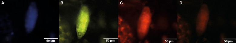 Fig. 1
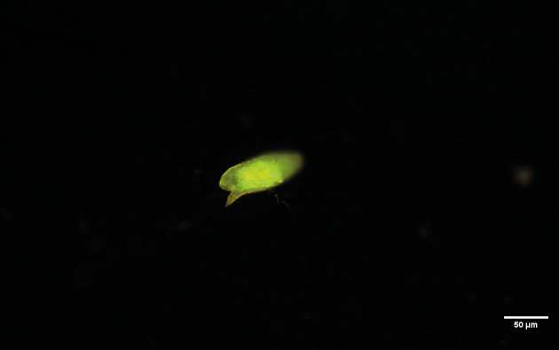 Fig. 2
| Reference | Egg sources | Main fluorescent structure | ||
| Mice tissue |
| Human tissue | ||
| Domingo 1968 | rectum / liver | rectum | eggshell | |
| Edwards 2015 | inside the worm | eggshell / miracidia (?) | ||
| Wang 2018 | released by worms | eggshell | ||
| Knhur 2018 | gut / liver | eggshell | ||
| Peterkova 2024 | liver | eggshell | ||
- —FAPES-DECIT
- —CNPq
Peer Reviews
No public reviews on file for this paper yet. If you reviewed it on a platform where reviews are public (OpenReview, ICLR, NeurIPS, ICML), you can paste yours below so the community can read it here.
Videos
No videos yet. Explain this paper in a talk, walkthrough, or lecture? Add one.
Taxonomy
TopicsParasites and Host Interactions · Parasite Biology and Host Interactions · Cancer Research and Treatment
Although the sensitivity limitations of egg detection–based methods for the diagnosis of parasitic infections are well known, the Kato-Katz fecal thick smear is still used widely in many endemic areas for the diagnosis of schistosomiasis. Due to eggs’ peculiar size and morphology,1 their identification in fecal samples enables highly specific classification, what is valuable for the establishment of reliable reference data for the assessment of other diagnostic tools’ accuracy. The Helmintex (HTX) method involves the use of magnetism to isolate Schistosoma mansoni eggs from feces; it has 100% sensitivity for egg burdens > 1.3 eggs per gram.2 Despite its accuracy, this method is labor intensive, and several improvements are currently underway. One promising possibility is a detection step using a flow system coupled with morphometric and fluorescent detectors. We thus investigated the autofluorescence of S. mansoni eggs to aid their identification in ongoing flow system detector development.
MATERIALS AND METHODS
Schistosoma mansoni eggs were obtained from (i) experimentally infected BALB/c mouse livers (ML) (CEUA-UFMG ethical approval 41/2024) and (ii) sediment pools from naturally infected human feces (HF) from a biorepository under proper ethical clearance (CEP-CCS-UFES CAAE 71027323.7.0000.5060). Eggs isolated from ML after seven-eight weeks post infection were kept in 10% formalin solution,3 and those isolated from HF were fixed in 50% alcohol and stored at 10ºC.
Eggs from MLs (n = 20) were inoculated into 30 µL uninfected HF sediment produced by the HTX method.4 Briefly, the HTX steps were sequential passage through 500-, 150- and 45-µm sieves and centrifugation with ethyl-acetate in water. The sediment with eggs was spread onto microscope slides and left to dry at room temperature. The same procedure was performed with three sets containing 12 to 14 HF eggs. The slides were examined under a fluorescence microscope (Eclipse 80i, LED light source; Nikon Corporation, Tokyo, Japan) with the filter sets indicated in Fig. 1.
autofluorescence of Schistosoma mansoni eggs from human fecal samples under microscope filters: (A) UV-2E (330-380 nm), Dichroic Mirror (DM) 400 nm, emission: > 420 longpass (LP), Blue; (B) B-2A (FITC, 420-495 nm), DM 505 nm, emission: > 515 LP, Green/Yellow; (C) G-2A(TRITC, 510-560 nm), 575 nm, emission: > 590 LP, Orange/Red; and (D) Y-2E/C (540-580 nm), 595 nm, emission: > 630 LP, Red.
RESULTS
All eggs from MLs and HF exhibited visible autofluorescence under the B-2A filter, with the miracidium having the most intense signal and the eggshell having a weaker signal (Fig. 2). Less-intense fluorescence was observed with the other filters (Fig. 1). These findings suggest that S. mansoni egg autofluorescence originates primarily from functional molecules in the miracidium, rather than from structural components such as the eggshell.
Schistosoma mansoni egg obtained from the liver of an experimentally infected mouse under B-2A (FITC) filter (420-495 nm, blue; emission: > 515 nm longpass) showing miracidium as the source of a more intense autofluorescence.
DISCUSSION
Autofluorescence is a natural optical property observed in the tissues and cellular organelles of organisms in various taxonomic groups.5 6 The autofluorescence of S. mansoni eggs in murine and human tissues (e.g., liver and gut) and eggshells released by or located in the reproductive organs of in vitro-cultivated worms have been reported7 , 8 , 9 , 10 , 11 (Table). In all these reports, the eggshell is identified as the source of fluorescence, although compelling evidence is lacking and some images do not clearly show fluorescent structures. In addition, these studies did not focus on the examination of parasite structure autofluorescence, which was reported as an undesired side effect in some cases.12 13
Sites of structural protein cross-linkage have been considered to be the main sites of autofluorescence in trematode eggshells.14 We observed prominent autofluorescence signals from S. mansoni egg miracidia, suggesting that metabolically active or intact miracidia are the main contributors (Fig. 2). Contrary to our findings, most reports describe autofluorescence only in S. mansoni eggshells, which may be explained by the performance of procedures with whole worms or tissues that remove miracidia contents or prevent their autofluorescence. The similarity of our findings for formaldehyde-fixed eggs from MLs and alcohol-fixed eggs from HF does not support the potential for the degradation or removal of the molecular source of autofluorescence by fixatives or reduced pH.15
A distinctive noise-to-signal ratio is important when using autofluorescence to detect infectious agents, as the present results demonstrate.13 The distinct fluorescence signature of the miracidium, especially under the B-2A filter, is a reliable marker that stands out among fecal debris. Such markers are essential for the development of automated detection systems, but they may also improve detection by conventional microscopy. Autofluorescence is not commonly used to detect parasites amid very diverse fecal debris. Sakurai and collaborators16 submitted a patent for a procedure involving the use of autofluorescence to detect Schistosoma japonicum eggs.
In conclusion, we report that S. mansoni eggs autofluorescence in fecal debris, enabling their relatively clear distinction from the background. We provide compelling evidence that this autofluorescence originates mainly from miracidia, and not eggshells, contrasting with most reports in the literature. These findings support the hypothesis that functional, rather than structural, molecules are the primary source of autofluorescence in mature S. mansoni eggs.
The reference list from the paper itself. Each links out to its DOI / PubMed record.
- 1de Souza RP Favero V Pascoal VF Lindholz C Bittencourt HR Graeff-Teixeira C Criteria for identification of Schistosoma mansoni eggs in faecal sediments prepared with the Helmintex method and stained by ninhydrin Mem Inst Oswaldo Cruz 2019114 e 18052910.1590/0074-0276018052931166420 PMC 6543901 · doi ↗ · pubmed ↗
- 2Teixeira CF Neuhauss E Ben R Romanzini J Graeff-Teixeira C Detection of Schistosoma mansoni eggs in feces through their interaction with paramagnetic beads in a magnetic field P Lo S Negl Trop Dis 200712 e 7310.1371/journal.pntd.00007318060086 PMC 2100366 · doi ↗ · pubmed ↗
- 3Dunne DW Lucas S Bickle Q Pearson S Madgwick L Bain J Identification and partial purification of an antigen (ω1) from Schistosoma mansoni eggs which is putatively hepatotoxic in T-cell deprived mice Trans R Soc Trop Med Hyg 1981751547110.1016/0035-9203(81)90013-46973849 · doi ↗ · pubmed ↗
- 4Favero V Candido RF Verissimo CM Jones MK St. Pierre T Lindholz C Optimization of the Helmintex method for schistosomiasis diagnosis Exp Parasitol 201717710.1016/j.exppara.2017.04.00128431921 · doi ↗ · pubmed ↗
- 5Daugschies A Bialek R Joachim A Mundt HC Autofluorescence microscopy for the detection of nematode eggs and protozoa, in particular Isospora suis, in swine faeces Parasitol Res 200187409121140338510.1007/s 004360100378 · doi ↗ · pubmed ↗
- 6Croce AC Light and autofluorescence, multitasking features in living organisms Photochem 2021126712410.3390/photochem 1020007 · doi ↗
- 7Domingo M Mais RF Weiskopf R Fink S Detection of schistosome ova by dark field fluorescence microscopy Gastroenterol 19815488465652522 · pubmed ↗
- 8Edwards J Brown M Peak E Bartholomew B Nash RJ Hoffmann KF The Diterpenoid 7-Keto-Sempervirol, derived from Lycium chinense, displays anthelmintic activity against both Schistosoma mansoni and Fasciola hepatica P Lo S Negl Trop Dis 201593 e 000360410.1371/journal.pntd.000360425768432 PMC 4358835 · doi ↗ · pubmed ↗