Ultraviolet-induced fluorescence dermoscopy in the diagnosis of scabies: The “ball sign”
Varun H, Adarshlata Singh, Bhushan Madke, Ambika Kondalkar, Bhakti Sarda

Abstract
Genes, proteins, chemicals, diseases, species, mutations and cell lines named across the full text — each resolved to its canonical identifier and authoritative record.
Click any figure to enlarge with its caption.
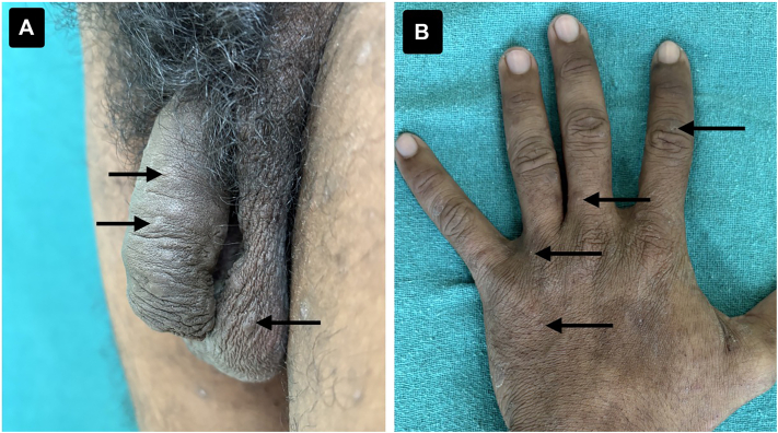 Figure 1
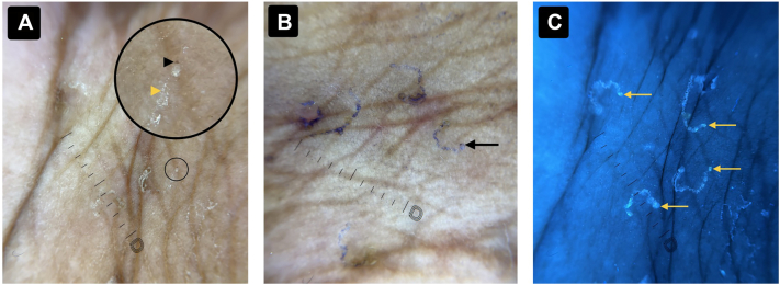 Figure 2
Peer Reviews
No public reviews on file for this paper yet. If you reviewed it on a platform where reviews are public (OpenReview, ICLR, NeurIPS, ICML), you can paste yours below so the community can read it here.
Videos
No videos yet. Explain this paper in a talk, walkthrough, or lecture? Add one.
Taxonomy
TopicsDermatological diseases and infestations · Nail Diseases and Treatments · Acne and Rosacea Treatments and Effects
Case description
A 35-year-old male agricultural worker presented with a 2-week history of generalized pruritus with marked nocturnal exacerbation. His wife and adolescent son reported similar symptoms. Pruritus predominantly involved the interdigital spaces, axillae, and groin. There was no history of fever, scaling, discharge, or bleeding.
Cutaneous examination revealed multiple ill-defined papules with healed excoriations. Burrows were noted over the interdigital spaces and genital skin (Fig 1).Fig 1A, Clinical image showing multiple well-defined papules and burrows over the penile and scrotal skin (black arrows). B, Clinical image of the left dorsum of the hand demonstrating multiple ill-defined papules over the interdigital spaces (black arrows).
Dermoscopy of interdigital papules demonstrated ill-defined S- and C-shaped gray-white curvilinear tracts. The classic ‘delta-wing jet’ sign was poorly visualized. Dermoscopic burrow ink test (D-BIT)enhanced visualization of the burrows and revealed a translucent mite at the terminal end.
Ultraviolet-induced fluorescence dermoscopy (UVFD), performed prior to D-BIT, demonstrated bright blue fluorescence of the burrows and a round blue-green fluorescent structure at the burrow tip, consistent with a live scabies mite—termed the “ball sign” (Fig 2). To validate this finding, a skin scraping was performed from the fluorescent area, confirming the presence of live, motile Sarcoptes scabiei mites and eggs. These findings confirmed the diagnosis of scabies.Fig 2A, Dermoscopic image (DermLite DL5 coupled with iPhone 16 camera) showing multiple ill-defined scabietic burrows with the classical ‘delta-wing jet with contrails’ appearance. The black arrowhead indicates the ‘delta-wing jet’ or ‘triangle sign’ (mite head), while the yellow arrowhead marks the ‘contrail’ (burrow behind the mite). The larger circle highlights the magnified view of the smaller circled area. B, D-BIT image of the same region showing multiple burrows filled with ink, with a scabies mite visible within the burrow (black arrow). C, UVFD image (DermLite Wood-Mode, 365 nm, coupled with iPhone 16 camera) demonstrating well-defined fluorescing scabietic burrows with the ‘ball sign’ (scabies mite) in each burrow (yellow arrows). D-BIT, Dermoscopic burrow ink test; UVFD, Ultraviolet-induced fluorescence dermoscopy.
The patient was treated with oral ivermectin (200 μg/kg) administered on an empty stomach, repeated after 1 week. Topical 5% permethrin cream was applied overnight from the neck down following a scrub bath, nail trimming, and subungual cleansing, and repeated after 1 week. Topical 2% fusidic acid was prescribed for excoriations, and oral antihistamines were given for symptomatic relief. All household contacts were treated simultaneously, with concurrent environmental decontamination of potentially infested fomites, including clothing, blankets, and bed linens.
Question: Which dermoscopic finding under ultraviolet-induced fluorescence dermoscopy is most suggestive of a live scabies mite?
- **A.**Delta-wing jet sign
- **B.**Triangle sign
- **C.**Serpiginous curvilinear tract
- **D.**Ball sign
- **E.**Ink-filled burrow
Discussion
Correct answer: D. Ball sign.
Scabies is caused by Sarcoptes scabiei var. hominis and is traditionally diagnosed based on clinical features such as nocturnal pruritus, involvement of the circle of Hebra, and identification of burrows or mites. Dermoscopy improves diagnostic accuracy by revealing characteristic burrows and mite-related signs, including the triangle sign and delta-wing jet sign. However, these features may be subtle or difficult to visualize, particularly in excoriated or inflamed skin.1^,^2
UVFD uses 365-nm ultraviolet light to enhance the visualization of skin structures based on fluorescence. In scabies, keratinous debris lining the burrow fluoresces bright blue due to interaction with UV light. Importantly, the body of a live mite emits a distinct round blue-green fluorescence, referred to as the “ball sign.” This sign provides a rapid and reliable method for identifying active infestations and may be easier to detect than conventional dermoscopic signs or ink-based techniques.3, 4, 5
Compared with the burrow ink test, UVFD is faster, nonmessy, and avoids excessive staining that can obscure burrow morphology. Incorporating UVFD into routine dermoscopic evaluation can improve diagnostic confidence, particularly in atypical or early presentations of scabies.3, 4, 5
Conflict of interest
None disclosed.
The reference list from the paper itself. Each links out to its DOI / PubMed record.
- 1Leung V.Miller M.Detection of scabies: a systematic review of diagnostic methods Can J Infect Dis Med Microbiol 22420111431462320502610.1155/2011/698494 PMC 3222761 · doi ↗ · pubmed ↗
- 2Fox G.Diagnosis of scabies by dermoscopy Case Rep 20092009 bcr 062008027910.1136/bcr.06.2008.0279 PMC 302954321686876 · doi ↗ · pubmed ↗
- 3Yürekli A.Musluİ.PektaşS.D.AlataşE.T.Aydoğdu C.T.Daşgin D.Using ultraviolet dermoscopy in diagnosing scabies Exp Dermatol 32112023199619993770226810.1111/exd.14930 · doi ↗ · pubmed ↗
- 4Pietkiewicz P.Navarette-Dechent C.Scabies mite is bright green under UV dermatoscopy Dermatol Pract Concept 1322023 e 202313510.5826/dpc.1302 a 135PMC 1018815136795853 · doi ↗ · pubmed ↗
- 5Kaliyadan F.Rafi S.Suresh S.The “ball sign” in UV dermoscopy of scabies Indian J Dermatol Venereol Leprol 91S 22025 S 18210.25259/IJDVL_999_202439635788 · doi ↗ · pubmed ↗