Anatomopathological Characterization of the Main Ocular Lesions in Green Turtles ( Chelonia mydas ) Along the Northern Coast of Bahia, Brazil
Danielle Nascimento Silva, José Luís Catão‐Dias, Wendell Marcelo de Souza Perinotto, Matheus Vilardo Loés Moreira, Nayone Lantyer‐Araujo, Pedro Enrique Navas Suárez, Gustavo Rodamilans Macedo, Thaís Pires, Arianne Pontes Oriá, Alessandra Estrela‐Lima

TL;DR
This study identifies common eye diseases in Green sea turtles along Brazil's northern coast, emphasizing their impact on conservation.
Contribution
The study reports the prevalence of ophthalmic lesions, particularly fibropapillomatosis and spirorchidiasis, in Green sea turtles.
Findings
Fibropapillomatosis was the most frequent ophthalmic lesion (58.9%) in Green sea turtles.
Spirorchidiasis was found in 46.8% of the evaluated samples.
Other notable lesions included mucopurulent conjunctivitis and corneal perforation.
Abstract
This study aimed to identify and report ophthalmic and adnexal diseases found in specimens of the Green sea turtle ( Chelonia mydas ). Thirty‐nine animals stranded on the beaches of the north coast of Bahia, Brazil were submitted to necropsy. A total of 158 samples of the visual system (eyelids, eyes, and salt glands) from females (71.8%; 28/39) and males (28.2%; 11/39) were analyzed. Samples without macro and microscopic changes counted as 30.4% (48/158) of the evaluated samples. Approximately 69.6% (110/158) had ophthalmic lesions; 92 were bilateral (24 eyelids, 44 eyes, and 24 salt glands), and 18 were unilateral. The anatomopathological evaluation of the specimens revealed predominantly neoplastic and inflammatory lesions, with fibropapillomatosis (FP) being the most frequent finding (58.9%; 93/158), followed by spirorchidiasis (46.8%; 74/158). Other ophthalmic lesions included…
Genes, proteins, chemicals, diseases, species, mutations and cell lines named across the full text — each resolved to its canonical identifier and authoritative record.
Click any figure to enlarge with its caption.
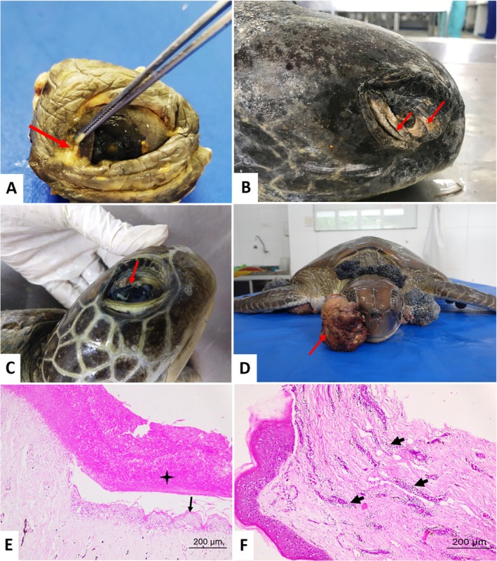 FIGURE 1
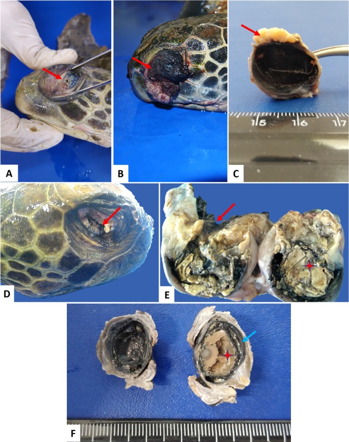 FIGURE 2
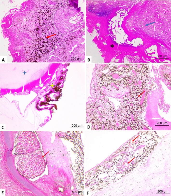 FIGURE 3
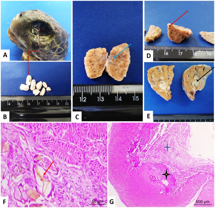 FIGURE 4
| Anatomical structures | Region | Morphological diagnosis | Total | (%) |
|
|---|---|---|---|---|---|
| Eyelids | Skin | Fibropapillomatosis | 15 | 46.9 | 9 |
| Scar tissue | 8 | 25 | 4 | ||
| Mucocutaneous junction | Fibropapillomatosis | 9 | 28.1 | 5 | |
| Conjunctiva | Fibropapillomatosis | 8 | 25 | 4 | |
| Mucopurulent conjunctivitis | 12 | 37.5 | 6 | ||
| Parasitic conjunctivitis | 8 | 25 | 4 | ||
| Eyes | Cornea | Fibropapillomatosis | 20 | 32.2 | 12 |
| Keratitis | 8 | 13 | 4 | ||
| Ulcerative keratitis | 2 | 3.2 | 2 | ||
| Limbus | Fibropapillomatosis | 3 | 4.8 | 2 | |
| Conjunctiva | Fibropapillomatosis | 38 | 61.3 | 23 | |
| Conjunctivitis | 12 | 19.3 | 6 | ||
| Sclera | Parasitic scleritis | 7 | 11.3 | 5 | |
| Uvea | Parasitic choroiditis | 16 | 25.8 | 10 | |
| Previous synechia | 3 | 4.8 | 3 | ||
| Closure of the iridocorneal angle (secondary glaucoma) | 4 | 6.4 | 4 | ||
| All structures | Panophthalmitis | 6 | 9.7 | 6 | |
| Phthisis bulbi | 12 | 19.3 | 12 | ||
| Salt gland | Centrilobular | Parasitic adenitis | 28 | 43.7 | 17 |
| Periglandular | Parasitic periadentitis | 12 | 18.7 | 7 | |
| Glandular duct | Lithiasis | 4 | 6.2 | 3 |
| Total | Number of animals | |
|---|---|---|
| Total study samples | 158 | 39 |
| Without changes | 48 | 20 |
| Fibropapillomatosis | 93 | 24 |
| Spirorchidiasis | 74 | 31 |
| Phthisis bulbi | 12 | 12 |
| Perforated eye | 4 | 4 |
| Lithiasis | 4 | 3 |
- —Fundação de Amparo à Pesquisa do Estado da Bahia10.13039/501100006181
- —Conselho Nacional de Desenvolvimento Científico e Tecnológico10.13039/501100003593
Peer Reviews
No public reviews on file for this paper yet. If you reviewed it on a platform where reviews are public (OpenReview, ICLR, NeurIPS, ICML), you can paste yours below so the community can read it here.
Videos
No videos yet. Explain this paper in a talk, walkthrough, or lecture? Add one.
Taxonomy
TopicsTurtle Biology and Conservation · Venomous Animal Envenomation and Studies · Traumatic Ocular and Foreign Body Injuries
Introduction
1
Maintaining good vision is paramount for sea turtles as they rely on it for migration and food intake. Most of the ophthalmic abnormalities described in sea turtles are located in the primary and accessory lacrimal glands, eyelids, and eyes, which are fundamental structures for the nutrition and normal behavior of these animals [1, 2, 3, 4, 5].
An understanding of the visual system pathology is essential for proper diagnosis and treatment as well as the rapid establishment of health in these animals [6, 7]. Changes in the eyes and adnexa can be caused by developmental disorders, infectious agents (viral, bacterial, fungal, and parasitological), neoplastic processes, nutritional imbalances, traumatic injuries, or even injuries that accompany systemic diseases [2, 3, 4, 5].
Few reports of eye disorders or injuries in sea turtles describe catarrhal and purulent conjunctivitis, keratitis, blepharitis, corneal perforation, and chemosis in Loggerhead sea turtle ( Caretta caretta ) [5]; eyelid necrosis, ulcerative keratitis, anterior uveitis, and hyphema in Green turtle ( Chelonia mydas ), Kemp's ridley sea turtle ( Lepidochelys kempii ), and Caretta caretta caused by low environmental temperatures [8].
The literature of free‐ranging sea turtles also describes a few reports of salt gland diseases, characterizing stone formation in severely dehydrated turtles [9]. Granulomatous adenitis by spirorchid fluke, of varying severity, is also common in the stroma surrounding the central canals of the lobules [9] and heterophilic adenitis associated with systemic lesions, or described as the sole lesion responsible for stranding sea turtles, is associated with infectious agents such as Vibrio spp., Citrobacter spp., Pseudomonas spp., Aeromonas hydrophila , Staphylococcus sp., and Aerococcus viridans [1, 3, 10, 11, 12, 13, 14].
In this context, the objective of this study was to identify and report ocular abnormalities observed in 39 turtles of the C. mydas species stranded on the beaches of the north coast of Bahia, Brazil, submitted to anatomopathological examination.
Materials and Methods
2
Ethical Considerations
2.1
The research protocols were approved by the Animal Welfare and Ethics Committee of the School of Veterinary Medicine and Animal Science of the Federal University of Bahia (protocol n°90/2018), in accordance with the Ministry's Biodiversity Information Authorization System of the Environment of Brazil—SISBIO (processes n°64518–1 and n°64518–3) and certified by the National System for the Management of Genetic Heritage and Associated Traditional Knowledge—SISGEN (registration n° A0F349F). In addition, the study was conducted according to the bioethics guidelines stated by the Association for Research in Vision and Ophthalmology (ARVO).
Animals and Samples
2.2
From February 2018 to September 2019, the eyes and adnexa (upper and lower eyelids and salt gland) of 39 specimens of C. mydas (38 juvenile animals and one adult animal) were collected. These were free‐living animals that washed ashore on the beaches of the northern coast of Bahia state, in the area monitored by the Fundação Projeto Tamar (Praia do Forte, latitude: −12.574743 and longitude: −38.0044715), found dead, or that died during treatment and rehabilitation at the Fundação Projeto Tamar.
Histopathological Processing and Analysis
2.3
After cleavage of the structures (the eyes were sectioned dorsoventrally, including the optic nerve, and the eyelids and salt glands were sectioned transversely), the fragments were placed in histological cassettes in a container with 10% phosphate‐buffered formalin solution for not less than 24 h. Tissues, after adequate fixation, were placed in cassettes for processing using a routine histological paraffin‐embedding technique. Hematoxylin and eosin (HE) staining was performed on 4‐μm sections [15].
Injuries were classified into six main categories: normal or without injury; fibropapillomatosis; spirorchidiasis; phthisis bulbi; perforated eye; and lithiasis. The animals were grouped into several categories based on the observed lesions.
The histopathological analysis was performed using a Leica optical microscope with an image capture system, using 4×, 10×, 20×, and 40× objectives. All captured photomicrographs were standardized in ImageJ software.
Results
3
Thirty‐nine specimens of C. mydas were evaluated; 71.8% (28/39) were female, and 28.2% (11/39) were male and a total of 158 samples from the eye and adnexa were analyzed for the present study, comprising 32 eyelid, 62 globes, and 64 Harderian gland samples.
Samples with no signs of abnormalities totaled 30.4% (48/158), comprising 30 bilateral (4 eyelids, 2 globes, and 24 Harderian glands) and 18 unilateral (2 eyelids, 8 globes, and 8 Harderian glands) cases. Ophthalmic lesions were observed in 69.6% (110/158), 92 bilateral (24 eyelids, 44 eyes, and 24 salt glands) and 18 unilateral cases.
All animals with fibropapillomas in the visual apparatus also had fibropapillomas in other parts of the body, such as fins, neck, plastron, and carapace. The macroscopic characteristics of these neoplasms ranged from nodules to sessile, pedunculated masses, sometimes with a verrucous appearance, and in some cases with the involvement of adjacent structures, such as the conjunctiva and eyelids.
Microscopic analysis of the neoplastic lesions showed proliferation of epithelial cells with papillomatous formation, and scarce fibrovascular or fibropapillomatous stroma, abundant fibrovascular stroma, with marked cellular hyperplasia, hyperkeratosis, acanthosis and, in some cases, intranuclear inclusion bodies that were observed in some epithelial cells, hydropic degeneration of the epidermis (external surface) and conjunctiva. In these lesions, there was a predominance of intratumoral spirorchid eggs, associated with predominantly lymphoplasmacytic inflammatory infiltrate in the stroma.
The most frequent lesion on the eyelids was fibropapilloma (20.2%; 32/158), followed by caseous discharge (7.6%; 12/158) and yellowish plaques or scar tissue on the skin and mucocutaneous junction (5%; 8/158).
The exudative lesions revealed parasitic infection by spirorchid eggs at microscopy, with perivasculitis, hyperemia, migration of melanomacrophages, lymphocytes, eosinophils, and heterophils. In the scar tissue, acanthosis, areas of fibrosis, and mixed inflammation were observed. In the bulbar conjunctiva, fibropapillomatosis was also the predominant lesion (24%; 38/158), followed by cases of conjunctivitis (7.6%; 12/158). The changes observed in the eyelids and bulbar conjunctiva are illustrated in Figure 1.
Lesions on the eyelids of Chelonia mydas . (A) Blepharitis. Note the caseous discharge. (arrow). (B) Scar tissue in the eyelid and conjunctiva of an adult C. mydas (arrows). (C, D) Fibropapillomas on eyelids. (C) Fibropapilloma in the upper eyelid (arrow). (D) Frontal view of the neoplasm with an irregular surface, exophytic growth, ulcerated (arrow). (E) Necrotic tissue (star) and hyperplasia of the conjunctival epithelium (arrow). (F) Photomicrograph of the eyelid of Chelonia mydas . Viral conjunctivitis. Perivasculitis. Moderate perivascular inflammatory infiltrate in the conjunctival stroma (arrows) and hyperplasia and hyperkeratosis of the epithelium (arrows). HE–Obj. 10×, 40×.
The evaluation of eye samples identified fibropapillomatous lesions (14.5%; 23/158) located in the cornea and limbus. Phthisis bulbi (7.6%; 12/158), panophthalmitis (3.8%; 6/158), caseous secretion and scar tissue in the bulbar conjunctiva and sclera (both 2.5%; 4/158), and a perforated eye (2.5%; 4/158).
In the histopathology of eyes with fibropapillomas, neoplastic proliferation with migration of melanocytes in the region was observed. Intratumoral parasite eggs, keratitis, and parasitic uveitis were observed in 6.9% (11/158) of the evaluated samples. Anterior synechiae and secondary glaucoma were also identified as lesions.
Parasitic infection was also observed in samples from eyes without fibropapillomatosis, characterized by a parasitic granulomatous reaction due to the migration of spirorchid eggs into the cornea, scleral cartilage, and uvea (parasitic keratitis, scleritis, and uveitis). An adult parasite compatible with a spirorchid was present in the space between the uvea and sclera. Secondary glaucoma was one of the lesions identified, as were anterior synechiae (Figures 2 and 3).
Morphological findings of ocular diseases in Chelonia mydas . (A) Anterior synechia and perforated eye (arrow). (B, C) Ocular fibropapilloma (arrow). (C) Longitudinal section of an eye with fibropapilloma attached to the cornea. (D, E) Necrotizing panophthalmitis. Perforated cornea (arrows), intraocular necrotic material (star). (F) Section in the sagittal plane of the eye with phthisis bulbi: Observe extensive caseous material in the anterior and posterior chambers with thickening of the uvea.
Photomicrographs of ophthalmic lesions in the eyes of Chelonia mydas . (A) Ocular fibropapillomatosis. Cornea—intratumoral melanocyte migration (arrow). (B) Necrotizing panophthalmitis. Necrotic tissue (asterisks) adhered to fibrovascular tissue (arrows). (C) Lens (blue star) and iris (yellow star) showing posterior synechia (arrow). (D) Migration of melanomacrophages (arrows) to the uveal tract. Ocular spirorchidiasis—Parasitic granulomas. (E) Sclera—granuloma with numerous spirorchid eggs (arrow). (F) Choroid parasitic granulomas, spirorchid eggs (arrow). HE staining–Obj. 4×, 10×.
In cases of phthisis bulbi, microscopy showed atrophy and disorganization of ocular structures, hyperplasia of the bulbar conjunctiva, keratitis, and hyperkeratosis. Eyes with panophthalmitis presented corneal ulceration, scleral hemorrhage, anterior and posterior synechia, hyperemia of the choroidal vessels; some cases had closure of the iridocorneal angle, characterizing a secondary glaucoma; and migration of inflammatory cells, melanomacrophages, melanocytes, lymphocytes, and heterophils (Figures 2 and 3).
Macroscopically, the salt glands showed on the cut surface, parasitic lesions characterized by multifocal blackened areas (19.6%; 31/158), purulent exudate, and calculi (both with 2.5%; 4/158). A juvenile female had dozens of white calculi of varying sizes located in the orbital cavity with an appearance similar to salt. Multifocal blackened areas were observed upon examination of this animal's Harderian gland on the surface and at the cut area, and millimetric calculi were possibly obstructing the glandular duct. This C. mydas had poor body condition, cachexia, hydroceloma, hydropericardium, and parasitic infection by spirorchids and specimens of Neoctangium sp. identified in the parasitological examination.
The lesions located at microscopy in the centrilobular and periglandular region were configured as parasitic adenitis and/or periadenitis (spirorchid eggs). This condition was also observed in glands without macroscopic alteration (6.3%; 10/158). The inflammatory process was mixed, consisting of melanomacrophages, lymphocytes, heterophils, plasmocytes, and fibrinocaseous exudate. Two cases (1.3%) with blackened multifocal areas did not have spirorchids, characterized by a mild inflammatory infiltrate of heterophils associated with melanomacrophages. In the glands with calculi, there were areas of multifocal mineralization (Figure 4).
Lesions observed in the visual apparatus of Chelonia mydas . (A, B) Lithiasis. Several salt calculi in the orbital cavity (double red arrow). (C–E) Salt gland cut surface. (C) Intraparenchymal stone (blue arrow). (D) Multifocal blackened areas (red arrow). (E) Purulent exudate (black arrow). Parasitic adenitis. (F) Parasitic granuloma in the centrilobular region showing the spirorchid eggs (arrow). Fibrinosuppurative adenitis. (G) Centrilobular region with large amounts of inflammatory exudate (blue star) and areas of fibrosis (yellow star). HE staining–Obj. 4× and 40×.
The spirorchid eggs observed in all parasitic lesions in the eyes and adnexa were predominantly type 1; in some cases, they were also associated with type 2 and 3 eggs.
Table 1 presents in detail the alterations according to the anatomical structures and the number of affected animals. A summary of the main changes is described in Table 2.
TABLE 1: Ophthalmopathies in Chelonia mydas stranded on the north coast of Bahia.
TABLE 2: Summary of changes in the visual apparatus of Chelonia mydas .
Discussion
4
Studies exploring and describing ocular diseases in sea turtles are infrequent. In this context, to our knowledge, the study presented here is the first to characterize ocular diseases in green turtles in Brazil and the second to correlate spirorchidiasis with ophthalmic lesions.
The results of this study show fibropapillomatosis as the main lesion observed in the visual apparatus of C. mydas . It is a debilitating disease of a multifactorial nature, with the main etiological agent being Chelonid Alphaherpesvirus 5 (ChHV5), characterized by the formation of single or multiple tumors, with a warty, smooth, or rough appearance, pigmented or not, in various regions of the body, especially in soft tissues [16, 17]. The presence of these tumors in this anatomical region was described in previous studies developed in other Brazilian states [18, 19].
Therefore, the high frequency of these neoformations in animals found in the northeast of Brazil is probably associated with several predisposing factors, such as viral and parasitic infections and exposure to pollutants in the marine environment, possibilities also considered in other regions of the country [19, 20], It is noteworthy that ChHV5 viral particles can be detected in blood, plasma, urine, cloacal, oral and ocular secretions [21, 22, 23]. In the region where the study was conducted, a case of viral infection was reported in a loggerhead turtle with bilateral mucoid secretion, chemosis, conjunctival hyperemia, and bilateral eyeball retraction [24]. The macro and microscopic characteristics observed in fibropapillomas corroborate the data described in the literature [9, 17, 25, 26]. All turtles affected by fibropapillomatosis were juveniles, confirming the study developed by Jones et al. [27].
Animals with ocular fibropapillomatosis may have reduced or total loss of vision, compromising their health conditions, making them vulnerable to the action of predators, and reducing their ability to feed [9, 28]. Despite the lack of data on the clinical history of these animals, particularly those with ophthalmic abnormalities, it is believed that there is a strong correlation between visual loss and the systemic condition. This is largely due to the presence of large neoplasms. A study on ophthalmic lesions in turtles stranded due to the cold in North Carolina, United States of America, revealed ocular and periocular lesions, such as bilateral superficial ulcerative keratitis, perforating lesions, one case of phthisis bulbi, proliferative tissue in adnexa, and synechiae [8]. Interestingly, similar lesions were observed in the present study; however, with a different etiology, associated with parasitic and neoplastic processes and consequently nutritional disorders, such as anorexia and malnutrition.
Other important ophthalmic lesions are represented by inflammatory processes caused by various pathogenic agents such as bacteria, viruses, parasites, and protozoa. Isler et al. [5] evaluated loggerhead turtles for diseases and diagnosed five ocular lesions: catarrhal conjunctivitis, caseous conjunctivitis, keratitis, blepharitis, and corneal perforation. All the mentioned lesions were observed in the present study with C. mydas.
In addition to the mixed inflammatory processes, trematode eggs were observed in different structures of the visual apparatus, such as the palpebral conjunctiva, cornea, sclera, and choroid. Recently Jerdy et al. [29] reported the presence of spirorchid eggs in the choroid and optic nerve in sea turtles of the species C. mydas , C. caretta , and L. olivacea , and concluded that animals with ocular lesions caused by spirorchids are approximately 300% more likely to be thin or cachectic than animals with this parasite, but without ocular damage. In C. mydas from the north coast of Bahia, 12.8% (5/39) had poor body condition or cachexia.
Regarding the location of the parasitic lesions, similar to other studies, the choroid was one of the most affected regions [29, 30]. However, other anatomical sites were also involved, such as the sclera and cornea, with a predominance of type 1 eggs. The presence of eggs in unreported sites emphasizes the ability of these eggs to migrate within the vascular system of these animals [30, 31, 32, 33, 34].
The observed cases of lithiasis, particularly in the one case that demonstrated dozens of calculi within the orbit and the salt gland, indicate a failure in the solute dissolution function, an alteration that can occur in severely dehydrated sea turtles [9, 35].
Salt glands are important as they assist in osmoregulation by excreting solutes through the tear ducts. Active transport via the sodium‐potassium pump draws salt from the blood into the glandular parenchyma. This process is necessary because reptilian kidneys are significantly less efficient in osmoregulation when compared to mammalian kidneys [3, 36, 37, 38]. These stones present as hard, rough deposits associated with necrosis of surrounding tissues [9].
According to Flint et al. [9] granulomatous adenitis due to spirorchid eggs, as observed in most of the salt glands of the animals in this study, presents variable severity, although these are lesions commonly observed in the stroma that surrounds the central channels of the lobules and that can cause obstruction if spread to other regions.
Conclusion
5
The anatomohistopathological evaluation of the specimens revealed predominantly neoplastic lesions, with fibropapillomatosis being the most frequent, followed by inflammatory lesions, caused especially by spirorchidiasis.
Studies related specifically to ophthalmic changes in C. mydas are infrequent, and it is necessary to develop research focused on this topic.
Authors' Contributions
Danielle Nascimento Silva: conceptualization, formal analysis, investigation, methodology, project administration, validation, writing – original draft. José Luís Catão‐Dias: investigation, resources, writing – original draft, writing – review and editing. Marcelo de Souza Perinotto: investigation, resources, writing – original draft, writing – review and editing. Matheus Vilardo Loés Moreira: formal analysis, validation, writing – review and editing. Nayone Lantyer‐Araujo: formal analysis, writing – review and editing. Pedro Enrique Navas Suárez: formal analysis, writing – review and editing. Gustavo Rodamilans Macedo – formal analysis, visualization, writing – review and editing. Thaís Pires: formal analysis, visualization, writing – review and editing. Arianne Pontes Oriá: investigation, resources, writing – original draft, writing – review and editing. Alessandra Estrela‐Lima: conceptualization, investigation, project administration, supervision, validation, writing – original draft, writing – review and editing.
Disclosure
The authors have not used AI to generate any part of the manuscript. Bioethics and Biosecurity Committee Approval: The research protocols were approved by the Animal Welfare and Ethics Committee of the School of Veterinary Medicine and Animal Science of the Federal University of Bahia (protocol n°90/2018), in accordance with the Ministry's Biodiversity Information Authorization System of the Environment of Brazil—SISBIO (process n°64518‐1 and n°64518‐3) and certified by the National System for the Management of Genetic Heritage and Associated Traditional Knowledge—SISGEN (registration n° A0F349F) (Annexes).
Conflicts of Interest
The authors declare no conflicts of interest.
Supporting information
Table S1: General sample data.
The reference list from the paper itself. Each links out to its DOI / PubMed record.
- 1J. S. Glazebrook and R. S. F. Campbell , “A Survey of the Diseases of Marine Turtles in Northern Australia I. Farmed Turtles,” Diseases of Aquatic Organisms 9 (1990): 83–95.
- 2T. R. Kelly , W. Walton , B. Nadelstein , and G. A. Lewbart , “Phaco Emulsification of Bilateral Cataracts in a Loggerhead Sea Turtle (Caretta caretta),” Veterinary Record 156 (2005): 774–777.15951501 10.1136/vr.156.24.774 · doi ↗ · pubmed ↗
- 3E. R. Jacobson , Infectious Diseases and Pathology of Reptiles Color Atlas and Text (CRC Press, 2007), 731.
- 4D. Reavill and R. E. Schmidt , “Proceedings Association of Reptilian and Amphibian Veterinarians Pathology of the Reptile Eye and Ocular Adnexa,” Zoo/Exotic Pathology Service, 2825 KOVR Drive, West Sacramento, CA 95605 USA, 2012.
- 5C. T. Işler , M. Altuğ , Z. Cantekin , Ş. Y. Ozsoy , Z. Yurtal , and M. Z. Y. Deveci , “Evaluation of the Eye Diseases Seen in Loggerhead Sea Turtle (Caretta caretta),” Revue de Médecine Vétérinaire 165 (2014): 258–262.
- 6L. Avens and K. J. Lohmann , “Use of Multiple Orientation Cues by Juvenile Loggerhead Sea Turtles Caretta caretta ,” Journal of Experimental Biology 206, no. Pt 23 (2003): 4317–4325.14581601 10.1242/jeb.00657 · doi ↗ · pubmed ↗
- 7C. J. Innis and L. A. Staggs , “Cold‐Stunning,” in Sea Turtle Health & Rehabilitation, ed. C. A. Manire , T. M. Norton , B. Stacy , C. J. Innis , and C. Harms (Journal Ross Publishing, 2017), 676–705.
- 8M. J. Lively , H. D. Westermeyer , C. A. Harms , and E. F. Christiansen , “Ophthalmic Lesions in a Population of Cold‐Stunned Sea Turtles ( Chelonia mydas , Lepidochelys kempii , Caretta caretta ),” Veterinary Ophthalmology 22 (2019): 910–915, 10.1111/vop.12672.30983145 · doi ↗ · pubmed ↗