Use of a Cytobrush for Sampling the Ear Canal of Dogs With Otitis Externa
Nicoly Radaeli Atanasio, Gabriela Reis Ledur, Danilo Marcondes Filho, Daniel Guimarães Gerardi

TL;DR
This study compares a cytobrush to a swab for collecting ear canal samples in dogs with otitis externa, finding both methods equally effective and safe.
Contribution
The study evaluates the cytobrush as a novel sampling tool for canine ear cytology, demonstrating its viability as an alternative to traditional swabs.
Findings
Cytobrush and swab methods showed no significant differences in cell and microorganism collection.
The cytobrush was found to be safe and well-tolerated by dogs.
High reproducibility was observed between two independent evaluators.
Abstract
Cytological examination of the ear canal is essential for evaluating dogs with otitis externa (OE). The conventional sampling method uses a swab. However, the cytobrush (gynaecological cervical brush), already used for cytological collection from other anatomical sites, has not been adequately investigated for this purpose in dogs with OE. To compare the cytobrush as a sampling tool for the ear canal of dogs with OE and compare it with the swab technique. Thirty ears from 17 dogs with OE, presented at a veterinary teaching hospital, were included for sampling. Cytological samples were collected using both a cytobrush and a swab in random order. Two independent and blinded evaluators quantified micro‐organisms (cocci, bacilli, yeasts), mononuclear cells, polymorphonuclear cells and epithelial cells. Animal discomfort during sampling was assessed using a scoring system. No significant…
Genes, proteins, chemicals, diseases, species, mutations and cell lines named across the full text — each resolved to its canonical identifier and authoritative record.
Click any figure to enlarge with its caption.
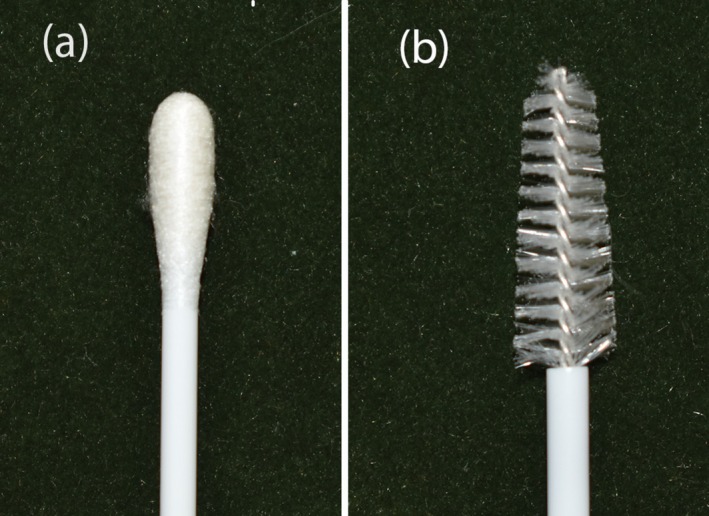 FIGURE 1
| Animal inclusion number | Ear inclusion number | Breed | Age (years) | Sex | Side affected |
|---|---|---|---|---|---|
| 1 | 1 & 2 | Dachshund | 2 | M | B |
| 2 | 3 & 4 | German Shepherd dog | 7 | F | B |
| 3 | 5 & 6 | Basset hound | 9 | M | B |
| 4 | 7 & 8 | Shih tzu | 8 | F | B |
| 5 | 9 | Shih tzu | 9 | F | U |
| 6 | 10 | Lhasa apso | 13 | F | U |
| 7 | 11 | Shih tzu | 10 | M | U |
| 8 | 12 & 13 | Golden retriever | 9 | F | B |
| 9 | 14 & 15 | Shih tzu | 8 | F | B |
| 10 | 16 & 17 | Shih tzu | 8 | F | B |
| 11 | 18 & 19 | Beagle | 10 | M | B |
| 12 | 20 & 21 | Mixed‐breed | 13 | F | B |
| 13 | 22 & 23 | Mixed‐breed | 4 | F | B |
| 14 | 24 & 25 | Pinscher | 10 | F | B |
| 15 | 26 | Mixed‐breed | 13 | M | U |
| 16 | 27 & 28 | Mixed‐breed | 3 | M | B |
| 17 | 29 & 30 | German spitz | 3 | F | B |
| Method | Median | Min. | Max. |
| |
|---|---|---|---|---|---|
| Cocci | S | 0.375 | 0.00 | 30.00 | 0.4455 |
| B | 0.250 | 0.00 | 29.40 | ||
| Bacilli | S | 4.5850 | 0.00 | 30.00 | ~1 |
| B | 4.693 | 0.00 | 30.00 | ||
|
| S | 11.650 | 0.00 | 29.90 | 0.1747 |
| B | 12.775 | 0.00 | 30.00 | ||
| Mononuclear cells | S | 0.02 | 0.00 | 0.60 | 0.3711 |
| B | 0.05167 | 0.00 | 0.85 | ||
| Polymorphonuclear cells | S | 0.0 | 0.00 | 0.60 | 0.1814 |
| B | 0.0 | 0.00 | 18.85 | ||
| Epithelial cells | S | 1.625 | 0.00 | 6.20 | 0.8693 |
| B | 1.750 | 0.00 | 6.40 |
| Method | ICC | IC 95% (ICC) | |
|---|---|---|---|
| Cocci | S | 0.99 | 0.980 < ICC < 0.995 |
| B | 0.982 | 0.963 < ICC < 0.991 | |
| Bacilli | S | 0.997 | 0.994 < ICC < 0.999 |
| B | 0.994 | 0.986 < ICC < 0.997 | |
|
| S | 0.997 | 0.987 < ICC < 0.999 |
| B | 0.997 | 0.981 < ICC < 0.999 | |
| Mononuclear cells | S | 1 | — |
| B | 0.527 | 0.214 < ICC < 0.742 | |
| Polymorphonuclear cells | S | 0.946 | 0.891 < ICC < 0.974 |
| B | 0.995 | 0.990 < ICC < 0.998 | |
| Epithelial cells | S | 0.93 | 0.859 < ICC < 0.966 |
| B | 0.978 | 0.954 < ICC < 0.989 |
| Collection method | Head movement | Bite attempt | Vocalisation | Oto‐podal reflex | Restlessness | Total score |
|
|---|---|---|---|---|---|---|---|
| Swab | 15 | 1 | 14 | 05 | 7 | 42 | 0.1096 |
| Brush | 15 | 1 | 14 | 10 | 7 | 47 |
Peer Reviews
No public reviews on file for this paper yet. If you reviewed it on a platform where reviews are public (OpenReview, ICLR, NeurIPS, ICML), you can paste yours below so the community can read it here.
Videos
No videos yet. Explain this paper in a talk, walkthrough, or lecture? Add one.
Taxonomy
TopicsVeterinary Oncology Research · Ear Surgery and Otitis Media · Human-Animal Interaction Studies
Introduction
1
Otitis externa (OE) is a highly prevalent condition in dogs, affecting approximately 15%–20% of the dogs presented to veterinary practice [1]. This inflammation of the external ear canal may have a wide range of primary and secondary causes, and is often associated with predisposing and perpetuating factors [2]. These elements frequently interact, resulting in clinical presentations that can vary from mild acute episodes to severe, chronic and recurrent cases [1, 3].
As a consequence of the complex aetiology of OE, its diagnosis and the identification of all contributing factors are challenging, and require precise and reliable methods to assess the severity of the disease and guide appropriate treatment [1, 4, 5]. Among the available diagnostic tools, cytological evaluation of the ear canal stands out as an accessible, rapid and indispensable technique for identifying microbial morphology (such as coccoid or rod‐shaped bacteria and yeasts), inflammatory and epithelial cells [4, 5, 6].
Cytological sampling is traditionally performed using cotton‐tipped swabs, a well‐established technique that is simple to execute and generally effective and inexpensive [1, 4, 5, 6]. However, alternative methods have been explored, including the use of urethral catheters to collect material from deeper regions of the ear canal [7]. Cytobrushes, commonly used in medical and veterinary contexts, have shown superior sampling capabilities, particularly in veterinary ophthalmology [8, 9] and in human gynaecological cytological investigation [10, 11]. These brushes are composed of nylon bristles that, when rotated or gently rubbed against epithelial surfaces, promote exfoliation and retention of cells. In dogs, brushes also have been used to obtain cytological samples from the respiratory tract [12, 13, 14, 15, 16], conjunctiva [9] and gastrointestinal tract [17]. Such evidence supports their potential as a sampling tool for the canine ear canal.
Despite this, no systematic studies have evaluated the comparative effectiveness of cytobrushes versus cotton swabs in the diagnosis of canine OE. The present study was therefore designed to prospectively and blindly assess whether the cytobrush is a viable and well‐tolerated alternative to the conventional swab for otic cytological investigation. Specifically, we aimed to compare microbial and cellular representativeness between techniques, including coccoid and rod‐shaped bacteria, yeasts, inflammatory infiltrates (mononuclear and polymorphonuclear cells) and epithelial cells, as well as to evaluate animal discomfort during sample collection. We hypothesised that the cytobrush would be a viable and equally or more effective method than the cotton swab for obtaining representative samples from the ear canal of dogs with OE, and that the cytobrush sampling procedure would be as well‐tolerated as the traditional swab.
Materials and Methods
2
Ethical Considerations
2.1
This study was conducted at Federal University of Rio Grande do Sul (UFRGS, Brazil), following approval of the study protocol by the Institutional Animal Care and Use Committee (IACUC) of UFRGS (approval no. 46456). Informed consent was obtained from all owners before inclusion through the signing of a written consent form.
Animals
2.2
This was a prospective, randomised, blinded study including dogs diagnosed with OE. All dogs were recruited from the clinical caseload of the Veterinary Clinical Hospital at UFRGS School of Veterinary Medicine.
Dogs were included if they presented with a clinical history and signs consistent with OE, such as head shaking, otic pruritus, pain, otorrhea, erythema of the concave pinna and external auditory meatus, hyperplasia and/or oedema. Each dog underwent a physical examination, otoscopic inspection and cytological evaluation of the external ear canal. In cases of bilateral otitis, each ear was assessed and included independently. Dogs were enrolled regardless of age, sex, reproductive status, breed or the primary cause of the otological condition. Dogs were excluded if, within the 4 weeks before inclusion or at the time of enrollment, they had received ear cleaners, ceruminolytic/ceruminosolvent products or topical or systemic medications for the treatment of OE.
Sample Collection Techniques and Slide Preparation
2.3
Cytological samples were collected from each ear using both a cotton‐tipped swab and a cytobrush. The order of sampling was randomised for each ear by a simple coin toss to ensure unbiased sequencing.
For the swab technique, a sterile cotton‐tipped applicator with a plastic shaft (15 mm length, model K41‐0201B, Olen; Figure 1a) was inserted into the lumen of the ear canal until the end of the vertical canal and rotated clockwise three times (360°). The swab tip was then rolled once along the longitudinal axis of a microscope slide to transfer the sample. The cytobrush technique followed the same guidelines yet used a sterile gynaecological cervical brush (ABSORVE) composed of a 180 mm plastic shaft with a 20 mm nylon bristle brush at the tip (Figure 1b).
Instruments used for cytological sampling of the external ear canal in dogs with otitis externa. (a) Cotton swab and (b) gynaecological cervical cytobrush.
All slides were stained using a Romanowsky‐type rapid stain (Panoptic; NEWPROV). Fixation was intentionally omitted to avoid removal of the lipid content. Therefore, slides were analysed and photographed immediately after preparation.
Cytological Evaluation
2.4
All slides were initially examined by a single investigator (N.R.A). Slides were first screened at low magnification (×40 or ×100) to identify areas with visible cellularity. One region was then selected for high‐power evaluation (×1000, oil immersion). Selection criteria included the presence of inflammatory cells, nuclear debris and micro‐organisms; in their absence, areas containing keratinocytes or generalised basophilic debris were chosen. Regions with excessive overlapping were excluded to minimise errors in structure identification [7, 18]. Each selected ×1000 oil immersion field was photographed using ZEN 2.3 software (Carl Zeiss). After assessing one region, scanning at low power was resumed in a continuous horizontal movement, from the groove to the opposite edge, until a total of 10 non‐overlapping fields had been captured [18].
The microphotographs were subsequently analysed independently by two blinded investigators (D.G.G and G.R.L). The number of yeasts, cocci, rods, polymorphonuclear and mononuclear cells, and epithelial cells was counted. The final score for each category was obtained by calculating the mean count across the 10 fields [7, 19]. A maximum of 30 micro‐organisms and 20 cells per field was counted, as established in previous studies [7, 18].
Assessment of Discomfort
2.5
In order to evaluate the discomfort shown by dogs during sampling, behavioural responses were recorded from the moment the swab or brush was introduced into the ear canal until its complete removal. A numeric scoring system was developed specifically for this study. Five behaviours indicative of discomfort—vocalisation, head shaking, attempt to bite the examiner, restlessness requiring physical restraint and the presence of an oto‐podal reflex—were scored individually, with one point assigned for each behaviour observed. Thus, the total discomfort score for each procedure ranged from 0 (no signs of discomfort) to 5 (all signs present). The overall discomfort score for each technique was calculated by summing the individual scores from all ears.
Statistical Methods
2.6
The sample size determination considered a significance level of 5% and an 82% probability of detecting moderate differences between the two methods with respect to the population means of the variables analysed [20].
Inter‐rater agreement between the two evaluators was calculated using the intraclass correlation coefficient (ICC). Agreement levels were interpreted as follows: < 0.5 = poor, 0.5–0.75 = moderate, 0.75–0.9 = good and > 0.9 = excellent [21].
The Wilcoxon signed‐rank test was applied to assess statistically significant differences between the two sampling techniques for each cytological finding and discomfort score. A p‐value < 0.05 was considered significant. All analyses were performed using the open‐source software R.
Results
3
Animals
3.1
Of the 17 dogs included in the study, 13 had bilateral otitis and four had unilateral otitis, totaling 30 sampled ears. Six male and 11 female dogs were included with a mean age of 8.1 years, range 2–13 years (Table 1).
Comparison Between Collection Techniques
3.2
No statistically significant differences were observed between the cytobrush and swab methods for any of the cytological findings assessed (p > 0.05), indicating that both methods were equivalent (Table 2).
Inter‐Rater Reliability
3.3
A high degree of similarity between results from the two collection methods was obtained by the two evaluators for the quantification of cocci, rods, Malassezia spp., polymorphonuclear cells and epithelial cells (Table 3). The ICC values ranged from 0.93 to 0.997, indicating excellent agreement levels. However, for mononuclear cells, the agreement was perfect with the swab method (ICC = 1), yet only moderate with the cytobrush (ICC = 0.527).
Discomfort Assessment
3.4
No significant differences were found in discomfort scores between the swab and cytobrush techniques (p = 0.1096) (Table 4).
Discussion
4
In our study, although not statistically significant, there was a trend toward higher median epithelial cell counts in samples collected with the cytobrush compared to swabs. Previous studies have shown better recovery of epithelial cells with cytobrushes than with cotton swabs in human Pap smear tests [11] and feline conjunctival cytological investigation [22], which is likely to be a consequence of the greater exfoliation caused by brush bristles [23]. However, because the auditory canal is not lined with mucosa, unlike the uterus or conjunctiva, epithelial exfoliation may be limited, explaining the absence of a significant difference between techniques.
Likewise, nonsignificant trends toward higher counts of bacilli, Malassezia and inflammatory cells with the brush may be related to the clinical presentation of OE and sampling depth. A previous study comparing urethral catheter and swab collection in dogs with OE also found no significant differences, except for polymorphonuclear cells, which were more numerous in catheter samples owing to deeper collection within the horizontal canal in suppurative otitis [7]. In our study, both instruments were inserted only up to the junction between the vertical and horizontal canals, which may explain the absence of differences.
The ICC between evaluators was excellent for most variables with both methods, contrasting with previous studies on ear and skin cytological investigation that reported ICC values ranging from moderate to good [7, 18, 24]. The high level of agreement observed is likely to have resulted from the use of digitised images, which ensured that both observers examined identical microscopic fields, thereby reducing variability. A previous study comparing the evaluation of whole slides with digitised images also demonstrated greater reproducibility with the latter [25]. Additionally, the adoption of standardised counting limits of 30 micro‐organisms [7, 18, 24] and 20 cells [7, 18] per field, as described in the literature, probably minimised subjectivity and contributed to the overall reliability.
The high ICC values (0.93–0.997) indicate excellent agreement between evaluators for cocci, rods, Malassezia, polymorphonuclear cells and epithelial cells. By contrast, the lower ICC for mononuclear cells in brush samples (0.527) and the perfect agreement with swabs (1.0) were likely to have been a consequence of the rarity of this cell type, identified in only two of the 30 ears examined, which limited data representativeness and reduced agreement [18, 19].
During sampling, the cytobrush occasionally became entangled in the hair of dogs with dense coats, making removal more difficult, particularly for less experienced clinicians, owing to hair traction. However, discomfort scores were similar between instruments. Sample collection was generally well tolerated, with minimal behavioural discomfort observed for both methods. The pinnal–pedal reflex occurred more frequently with the brush, possibly owing to greater friction of its bristles against the epithelium, inducing a pruritic sensation. No additional discomfort or trauma was observed immediately after sampling with either method, except in one dog with suppurative otitis, which is likely to have been related to the combined manipulation.
Although both methods showed similar cytological representativeness and tolerance, some distinctions were observed. The brush proved more adaptable in stenotic ear canals, probably owing to the flexibility of its bristles, which conform to the lumen, and appeared to remove greater amounts of dense cerumen, while it may be more traumatic in suppurative otitis. Future studies should compare sampling performance according to the clinical form of OE.
Given the comparable diagnostic information and minimal discomfort, the routine replacement of cotton swabs by cytobrushes may not be justified, especially considering their low cost and wide availability. However, patient anatomy, clinician experience and training context may influence instrument choice. From an environmental standpoint, both are single‐use devices with similar ecological impact and should be discarded as infectious waste. A limitation of this study was the lack of correlation between cytological findings and clinical severity (OTIS‐3 [0–3 Otitis Index Score] scoring), which could have provided a more comprehensive assessment, particularly for inflammatory cells which were observed at low frequency in this study.
The results of this study indicate that the cytobrush is a viable instrument for collecting cytological samples from the ear canal of dogs with OE. Microbial and cellular representativeness, as well as comfort during sampling, were similar to those obtained with the traditional cotton swab method. Future studies considering different clinical presentations of OE may provide further insights into the applicability of the cytobrush, contributing to the optimisation of otic cytological techniques in veterinary patients.
Author Contributions
Nicoly Radaeli Atanasio: investigation (lead), writing – original draft, writing – review and editing. Danilo Marcondes Filho: formal analysis, methodology (supporting), investigation, writing – review and editing. Gabriela Reis Ledur: investigation (equal), writing – original draft, writing – review and editing. Daniel Guimarães Gerardi: conceptualization (lead), methodology (lead), investigation (equal), writing – review and editing, supervision.
Conflicts of Interest
The authors declare no conflicts of interest.
The reference list from the paper itself. Each links out to its DOI / PubMed record.
- 1E. Bensignor , “An Approach to Otitis,” in BSAVA Manual of Canine and Feline Dermatology, 4th ed., ed. H. Jackson and R. Marsella (BSAVA, 2021), 143–150.
- 2W. H. Miller , C. E. Griffin , and K. L. Campbell , Muller and Kirk's Small Animal Dermatology, 7th ed. (Elsevier Mosby, 2013), 714–773.
- 3T. Nuttall , “Managing Recurrent Otitis Externa in Dogs: What Have We Learned and What Can We Do Better?,” Journal of the American Veterinary Medical Association 261, no. S 1 (2023): S 10–S 22.37019436 10.2460/javma.23.01.0002 · doi ↗ · pubmed ↗
- 4J. C. Angus , “Otic Cytology in Health and Disease,” Veterinary Clinics of North America. Small Animal Practice 34 (2004): 411–424.15062616 10.1016/j.cvsm.2003.10.005 · doi ↗ · pubmed ↗
- 5K. A. Walsh , H. L. De Heer , and R. Patel , “The External Ear Canal,” in Cowell and Tyler's Diagnostic Cytology and Hematology of the Dog and Cat, 5th ed., ed. A. C. Valenciano and R. L. Cowell (Elsevier, 2020), 162–170.
- 6T. Nuttall , R. G. Harvey , and P. J. Mc Keever , A Colour Handbook of Skin Diseases of the Dog and Cat, 2nd ed. (Manson Publishing, 2009).
- 7N. Choi , H. D. Edgington , C. E. Griffin , and J. C. Angus , “Comparison of Two Ear Cytological Collection Techniques in Dogs With Otitis Externa,” Veterinary Dermatology 29 (2018): 413‐e 136.30109742 10.1111/vde.12664 · doi ↗ · pubmed ↗
- 8L. V. Athanasiou , D. E. Psemmas , and N. Papaioannou , “Conjunctival Cytology Assessment in Dogs and Cats: Sampling, Diagnostic Techniques and Findings,” Journal of the Hellenic Veterinary Medical Society 69 (2018): 701–710.