Exploring the Role of Environmental Factors on Chromosomal Translocations Associated With Childhood Leukaemia
Jessica R. Saville, Lisa J. Russell, Kay Padget, Jill A. McKay

TL;DR
This study explores how environmental factors like caffeine, benzene, and cotinine may cause chromosomal changes linked to childhood leukaemia.
Contribution
It provides preliminary evidence that these factors can induce specific translocations in cells at physiologically relevant concentrations.
Findings
Benzene and cotinine exposure induced TCF3::PBX1 and RUNX1::RUNX1T1 translocations in cells.
Caffeine exposure at pregnancy-related concentrations also induced TCF3::PBX1 translocations.
Folic acid levels within normal ranges were associated with translocation induction.
Abstract
Leukaemia is the most common cancer in children, with incidence rates increasing. Chromosomal translocations are considered one of the leukaemia‐initiating events; however, the causes of many translocations remain unknown. Epidemiological studies have identified environmental exposures associated with altered risk of childhood leukaemia; however, there is little understanding of the molecular role they play in the aetiology of leukaemia. It is plausible that they contribute to the induction of translocations. In this exploratory study, in vitro techniques were used to screen for the induction of translocations in response to environmental exposures suggested to be associated with childhood leukaemia risk that is, caffeine, benzene (smoking/air pollution), cotinine (smoking) and folate. Using physiologically relevant concentrations, NALM6 cells were exposed to each risk factor for up to…
Genes, proteins, chemicals, diseases, species, mutations and cell lines named across the full text — each resolved to its canonical identifier and authoritative record.
Click any figure to enlarge with its caption.
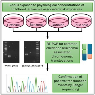 Figure 1
| Exposure | Conc. | Physiological range |
|
| ||
|---|---|---|---|---|---|---|
| Day 2 | Day 4 | Day 2 | Day 4 | |||
| DMSO | 0.01% | Control | ||||
| Benzene | 1.5 nM | Non‐smoker | 1 | 1 | ||
| 3 nM | Smoker | 1 | ||||
| 6 nM | Benzene worker | |||||
| 12 nM | Heavy smoker | 1 | ||||
| 24 nM | Benzene worker smoker | 1 | ||||
| 48 nM | Extreme | 2 | 1 | |||
| Cotinine | 10 nM | Non‐smoker | 1 | |||
| 100 nM | Second‐hand smoke | 1 | ||||
| 1 μM | Average smoker | |||||
| 5 μM | Very heavy smoker | 1 | ||||
| 10 μM | Extreme exposure | |||||
| Caffeine | 2 μM | Low intake | ||||
| 10 μM | Medium intake/average 1st trimester | 1 | ||||
| 20 μM | Average 3rd trimester | |||||
| 40 μM | Very high 3rd trimester | |||||
| 80 μM | Extreme | 1 | ||||
| Folic acid | 2000 μM | Normal TC media (control) | ||||
| 200 nM | Very high | |||||
| 100 nM | High | 1 | ||||
| 50 nM | High‐end normal range | |||||
| 10 nM | Low‐end normal range | 1 | ||||
| 5 nM | Low | |||||
| 1 nM | Depleted | 1 | ||||
| 0.1 nM | Deficient | |||||
| Sample | Sequence of translocation junction | Genes identified in BLAST |
|---|---|---|
| Pre‐B 697 positive cell line control |
|
|
| Benzene 1.5 nM Day 2 |
|
|
| Benzene 1.5 nM Day 4 |
|
|
| Cotinine 100 nM Day 2 ( |
|
|
| Cotinine 10 nM Day 2 ( |
|
|
| Caffeine 80 μM Day 4 |
|
|
| Kasumi‐1 positive cell line control |
|
|
| Benzene 48 nM Day 2 |
|
|
| Benzene 3 nM Day 2 |
|
|
| Cotinine 5 μM Day 2 ( |
|
|
| Exposure | Concentration |
|
| ||
|---|---|---|---|---|---|
| 2 day | 4 day | 2 day | 4 day | ||
| DMSO | 0.01% | 2/20 | 2/20 | 0/20 | 0/20 |
| Benzene | 1.5 nM | 1/20 (> 0.9999) | 5/20 (0.047) | ||
| 48 nM | 6/20 (0.020) | ||||
| Cotinine | 5 μM | 7/20 (0.0083) | |||
| Caffeine | 80 μM | 13/20 (< 0.0001) | |||
| Folic acid | 1 nM | 7/10 (0.0017) | |||
- —CHILDREN with CANCER UK10.13039/501100001273
- —World Cancer Research Fund10.13039/501100000321
- —Northumbria University10.13039/100010052
Peer Reviews
No public reviews on file for this paper yet. If you reviewed it on a platform where reviews are public (OpenReview, ICLR, NeurIPS, ICML), you can paste yours below so the community can read it here.
Videos
No videos yet. Explain this paper in a talk, walkthrough, or lecture? Add one.
Taxonomy
TopicsAcute Lymphoblastic Leukemia research · Carcinogens and Genotoxicity Assessment · Health, Environment, Cognitive Aging
Introduction
1
Childhood leukaemia is the most common cancer in children, accounting for nearly a third of all childhood cancers [1]. Incidence of childhood leukaemia shows a 1% increase each year [2], suggesting an environmental contribution to risk. Epidemiological studies have identified various in utero environmental exposures as potential risk factors for childhood leukaemia. Although the magnitude of the observed associations is variable and sometimes inconsistent, factors that have been implicated to increase the risk of childhood leukaemia include maternal and paternal smoking [3] and maternal caffeine intake [4, 5, 6]. Maternal supplementation with folic acid during pregnancy has been suggested to decrease risk [7, 8]. The underlying mechanisms through which these in utero exposures may infer risk are poorly understood; therefore, further understanding of the biological mechanisms through which they may contribute to childhood leukaemia development will be important to reduce incidence rates.
Using cord blood and blood from newborn heel pricks, multiple studies have identified the presence of leukaemia‐associated chromosomal translocations at birth, indicating that these events occurred in utero [9, 10]. Chromosomal translocations occur when there are two breaks on different chromosomes, which occur in spatial and temporal proximity, such that incorrect ends are joined together, which can result in fusion gene products and misregulation of genes [11]. It is plausible that some exposures suggested to influence childhood leukaemia risk may do so by contributing to the incidence of chromosomal translocations, as some exposures are known to influence DNA structure.
Benzene, a major carcinogen of cigarette smoke, causes DNA damage both in vitro and in animal models [12, 13]. It can travel across the placenta, exposing offspring in utero at concentrations equal to or higher than maternal concentrations [14], and therefore could potentially influence the developing foetal genome. Another component of cigarette smoking is nicotine, largely metabolised to cotinine, with offspring exposed in utero at levels observed in active smokers [15] and evidence of DNA damage has been observed in vitro [16]. In addition, in vitro studies have shown that caffeine exposure can impact DNA, including altering DNA repair and cell cycle checkpoints [17]. Finally, folate is key in DNA replication due to its role in one‐carbon metabolism and the synthesis of purines and pyrimidines [18] and under deficient conditions, DNA damage, including double‐strand breaks, has been observed both in vivo and in vitro [19, 20].
As DNA damage can lead to DNA double‐strand breaks [21], known to be potent inducers of chromosomal translocations [22], we hypothesised that it is plausible that environmental factors associated with increased risk of childhood leukaemia, and are known to cause DNA damage, may influence chromosomal translocation events and thus be one potential mechanism driving those associations. In a highly exploratory study, we exposed NALM6 cells, a pre‐B lymphoblastic cell line with compromised mismatch repair response, to a range of physiological levels of selected exposures to investigate the role of these exposures on the potential formation of chromosomal translocations that are commonly associated with childhood leukaemia. Given the potential to influence DNA damage, we selected benzene and cotinine as proxy exposures for smoking and caffeine and folic acid as exposures for investigation.
Methods
2
Cell Culture
2.1
NALM6 cells (DSMZ, Germany, RRID:CVCL_0092), were expanded in RPMI‐1640 (Gibco, Fisher Scientific, UK) with 10% foetal bovine serum (FBS, Gibco, Fisher Scientific, UK) and 1% penicillin/streptomycin (P/S, Gibco, Fisher Scientific, UK), incubated at 37°C and 5% CO_2_. The NALM6 cell line used in this study was authenticated by STR profiling, performed by Eurofins Genomics (Eurofins, Germany), and compared with the CLASTR 1.4.4 Cellosaurus STR Similarity Search Tool [23] (https://www.cellosaurus.org/str‐search/) with a score of 81.08% indicating a good match with NALM6. The NALM6 cell line was confirmed as mycoplasma free through routine mycoplasma testing using VenorGeM OneStep (Minerva Biolabs, Berlin, Germany) following the manufacturer's instructions. Cell counts and viability were assessed every 24 h using a haemocytometer and trypan blue staining [24]. Cells were seeded at 0.5 × 10^6^ cells/mL in RPMI‐1640, treated with benzene (Scientific Laboratory Supplies, UK), cotinine (Fisher Scientific, UK), caffeine (Scientific Laboratory Supplies, UK) or dimethylsulfoxide (DMSO, Fisher Scientific, UK), (Table S1), and harvested after 2 and 4 days. For folic acid treatment, cells were washed and seeded in folic acid free RPMI‐1640 (Gibco, Fisher Scientific, UK, supplemented with 10% FBS and 1% P/S), at 0.5 × 10^6^ cells/mL, treated with folic acid (Fisher Scientific, UK) dissolved in DMSO, (Table S1), and harvested after 4 days (suggested to deplete endogenous folate whist maintaining cell viability [25]). Three biological replicates were performed per exposure and 10–20 replicates for reproducibility experiments.
RT‐PCR
2.2
RNA was isolated using EZNA Total RNA Isolation Kit I (Omega Bio‐Tek, USA), treated with DNase (Primer Design, UK) and reverse transcribed using Precision nanoScript2 Reverse Transcription Kit (Primer Design, UK). PCR was performed on a 6331 Nexus Gradient MasterCycler Thermal Cycler (Eppendorf, Germany) with a master‐mix of PCRBIO 2X HS Taq Mix (PCR Biosystems, UK) and 10 pmol of primers (Eurofins, Germany). TCF3::PBX1 primers, forward: GGCCTGCAGAGTAAGATAG, and reverse: CACGCCTTCCGCTAACAG. RUNX1::RUNX1T1 primers, forward: GAGGGAAAAGCTTCACTCTG and reverse: GCGAACTCTTTCTCCTATC. Translocation‐positive cell lines pre‐B 697 (TCF3::PBX1) and Kasumi‐1 (RUNX1::RUNX1T1) were used as a positive control, with 5 ng/μl of cDNA. Cycling conditions included denaturation (95°C, 2 min), 40 cycles of denaturation (95°C, 15 s), annealing (60°C, 15 s), and extension (72°C, 30 s), and a final extension (72°C, 2 min). Visualisation with gel electrophoresis was performed with 1% agarose gel containing Sybr Safe (Invitrogen, USA).
Sanger sequencing was performed on positive RT‐PCR controls and a sub‐sample of translocation‐positive exposure cells to confirm alignment with expected translocations. RT‐PCR products were purified using GeneJET PCR purification/gel extraction kit (Fisher Scientific, UK) and sent to DBS Genomics (Durham University, UK) for Sanger sequencing using Applied Biosciences 3730 capillary instrument. Forward and reverse sequencing reactions used 3.2 pmol/μl primer with > 10 ng/μl DNA. Sequencing data were submitted to the Basic Local Alignment Search Tool (BLAST, National Center for Biotechnology Information) for sequence comparison.
Results
3
Exposure to Benzene, Cotinine, Caffeine and Folic Acid Induces Chromosomal Translocations in NALM6
3.1
NALM6 were incubated for 2 or 4 days with physiologically relevant concentrations of benzene, cotinine, caffeine and folic acid (Table 1). Exposure to all risk factors resulted in decreased cell viability over time (Figure S1). However, for all exposures, over 50% of cells were viable at the time of harvesting.
RT‐PCR on cDNA synthesised from DMSO treated and 2000 nM folic acid treated (to mimic standard media) cells, that is, unexposed controls, resulted in no amplification of the target translocations TCF3::PBX1 and RUNX1::RUN1T1 (Table 1). The highest number of translocations were observed in response to benzene, n = 8, followed by folate depletion, n = 3, whilst cotinine and caffeine exposure were at similar levels, that is, n = 3 and n = 2 respectively (Table 1).
In benzene‐exposed cells, at least one translocation was observed for most concentrations tested, and an equal number of translocations were observed at 2 and 4 days (n = 4). There was some evidence of recurrent events that is, TCF3::PBX1 observed in response to 1.5 nM benzene after both 2 and 4 days exposure, and two RUNX1::RUNX1T1 events observed after 2 days exposure of 48 nm benzene and one observed after 4 days.
In cotinine‐exposed cells, a single event was observed for three of five concentrations tested. There was some evidence to suggest that different translocation events occur in response to high and low cotinine concentrations with TCF3::PBX1 observed at the lowest concentrations of cotinine exposure that is, 10 nM and 100 nM after 2 days, and RUNX1::RUNX1T1 occurring following exposure to a high cotinine concentration, that is, 5 μM after 2 days.
Translocations were only observed for two of five caffeine concentrations tested, with a single TCF3::PBX1 translocation at 10 μM after 2 days, and at 80 μM after 4 days.
A single translocation event was observed for three of the seven folic acid concentrations. Of these, two TCF3::PBX1 events were observed at folic acid concentrations that are considered physiologically low, that is, 10 nM and 1 nM. A single RUNX1::RUNX1T1 event was observed at 100 nM folic acid, considered a high physiological concentration, although low in comparison to standard tissue culture media.
Sanger sequencing for TCF3::PBX1 positive samples (Table 2) show identical breakpoints to those observed in the pre‐B 697 control. For RUNX1::RUNX1T1 positive samples, one sample showed an identical breakpoint to the Kasumi‐1 control, and two further samples showed the correct genes (i.e., RUNX1 and/or RUNX1T1) with sequencing noise that did not match the positive breakpoint (Table 2).
Replicability of Translocation Events in NALM6 in Response to Benzene, Cotinine, Caffeine and Folic Acid Exposure
3.2
To understand the replicability of translocation events, experiments were repeated up to 20 times for a subset of exposures where translocation events were previously observed.
Significantly more, TCF3::PBX1 translocations were observed in response to 80 μM caffeine for 4 days (13/20 replicates), compared with DMSO controls (2/20; Fisher's exact test, p =< 0.0001) (Table 3). This gives an expected translocation frequency of 65% in response to caffeine exposure, suggesting that 1–2 translocation events should be observed in every three replicates, as previously observed (Table 1). Similarly, seven TCF3::PBX1 translocations were observed in 10 replicates for 1 nM folic acid, significantly higher than the DMSO controls (Fisher's exact test, p = 0.0017), giving an expected translocation frequency of 70%, suggesting two events in every three replicates, whilst only one event was observed previously (Table 1). For cells exposed to 1.5 nM benzene for 2 days, only one TCF3::PBX1 translocation was observed across 20 replicates, which was not significant compared to DMSO controls (Fisher's exact test, p => 0.9999).
A significant number of RUNX1::RUNX1T1 events were observed for benzene exposure for 2 days, including five out of 20 replicates at 1.5 nM and six out of 20 replicates at 48 nM, compared to the DMSO controls, n = 0 (Table 3) (Fisher's exact test, p = 0.047 and p = 0.020 respectively). This gives an expected frequency of 25%–30% for translocation events, suggesting one event in every three replicates, which is lower than the highest number of events observed in initial experiments, n = 2 (Table 1). A significant number of RUNX1::RUNX1T1 events were also observed in 5 μM cotinine for 2 days that is, 7/20 replicates (Fisher's exact test, p = 0.0083). This gave an expected frequency of 35%, suggesting one translocation event in every three replicates, which was observed in our initial experiment (Table 1).
Discussion
4
Several epidemiological studies have reported associations between parental smoking, maternal caffeine intake and folic acid supplementation and childhood leukaemia risk [3, 5, 7, 26]. As these risk exposures have previously been shown to induce DNA damage and double‐strand breaks [12, 13, 16, 17, 19, 20] we hypothesised they have the potential to induce chromosomal translocations associated with childhood leukaemia. In this preliminary study, we report for the first time that exposure to physiological levels of benzene, cotinine (as components of cigarette smoke), caffeine and folic acid in vitro shows detectable chromosomal translocations associated with childhood leukaemia via RT‐PCR. These preliminary results will require further in‐depth follow‐up studies to confirm these findings.
In initial experiments, RUNX1::RUNX1T1 events were most frequent at high benzene concentrations, associated with levels in smokers and extreme exposure, whilst TCF3::PBX1 events were found at lower benzene concentrations. RUNX1::RUNX1T1 events were significantly enriched across 20 additional replicates at low and high benzene concentrations, whilst no TCF3::PBX1 events were observed. This may suggest that translocation induction in response to benzene exposure may therefore be targeted and more likely to appear in specific genomic locations. Indeed, in support of this, the RUNX1::RUNX1T1 translocation is observed at a higher incidence in benzene‐exposed patients compared to de novo and therapy related AML [27]. It is important to highlight the different cell lineages examined between these findings, which may demonstrate the effect of benzene on the genome‐specific location, regardless of specific cell type.
Benzene toxicity is generally believed to be caused by benzene metabolites; however, here we suggest that exposure to benzene itself may exert genotoxic effects since NALM6 are not considered capable of benzene metabolism [28, 29]. Further investigation is warranted into the effect of benzene, as well as its metabolites, on toxicity. Benzene may contribute to leukemogenesis through mechanisms including disruption of signalling pathways, epigenetic alterations, and oxidative stress [30]. Benzene produces ROS, shown to increase double‐strand breaks and impede DNA repair [31, 32]. Measuring ROS production and DNA double‐strand break occurrence in future work will aid mechanistic understanding of how benzene exposure may induce childhood leukaemia‐associated translocations.
Cigarette smoking has well categorised carcinogens and oxidative stress inducers, known to contribute to cancer development [33], but the impact of cotinine is less known. We observed RUNX1::RUNX1T1 translocations at cotinine concentrations associated with very heavy smokers, whereas TCF3::PBX1 events were observed at low cotinine concentrations associated with non‐smokers. The RUNX1::RUNX1T1 translocation is primarily found in childhood AML, which is rarer than childhood ALL, and may require a higher level of exposure to induce translocations [34]. Cotinine exposure in neuroblastoma cells observed significant differences in DNA damage index and frequency, and found increased oxidative damage in cotinine‐exposed cells [16]. This suggests that cotinine may induce double‐strand breaks through oxidative damage, and as such could lead to translocation formation. However, further investigation would be required to assess the impact of cotinine on oxidative and DNA damage in NALM6 cells.
Studies have observed associations between increased risk of ALL and consumption of two or more cups of coffee per day, equivalent to 10 μM caffeine, at which we observed translocations in our exploratory experiments [4, 6, 35, 36]. Caffeine has been shown to be a topoisomerase inhibitor, a mechanism through which chemotherapy agents such as etoposide are known to induce double‐strand breaks and translocations [37, 38]. Furthermore, through its involvement as an adenosine receptor antagonist and phosphodiesterase inhibitor, caffeine can affect DNA repair and checkpoint pathways [39]. These mechanisms should be explored in future research.
The translocation events observed in this study support the hypothesis that folate deficient conditions may plausibly induce translocations. The folate cycle is required for nucleotide synthesis with reduced folate metabolism leading to accumulation of excess uracil, which can be incorporated into DNA leading to double‐strand breaks [18, 40, 41]. One‐carbon metabolism provides methyl donors for a range of cellular processes, including the epigenome. DNA methylation is important in regulating genes involved in DNA repair, chromosome stability, and therefore, altered methylation could contribute to translocation induction [42], which should be investigated in future studies.
A strength of this exploratory study was in screening a range of exposures associated with childhood leukaemia; however, this was limited by only measuring three replicates in one cell line. Future studies should extend to test a range of cell lines and potentially organoid model systems, with larger replicate experiments for robustness of these associations.
Studies have shown some associations between risk factors and specific translocations not measured in this study, such as ETV6::RUNX1 with caffeine and smoking [3, 43], therefore future work should also include the development of further assays to evaluate additional childhood leukaemia‐associated translocations. Furthermore, as RUNX1::RUNX1T1 is commonly associated with AML and myeloid progenitors rather than pre‐B cells (NALM6), future validation in myeloid cell lines or CD34^+^ progenitors is required to confirm the biological relevance of these translocations [44].
Fluorescent in situ hybridisation (FISH) is the gold standard for identifying translocated cells; however, cost and time make it unrealistic as a screening tool [45]. The RT‐PCR assays used here have a detection rate of 1% for translocated cells in line with FISH detection rates (Figure S2), and so are suitable for this screening study. However, in future experiments, using digital droplet PCR would allow quantification of the number of translocated cells within each exposure sample, which would aid detection of the frequency of events. To further understand the mechanistic pathways of translocation induction by these exposures, further experimental validation should be performed including testing of intermediate DNA damage using COMET assays, oxidative stress measurements and topoisomerase complex identification using TARDIS assays [46, 47].
This exploratory study provides proof of principle that caffeine, benzene, cotinine and folic acid may impact chromosomal translocation induction, linked to key exposures associated with childhood leukaemia risk. Few prior studies have directly investigated if exposures could influence the occurrence of translocations [48, 49, 50], with no studies focusing on the specific exposures examined here. Additionally, our study is novel in addressing exposure levels of physiological relevance. Whilst this study was not designed to estimate in vivo risk or translocation frequency, it has allowed us to demonstrate a biologically plausible mechanism for epidemiological associations linking environmental exposures with childhood leukaemia risk. Whilst further in‐depth investigation is needed, this study has contributed novel data demonstrating that exposures associated with childhood leukaemia risk have the potential to cause leukaemia‐associated chromosomal translocations. Understanding the environmental contribution to leukaemia‐initiating chromosomal translocations will be important to develop preventative interventions to reduce the incidence of childhood leukaemia.
Author Contributions
Jessica R. Saville: data curation (lead), formal analysis (equal), methodology (lead), writing – original draft (equal), writing – review and editing (equal). Lisa J. Russell: conceptualization (equal), formal analysis (equal), supervision (supporting), writing – review and editing (equal). Kay Padget: conceptualization (equal), formal analysis (equal), supervision (supporting), writing – review and editing (equal). Jill A. McKay: conceptualization (equal), formal analysis (equal), resources (lead), supervision (lead), writing – original draft (supporting), writing – review and editing (equal).
Funding
This work was supported by CHILDREN with CANCER UK, 19‐316. World Cancer Research Fund, IIG_FULL_2022_007. Northumbria University, RDF Studentship.
Ethics Statement
This work has been approved by Northumbria University Ethics Committee.
Conflicts of Interest
The authors declare no conflicts of interest.
Supporting information
Figure S1: Viability of NALM6 cells. Percentage cell viability was determined by the ratio of live to dead cells using trypan blue exclusion counted every 24 h following exposure to various physiological concentrations of (A) benzene, (B) cotinine, (C) folic acid and (D) caffeine. DMSO was used as a vehicle control for benzene, cotinine and caffeine, with 2000 nM folic acid (+DMSO as a vehicle control) was used as a standard media control for folic acid. Two replicates are reported for cotinine, folic acid and caffeine, showing standard error. One replicate is reported for benzene. Figure S2: Gel electrophoresis image for RT‐PCR amplification of (A) TCF3::PBX1 and (B) RUNX1::RUNX1T1. The positive cell line for each translocation was diluted with a negative cell line at different percentages before RNA extraction and reverse transcription. The final cDNA concentration used was 5 ng/μl. MWM = molecular weight marker (New England Biosciences 100 bp DNA ladder). Table S1: List of concentrations used in exposure experiments and corresponding literature used to reflect physiological levels.
The reference list from the paper itself. Each links out to its DOI / PubMed record.
- 1UK Cw C , “Childhood Cancer Facts & Figures,” (2018), https://www.childrenwithcancer.org.uk/childhood‐cancer‐info/childhood‐cancer‐facts‐figures/.
- 2WHO , “Incidence of Childhood Leukaemia, an ENHIS Fact Sheet,” (2020), https://www.euro.who.int/en/health‐topics/noncommunicable‐diseases/cancer/publications/2009/4.1‐incidence‐of‐childhood‐leukaemia,‐an‐enhis‐fact‐sheet.
- 3C. Metayer , L. Zhang , J. L. Wiemels , et al., “Tobacco Smoke Exposure and the Risk of Childhood Acute Lymphoblastic and Myeloid Leukemias by Cytogenetic Subtype,” Cancer Epidemiology, Biomarkers & Prevention 22, no. 9 (2013): 1600–1611, 10.1158/1055-9965.EPI-13-0350.PMC 376947823853208 · doi ↗ · pubmed ↗
- 4F. Menegaux , C. Steffen , S. Bellec , et al., “Maternal Coffee and Alcohol Consumption During Pregnancy, Parental Smoking and Risk of Childhood Acute Leukaemia,” Cancer Detection and Prevention 29, no. 6 (2005): 487–493, 10.1016/j.cdp.2005.06.008.16289502 · doi ↗ · pubmed ↗
- 5E. Milne , K. R. Greenop , E. Petridou , et al., “Maternal Consumption of Coffee and Tea During Pregnancy and Risk of Childhood ALL: A Pooled Analysis From the Childhood Leukemia International Consortium,” Cancer Causes & Control 29, no. 6 (2018): 539–550, 10.1007/s 10552-018-1024-1.29600472 · doi ↗ · pubmed ↗
- 6J. Blanco‐Lopez , I. Iguacel , S. Pisanu , et al., “Role of Maternal Diet in the Risk of Childhood Acute Leukemia: A Systematic Review and Meta‐Analysis,” International Journal of Environmental Research and Public Health 20, no. 7 (2023) 5428, 10.3390/ijerph 20075428.37048042 PMC 10093835 · doi ↗ · pubmed ↗
- 7A. Amigou , J. Rudant , L. Orsi , et al., “Folic Acid Supplementation, MTHFR and MTRR Polymorphisms, and the Risk of Childhood Leukemia: The ESCALE Study (SFCE),” Cancer Causes & Control 23, no. 8 (2012): 1265–1277, 10.1007/s 10552-012-0004-0.22706675 · doi ↗ · pubmed ↗
- 8W. R. Wan Ismail , R. Abdul Rahman , N. A. A. Rahman , A. Atil , and A. M. Nawi , “The Protective Effect of Maternal Folic Acid Supplementation on Childhood Cancer: A Systematic Review and Meta‐Analysis of Case‐Control Studies,” Journal of Preventive Medicine and Public Health 52, no. 4 (Jul 2019): 205–213, 10.3961/jpmph.19.020.31390683 PMC 6686110 · doi ↗ · pubmed ↗