Challenges of dermoscopic assessment of basal cell carcinoma on tattooed skin
Joana Margarida Reis, José Carlos Cardoso, André Oliveira

Abstract
Genes, proteins, chemicals, diseases, species, mutations and cell lines named across the full text — each resolved to its canonical identifier and authoritative record.
Click any figure to enlarge with its caption.
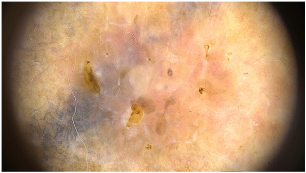 Figure 1
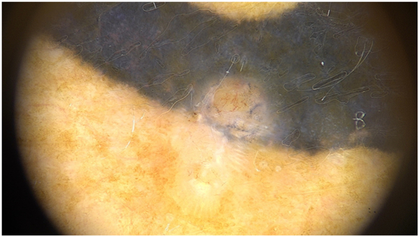 Figure 2
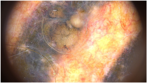 Figure 3
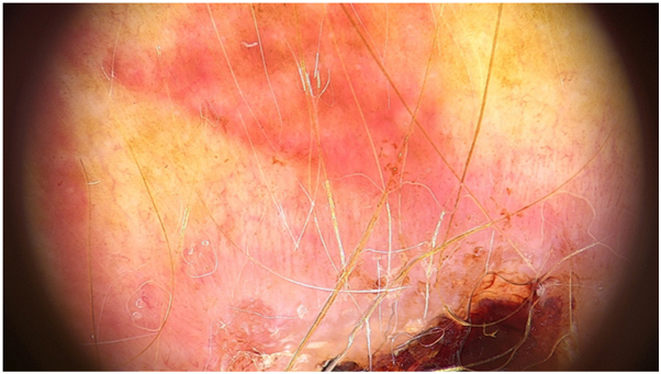 Figure 4
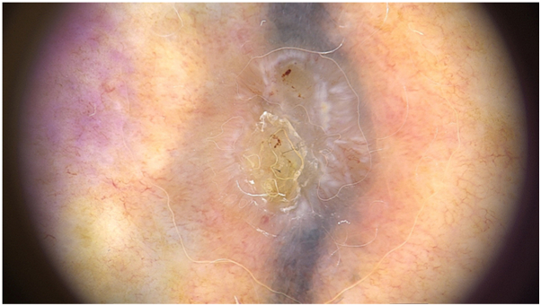 Figure 5
Peer Reviews
No public reviews on file for this paper yet. If you reviewed it on a platform where reviews are public (OpenReview, ICLR, NeurIPS, ICML), you can paste yours below so the community can read it here.
Videos
No videos yet. Explain this paper in a talk, walkthrough, or lecture? Add one.
Taxonomy
TopicsTattoo and Body Piercing Complications · Nonmelanoma Skin Cancer Studies · Dermatologic Treatments and Research
Introduction
Tattoos are becoming increasingly popular as a form of body art, with prevalence estimates of 35% to 48% in Europe.1 Although infectious and allergic complications are frequent, malignant tumors have been less described.2^,^3 This is remarkable in basal cell carcinoma (BCC), because its high incidence contrasts with rare literature reports occurring on tattooed skin. Diagnosing skin tumors in tattooed skin can be challenging, as exogenous pigment may mask or mimic key structures, complicating clinical examination and dermoscopic interpretation.4^,^5 In our series, all BCCs were associated with tattoo pigment, which was histologically identifiable in every case. We present a series of 5 tattoo-associated BCCs, emphasizing their dermoscopic features. Clinical and histological characteristics of tattoo-associated BCC are represented in Table I and dermoscopic features are described in Table II.Table IClinical and histologic characteristics of tattoo associated BCCCase nrAge/sexFototypeTattoo duration (yr)Tatto locationPigment colorDimension (mm)Evolution time (mo)Lesion typeHistopathology145/FII15Scapular regionBlack712PlaqueSuperficial BCC247/FI2ThighBlack76PapuleNodular BCC355/MI17ChestBlack5UnknownPapuleNodular BCC464/MII20ArmRed1724PlaqueNodular BCC558/FI18ShoulderBlack712PapuleNodular BCCClinical and histologic features of the 5 BCCs arising in tattooed skin: 5 patients (3 women), aged between 45 and 64 years; tattoo duration ranged from 2 to 20 years, and clinical evolution from 6 to 24 months. Histopathology showed 4 nodular BCCs and 1 superficial.Table IIDermoscopic features of tattoo-associated BCCCase nrVascular structuresPigmented structuresShiny white structuresArborizing vesselsIrregular linear vesselsCorkscrew vesselsHairpin vesselsDotted vesselsVascular polymorphism1-+--++Gray ovoid nests+2-++--+-+3-+--++--4-+--++-+5-+-+++-+Pigmented dermoscopic criteria were frequently obscured by exogenous tattoo pigment, whereas vascular patterns and shiny white structures remained consistently identifiable.+, Present; -, absent.
Case 1
A 45-year-old woman with a 15-year-old tattoo presented with an associated lesion on the scapular area with 12 months of evolution. Dermoscopy revealed vascular polymorphism with irregular linear and dotted vessels. Shiny white structures, large globules, grey ovoid nests and erosions were also observed (Fig 1). Histopathology confirmed superficial BCC.Fig 1. Dermoscopy of BCC revealing irregular linear and dotted vessels. Shiny white structures, concentric structures, gray ovoid nests and erosions are also seen.
Case 2
A 47-year-old woman with a 2-year-old tattoo developed an associated lesion on the thigh with a 6 month history of growth. Dermoscopy showed vascular polymorphism with irregular linear and corkscrew vessels. Shiny white structures were also present (Fig 2). Histopathology revealed nodular BCC.Fig 2BCC revealing irregular linear vessels, corkscrew vessels, and shiny white structures.
Case 3
A 55-year-old man with a 17-year-old tattoo presented with a lesion on the chest of uncertain duration. Dermoscopy revealed vascular polymorphism with irregular linear and dotted vessels (Fig 3). Histopathology confirmed nodular BCC.Fig 3BCC dermoscopy with evident vascular polymorphism, with irregular linear and dotted vessels.
Case 4
A 64-year-old man with a 20-year-old tattoo presented with a concomitant lesion on his arm with 2 years of evolution. Dermoscopy demonstrated vascular polymorphism with irregular linear and dotted vessels. Ulceration and shiny white structures were also present (Fig 4). Histopathology confirmed nodular BCC.Fig 4BCC dermoscopy revealing vascular polymorphism, with irregular linear vessels and dotted vessels. Ulceration and shiny white structures are also present.
Case 5
A 58-year-old woman with an 18-year-old tattoo presented with a lesion on her shoulder with a 12 months of evolution. Dermoscopy revealed vascular polymorphism with irregular linear, dotted, hairpin, and corkscrew vessels. Shiny white structures and erosions were also present (Fig 5). Histopathology confirmed nodular BCC.Fig 5BCC dermoscopy with vascular polymorphism, with irregular linear vessels, hairpin and dotted vessels. Shiny white structures and erosions are also seen.
Discussion
Forty-one cases of BCC on tattooed skin have been reported to date,1 yet only 3 included dermoscopic descriptions.6^,^7 Our series suggests that classical pigmented dermoscopic criteria of basal cell carcinoma, such as ovoid nests, spoke-wheel structures, and maple-leaf-like areas may be obscured by exogenous tattoo pigment. This masking effect can reduce the sensitivity of dermoscopy and contribute to diagnostic challenges. Otherwise, in all cases, vascular structures were readily identifiable, with a predominance of polymorphous vessels, including linear-irregular vessels, despite the consistent absence of arborizing vessels typically associated with non-ulcerated nodular BCC. These vascular patterns, usually seen in amelanotic or hypomelanotic melanoma and aggressive fast-growing palpable tumors, may reflect optical artifacts induced by tattoo ink, as well as local inflammatory or elastotic changes. In addition, shiny white structures, another key dermoscopic feature of BCC, appear to maintain diagnostic relevance across most cases. Taken together, these findings support a dermoscopy-first approach focused on vascular morphology and shiny white structures when evaluating suspicious lesions on tattooed skin, rather than classical pigmented criteria. Our findings are intended to be descriptive and to emphasize practical diagnostic considerations when evaluating suspicious lesions on tattooed skin. Careful and systematic dermoscopic assessment of tattooed skin therefore remains essential.
Conflicts of interest
None disclosed.
The reference list from the paper itself. Each links out to its DOI / PubMed record.
- 1Lebhar J.Jacobs J.Rundle C.Kaplan S.J.Mosca P.J.Skin cancers arising within tattoos: a systematic review JAAD Int 16202413314310.1016/j.jdin.2024.03.01538957835 PMC 11217691 · doi ↗ · pubmed ↗
- 2Cohen P.R.Erickson C.P.Uebelhoer N.S.Calame A.Tattoo-associated basal cell carcinoma: coincident or coincidence Biomed Hub 5220201810.1159/000508208 PMC 744365132884932 · doi ↗ · pubmed ↗
- 3Abudu B.Erickson C.P.Calame A.Cohen P.R.Basal cell carcinoma originating in a tattoo: case report and review of an uncommon complication in tattoo recipients Dermatol Pract Concept 94201926527010.5826/dpc.0904 a 0331723458 PMC 6830564 · doi ↗ · pubmed ↗
- 4Leijs M.Schaefer H.Rübben A.Cacchi C.Rustemeyer T.van der Bent S.Cutaneous malignancies in tattoos, a case series of six patients Curr Oncol 28620214721473710.3390/curroncol 2806039834898571 PMC 8628776 · doi ↗ · pubmed ↗
- 5Kluger N.Cutaneous complications related to tattoos: 31 cases from Finland Dermatology 2331201710010910.1159/00046853628441655 · doi ↗ · pubmed ↗
- 6Padoveze E.H.Di Chiacchio N.G.Pinto T.C.Lima A.A.N.Santana G.L.Basal cell carcinoma originating in a tattoo: report of two cases Surg Cosmet Dermatol 124202036636810.5935/scd 1984-8773.20201243688 · doi ↗
- 7Verdaguer-Faja J.Mora-Fernandez V.Fabregat-Pratdepadua M.Jaka A.“A strange flower in the garden”: the importance of the dermatoscope in the assessment of tattoos Dermatol Pract Concept 1412024 e 202401110.5826/dpc.1401 a 11PMC 1086889238364428 · doi ↗ · pubmed ↗