Photon-counting CT for radiotherapy: Qualitative assessment of potential clinical value by semi-structured expert interviews
Thiele Kroes-Kobus, Linda Rossi, Joost J.M.E. Nuyttens, Dirk K.M. de Ruysscher, Arlette E. Odink, Manouk J.J. Olofsen-van Acht, Ilse M.N. de Pree, Edwin H.G. Oei, Joris B.W. Elbers, Michiel Kroesen, Anke W. van der Eerden, Jan Willem M. Mens, Steven H.J. Nagtegaal

TL;DR
This study explores how photon-counting CT could improve radiotherapy, finding it most valuable for head and neck, bone, and lung tumors due to spectral data and high-resolution imaging.
Contribution
Novel insights into clinical applications of photon-counting CT in radiotherapy through expert interviews.
Findings
Spectral data and high-resolution imaging are most valuable for clinical applications.
Head and neck, bone, and lung tumors scored highest for added clinical value.
Prostate tumors showed no added value from photon-counting CT.
Abstract
•Semi-structured interviews provided insight in the potential added value of PCCT for radiotherapy.•Head and neck, bone and lung tumors had the highest potential of having added clinical value.•Availability of spectral data and high-resolution imaging contributed most to the added clinical value. Semi-structured interviews provided insight in the potential added value of PCCT for radiotherapy. Head and neck, bone and lung tumors had the highest potential of having added clinical value. Availability of spectral data and high-resolution imaging contributed most to the added clinical value. Photon-counting CT (PCCT) has several potential benefits that can considerably improve the radiotherapy workflow, like availability of intrinsic spectral data and high resolution imaging. It is unknown which treatment sites in radiotherapy will benefit the most from PCCT. This study aimed to…
Genes, proteins, chemicals, diseases, species, mutations and cell lines named across the full text — each resolved to its canonical identifier and authoritative record.
Click any figure to enlarge with its caption.
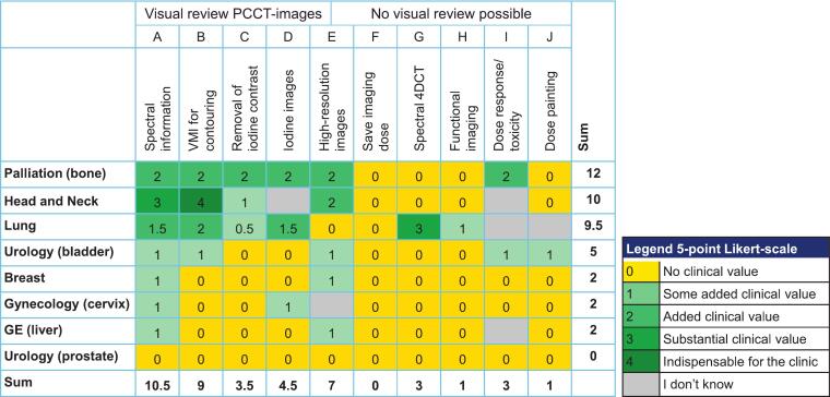 Figure 1
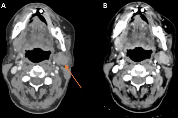 Figure 2
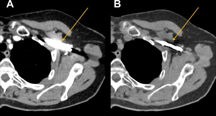 Figure 3
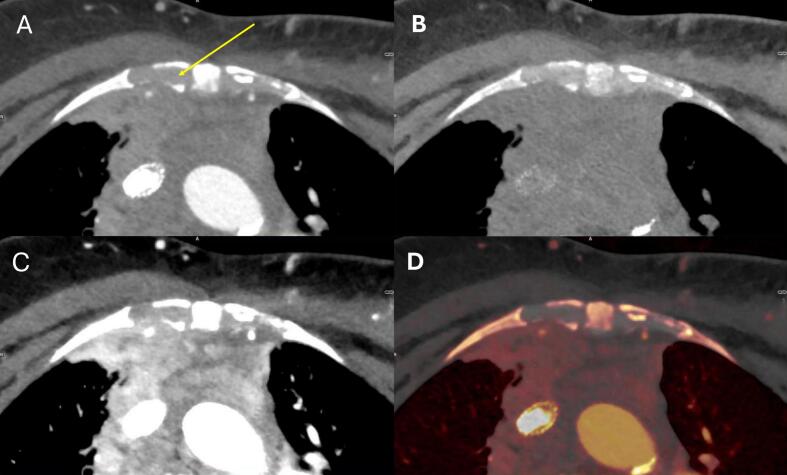 Figure 4
Peer Reviews
No public reviews on file for this paper yet. If you reviewed it on a platform where reviews are public (OpenReview, ICLR, NeurIPS, ICML), you can paste yours below so the community can read it here.
Videos
No videos yet. Explain this paper in a talk, walkthrough, or lecture? Add one.
Taxonomy
TopicsAdvanced X-ray and CT Imaging · Radiation Dose and Imaging · Medical Imaging Techniques and Applications