CBCT‐Assisted Clinic and Radiographic Diagnosis of an Atypically Erupted Odontoma Associated With Supernumerary Tooth
Marina Nunes de Faria Corrêa, Pedro Schwartz Kalil Pereira, Marcos Antonio Torriani, Josué Martos, Melissa Feres Damian

TL;DR
A rare case of an erupted odontoma linked to a supernumerary tooth is diagnosed using advanced imaging, highlighting the importance of 3D scans in complex dental diagnoses.
Contribution
Demonstrates the diagnostic and surgical challenges of an atypically erupted compound odontoma with a supernumerary tooth using CBCT.
Findings
CBCT provided critical 3D imaging for accurate diagnosis of an intraosseous compound odontoma.
Surgical removal of an embedded tooth was performed, while preserving the supernumerary tooth and odontogenic lesion.
Residual denticles remained due to anatomical limitations, emphasizing challenges in complete removal.
Abstract
Odontomas are common benign odontogenic tumors and are typically asymptomatic, often detected incidentally through imaging exams. However, in some instances, these lesions may erupt into the oral cavity and coexist with supernumerary teeth, creating diagnostic and therapeutic challenges and interfering with normal tooth eruption. This case reports an unusual and complex presentation of an odontogenic lesion with clinic and radiographic features consistent with a partially erupted compound odontoma, associated with a supernumerary tooth and an embedded permanent lateral incisor. A 28‐year‐old female was referred to a dental school for evaluation of a recurrent, asymptomatic whitish spicule in the anterior mandibular gingiva. Initial radiographic examination revealed multiple radiopaque, denticle‐like structures, in addition to a vertically oriented supernumerary tooth and an embedded…
Genes, proteins, chemicals, diseases, species, mutations and cell lines named across the full text — each resolved to its canonical identifier and authoritative record.
Click any figure to enlarge with its caption.
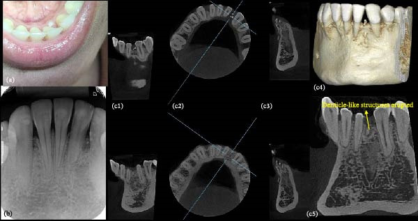 Figure 1
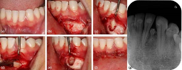 Figure 2
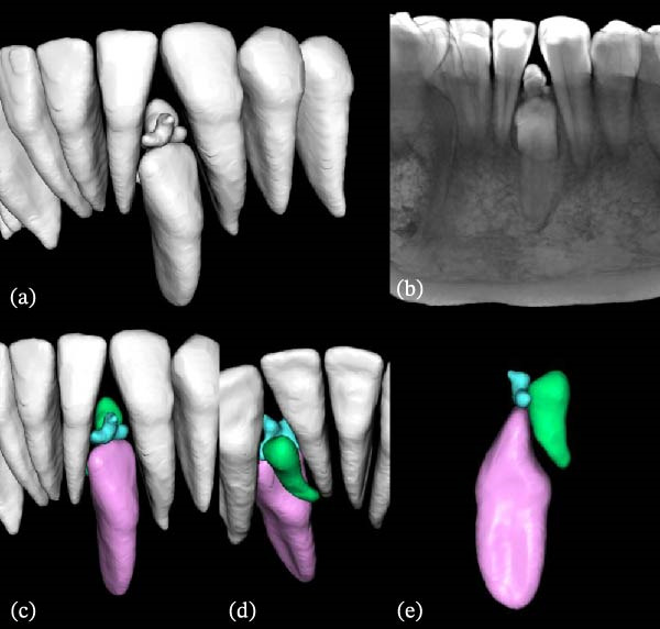 Figure 3
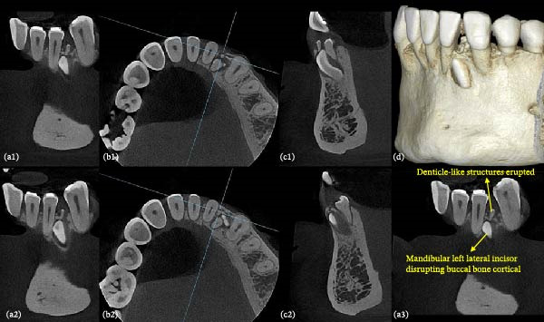 Figure 4
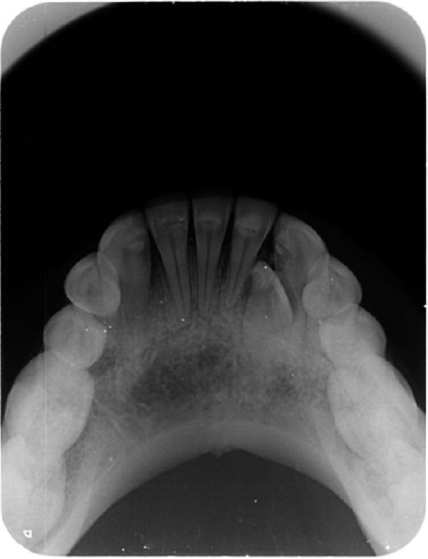 Figure 5
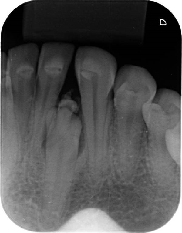 Figure 6
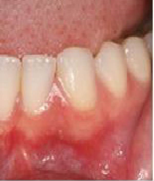 Figure 7
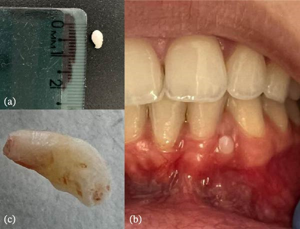 Figure 8
Peer Reviews
No public reviews on file for this paper yet. If you reviewed it on a platform where reviews are public (OpenReview, ICLR, NeurIPS, ICML), you can paste yours below so the community can read it here.
Videos
No videos yet. Explain this paper in a talk, walkthrough, or lecture? Add one.
Taxonomy
TopicsOral and Maxillofacial Pathology · dental development and anomalies · Endodontics and Root Canal Treatments