Quantification of dual-state 5-ALA-induced PpIX fluorescence: methodology and validation in tissue-mimicking phantoms
Silvére Ségaud, Charlie Budd, Matthew Elliot, Graeme J Stasiuk, Jonathan Shapey, Yijing Xie, Tom Vercauteren

TL;DR
This paper introduces a new method to accurately measure PpIX fluorescence in brain tumors using realistic tissue-like models, improving potential clinical applications.
Contribution
A novel pipeline for PpIX quantification that differentiates dual emission states and accounts for optical distortions in tissue-mimicking phantoms.
Findings
The pipeline achieves strong correlation with ground-truth PpIX concentrations (R² = 0.918 ± 0.002).
The method differentiates dual PpIX emission states without relying on prior spectral information.
Tissue-mimicking phantoms replicate glioma optical properties and PpIX fluorescence variability.
Abstract
Quantification of protoporphyrin IX (PpIX) fluorescence in human brain tumours has the potential to significantly improve patient outcomes in neuro-oncology, but represents a formidable imaging challenge. Protoporphyrin is a biological molecule which interacts with the tissue micro-environment to form two photochemical states in glioma. Each exhibits markedly different quantum efficiencies, with distinct but overlapping emission spectra that also overlap with tissue autofluorescence. Fluorescence emission is known to be distorted by the intrinsic optical properties of tissue, coupled with marked intra-tumoural heterogeneity as a hallmark of glioma tumours. Existing quantitative fluorescence systems are developed and validated using simplified phantoms that do not simultaneously mimic the complex interactions between fluorophores and tissue optical properties or micro-environment.…
Genes, proteins, chemicals, diseases, species, mutations and cell lines named across the full text — each resolved to its canonical identifier and authoritative record.
Click any figure to enlarge with its caption.
 Figure 1
 Figure 2
 Figure 3
 Figure 4
 Figure 5
 Figure 6
 Figure 7
 Figure 8
 Figure 9
 Figure 10
 Figure 11
 Figure 12
 Figure 13
 Figure 14
 Figure 15
 Figure 16
 Figure 17
 Figure 18
 Figure 19
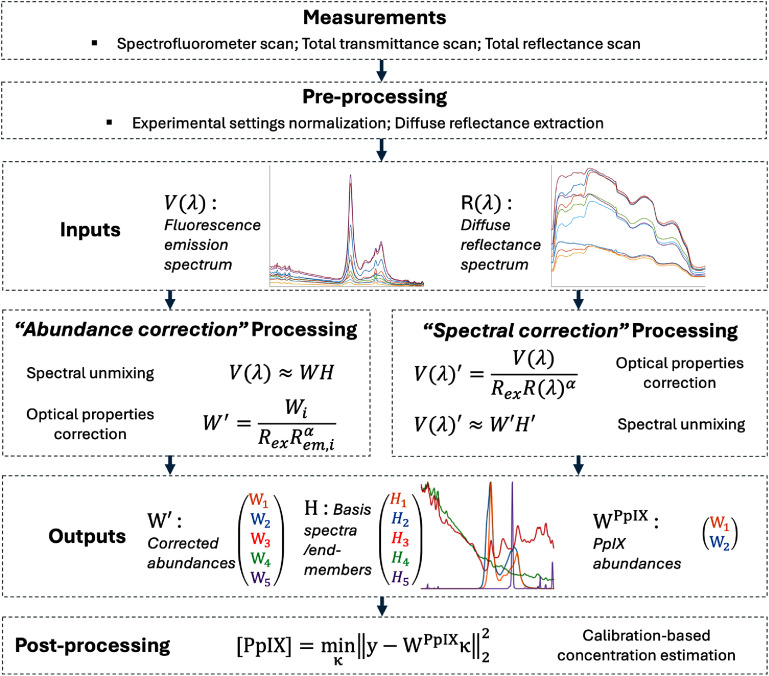 Figure 20
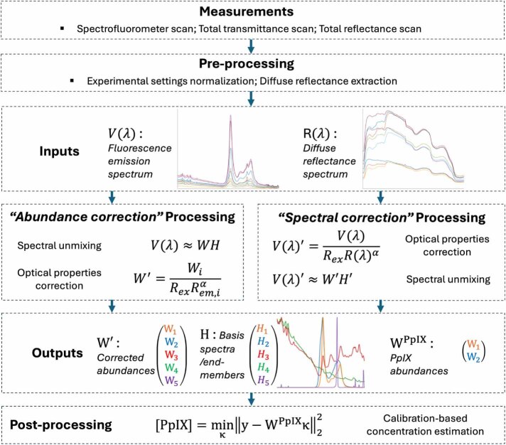 Figure 21
 Figure 22
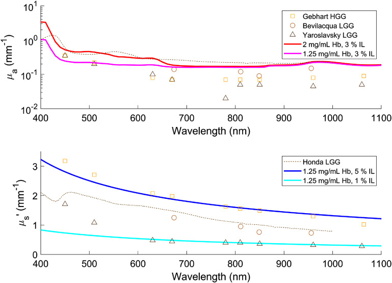 Figure 23
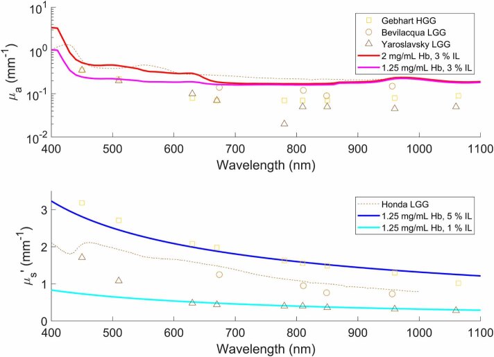 Figure 24
 Figure 25
 Figure 26
 Figure 27
 Figure 28
 Figure 29
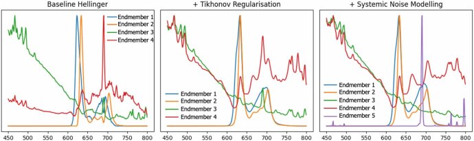 Figure 30
 Figure 31
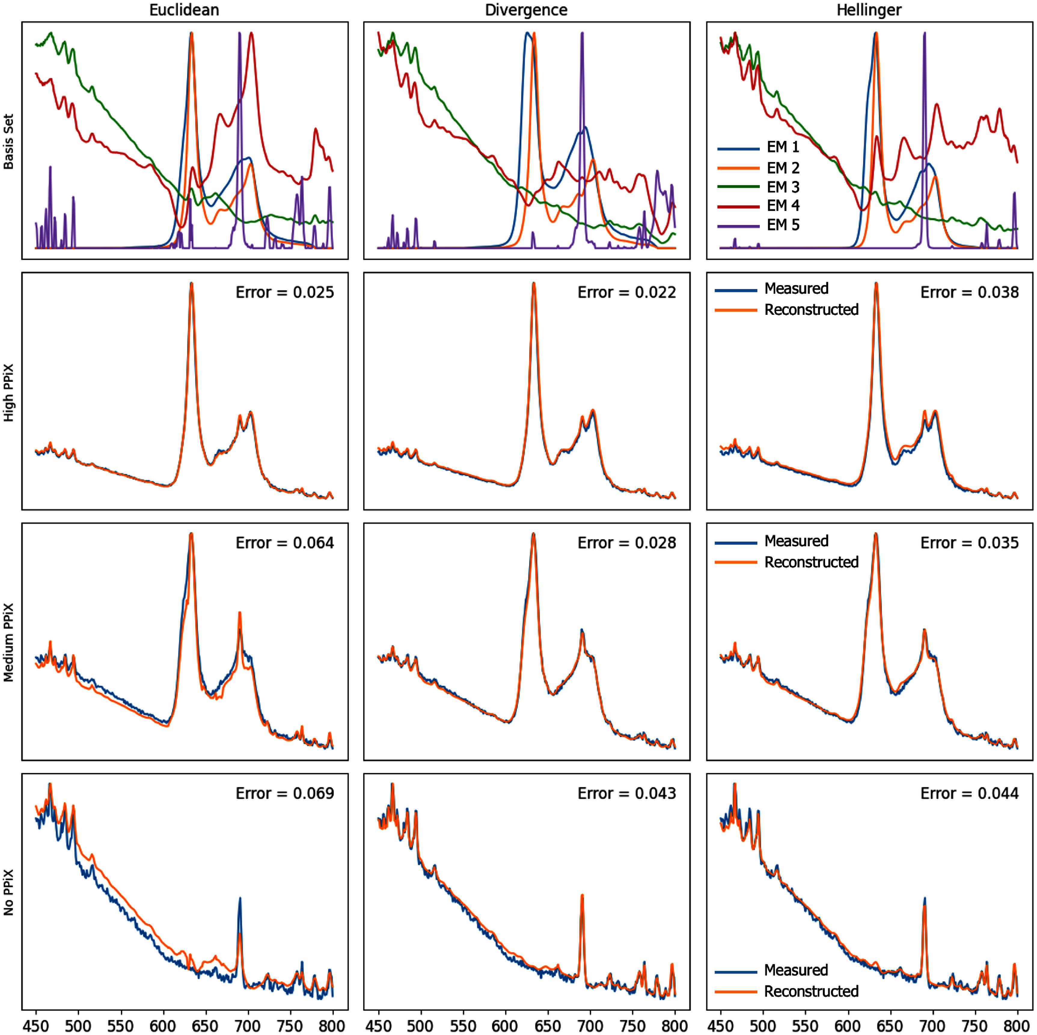 Figure 32
 Figure 33
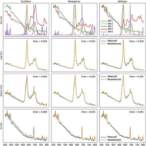 Figure 34
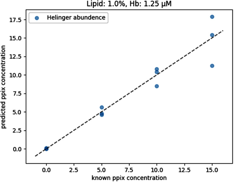 Figure 35
 Figure 36
 Figure 37
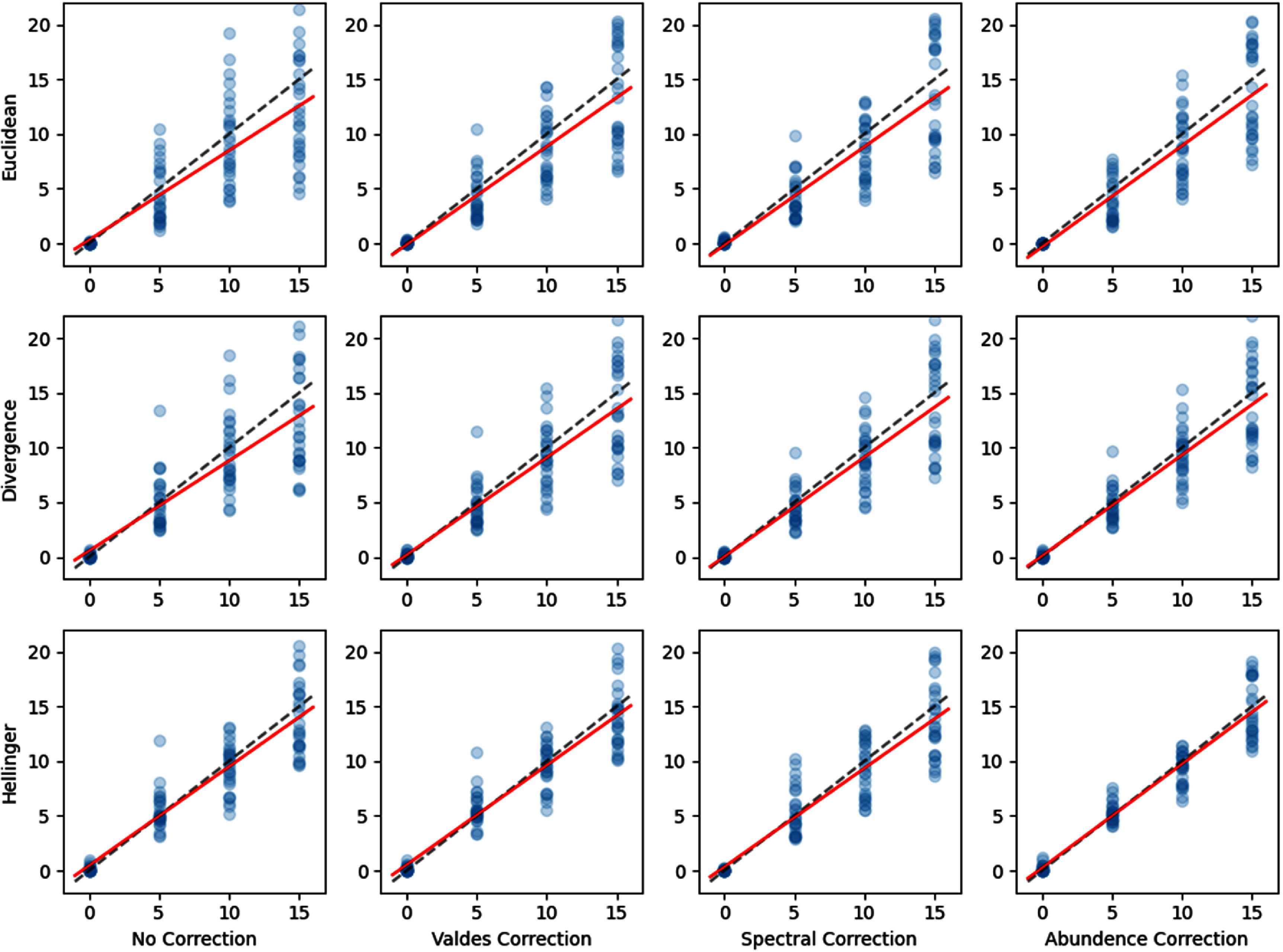 Figure 38
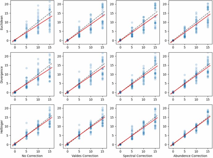 Figure 39
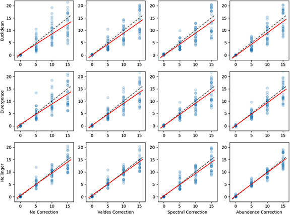 Figure 40
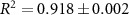 Figure 41
 Figure 42
 Figure 43
 Figure 44
 Figure 45
 Figure 46
 Figure 47
 Figure 48
 Figure 49
 Figure 50
Peer Reviews
No public reviews on file for this paper yet. If you reviewed it on a platform where reviews are public (OpenReview, ICLR, NeurIPS, ICML), you can paste yours below so the community can read it here.
Videos
No videos yet. Explain this paper in a talk, walkthrough, or lecture? Add one.
Taxonomy
TopicsPhotodynamic Therapy Research Studies · Nanoplatforms for cancer theranostics · Advanced Fluorescence Microscopy Techniques