Carrier of a pathogenic LAMB3 variant: Exploring the interface of genetic skin fragility and cutaneous autoimmunity
Ashley Jakubowicz, Emily R. Nadelmann, Michael A. Occidental, Tobi Klar

Abstract
Genes, proteins, chemicals, diseases, species, mutations and cell lines named across the full text — each resolved to its canonical identifier and authoritative record.
Click any figure to enlarge with its caption.
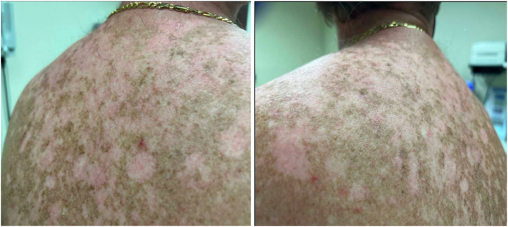 Figure 1
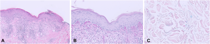 Figure 2
Peer Reviews
No public reviews on file for this paper yet. If you reviewed it on a platform where reviews are public (OpenReview, ICLR, NeurIPS, ICML), you can paste yours below so the community can read it here.
Videos
No videos yet. Explain this paper in a talk, walkthrough, or lecture? Add one.
Taxonomy
TopicsT-cell and B-cell Immunology · Cell Adhesion Molecules Research · Diabetes and associated disorders
Case description
A 59-year-old male was referred to dermatology for evaluation of new, persistent, pruritic lesions on the upper back that had developed over several months. He denied any prior history of skin fragility, blistering, or eruptions in that area or elsewhere on the body. There was no history of mucosal involvement, photosensitivity or increased ultraviolet exposure, smoking, medication exposures associated with cutaneous lupus erythematosus, or other known risk factors for cutaneous lupus, and no prior dermatologic diagnoses.
Genetic testing given his family history revealed a heterozygous pathogenic variant in LAMB3 (c.1903C>T, p.R635∗). His family history was notable for a sibling with a confirmed diagnosis of junctional epidermolysis bullosa (JEB).
On examination, the upper back showed several light pink, atrophic plaques, some with overlying scale and central erosion (Fig 1).Fig 1. Clinical examination revealed multiple light pink atrophic plaques, some eroded, on upper back.
Histopathologic evaluation revealed lichenoid interface dermatitis with basal vacuolar alteration and a band-like lymphocytic infiltrate (Fig 2, A). Periodic acid–Schiff with diastase staining showed thickening of the basement membrane zone (Fig 2, B), and Alcian Blue (pH 2.5) staining revealed interstitial dermal mucin deposition (Fig 2, C). Of note, there was a lack of subepidermal clefting and vesiculation.Fig 2. Punch biopsy demonstrated lichenoid lymphocytic inflammatory infiltrate and vacuolar interface changes (A). A PASD stain highlighted basement membrane thickening at the dermo-epidermal junction, (B), and Alcian Blue pH 2.5 stain highlighted mucin between collagen bundles in the dermis (C).
Laboratory work-up revealed a positive antinuclear antibody at 1:320 with a speckled pattern and an elevated rheumatoid factor IgM of 8.2 IU/mL. Following evaluation, including rheumatologic assessment, the patient did not meet criteria for systemic lupus erythematosus and had no evidence of systemic involvement.
Question 1: What is the diagnosis?
- **A.**Junctional epidermolysis bullosa
- **B.**Discoid lupus erythematosus
- **C.**Subacute cutaneous lupus erythematosus
- **D.**Granuloma annulare
- **E.**Polymorphic light eruption
Answer
- **B.**Discoid lupus erythematosus – Correct.
Discussion
JEB is a rare inherited blistering disorder caused by biallelic mutations in genes encoding components of the dermoepidermal basement membrane, most commonly LAMB3.1, 3^,^1, 2 While carriers of pathogenic LAMB3 variants are typically asymptomatic, emerging evidence suggests that heterozygous mutations may have subclinical or modulatory effects on skin integrity, wound healing, or immune response.3
This case is, to our knowledge, the first report of discoid lupus erythematosus (DLE) arising in a patient who is a carrier of a pathogenic LAMB3 variant, in the absence of clinical or histologic evidence of JEB. The patient had no prior history of skin fragility, blistering, or eruptions at the site of DLE involvement, raising the possibility of a unique interaction between genetic susceptibility and autoimmune skin disease.
This case raises important questions about the potential for genetic variations in LAMB3 to interact with environmental triggers, such as ultraviolet radiation, in the development of autoimmune skin conditions. Heterozygosity for a pathogenic variant in a gene associated with JEB may not only impact skin integrity but also subtly influence immune responses to endogenous or environmental stimuli.
The development of DLE at a site with no previous skin trauma or scarring distinguishes this case from those of secondary autoimmune processes that typically arise in chronically injured skin. These findings suggest that haploinsufficiency of LAMB3 may subtly compromise basement membrane integrity or immune surveillance in the skin, potentially increasing susceptibility to autoimmunity even in the absence of overt blistering.
Conflicts of interest
None disclosed.
The reference list from the paper itself. Each links out to its DOI / PubMed record.
- 1Pfendner E.G.Lucky A.W.Junctional Epidermolysis Bullosa MP Adam D Bick GM Mirzaa Gene Reviews [Internet]2018 University of Washington
- 2Medline Plus Genetics. LAMB 3 gene: laminin subunit beta 3. U.S. National Library of Medicine. Medline Plus genetics website. 2019. Accessed December 1, 2025. https://medlineplus.gov/genetics/gene/lamb 3/#resources
- 3Wen D.Hunjan M.Bardhan A.Genotype-phenotype correlation in junctional epidermolysis bullosa: signposts to severity J Invest Dermatol 144202413341343.e 143815793110.1016/j.jid.2023.11.021 · doi ↗ · pubmed ↗