Molecular detection of Mollicutes agents in reproductive system of cattle: a three-year study in Rio Grande do Sul, southern Brazil (2022–2024)
Helena Castro Alves Wessely, Evelyn Kaus Dotto, Alice Sampaio Moraes da Costa, Fernanda Silveira Flôres Vogel, Juliana Felipetto Cargnelutti, Alfredo Quites Antoniazzi

TL;DR
This study found that Mollicutes bacteria, including Ureaplasma and Mycoplasma species, are present in the reproductive systems of cattle in southern Brazil, even in animals without symptoms.
Contribution
The study provides the first detailed three-year molecular detection of Mollicutes agents in cattle reproductive systems in southern Brazil.
Findings
Ureaplasma diversum was detected in 32% of samples from cattle with and without reproductive issues.
Mollicutes were found in 23% of samples, indicating their presence in both symptomatic and asymptomatic cattle.
Mycoplasma spp. were detected in 18% of samples, highlighting the diversity of Mollicutes in the region.
Abstract
Rio Grande do Sul state, in southern Brazil, is one of the country’s largest cattle producers but reports few cases of genital infections caused by Mollicutes, specially Ureaplasma diversum and Mycoplasma bovis. The clinical signs associated with these bacteria can be easily confused with those caused by bovine herpesvirus (Varicellovirus bovinealpha1), Campylobacter fetus, and Tritrichomonas fetus, which complicates diagnosis. This study aimed to assess the occurrence of Mollicutes in cattle from Rio Grande do Sul between April 2022 and November 2024. Vulvo-vaginal and preputial lavage samples submitted for routine diagnosis at a Bacteriology Laboratory, were tested using PCR. Samples were first tested for U. diversum and M. bovis. Those testing negative were further examined for Mycoplasma bovigenitalium and Mycoplasma spp., and if still negative, for the Mollicutes class to detect…
Genes, proteins, chemicals, diseases, species, mutations and cell lines named across the full text — each resolved to its canonical identifier and authoritative record.
Click any figure to enlarge with its caption.
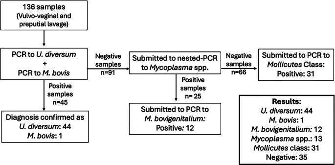 Figure 1
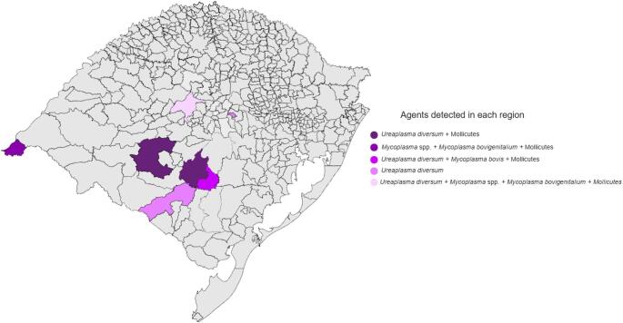 Figure 2
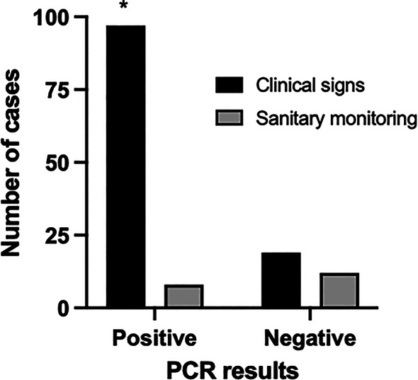 Figure 3
- —Universidade Federal De Santa Maria
Peer Reviews
No public reviews on file for this paper yet. If you reviewed it on a platform where reviews are public (OpenReview, ICLR, NeurIPS, ICML), you can paste yours below so the community can read it here.
Videos
No videos yet. Explain this paper in a talk, walkthrough, or lecture? Add one.
Taxonomy
TopicsMicrobial infections and disease research · Milk Quality and Mastitis in Dairy Cows · Reproductive tract infections research
Introduction
Beef cattle farming represents a significant component of Brazil’s national agribusiness sector. In 2024, it contributed approximately 8.4% to the country’s gross domestic product, generating an estimated R$ 987.36 billion through the production of 10.2 million tons of beef (ABIEC, 2025). In the same year, the national cattle herd was estimated at 238,180,757 head, with the state of Rio Grande do Sul accounting for 11,530,056 animals, approximately 3.2% of the national total, according to the most recent data from the Brazilian Institute of Geography and Statistics (IBGE) (BRASIL, 2024; SECRETARIA DE PLANEJAMENTO, GOVERNANÇA E GESTÃO, 2024). Sector productivity, however, is influenced by a range of factors across the production chain, among which reproductive performance plays a pivotal role. Within this context, reproductive health issues such as infertility, embryonic loss, and abortion have a direct impact on both productivity and farm profitability (Antoniassi et al. 2007). Then, the routine molecular diagnostics and sanitary measures (herd health monitoring) could be readily implemented to better manage reproductive issues and reduce economic losses across the beef production chain.
Infectious and parasitic diseases are the primary causes of reproductive losses in cattle (Alfieri and Alfieri 2017). Among the primary pathogens associated with reproductive disorders worldwide are Pestivirus bovis, Varicellovirus bovinealpha1, Brucella abortus, Leptospira interrogans, Campylobacter fetus, and Neospora caninum (Tramuta et al. 2011; Derdour et al. 2017). However, less commonly investigated microorganisms can also induce infections and/or lesions in the reproductive tract of cows and bulls, compromising reproductive performance and leading to infertility, abortion, reduced pregnancy rates, and return to estrus (Alfieri and Alfieri 2017).
Mollicutes is a bacteria class characterized by fastidious growth in culture media, small size, highly compact genomes, and lack of a rigid cell wall, which confers resistance to numerous antimicrobials (Kuppeveld et al. 1993). Members of this class, such as Mycoplasma bovis, Mycoplasma bovigenitalium, and U. diversum, are associated with reproductive infections in cattle (Karl-Erik Johansson & Bertil Pettersson, 2002; Azevedo et al. 2017; Parker et al. 2018). These opportunistic pathogens colonize the mucosa of the vulva, vagina, and udder, causing severe granular vulvovaginitis, salpingitis, endometritis, mastitis, placentitis, and fetal alveolitis, potentially resulting in temporary infertility, spontaneous abortion, or weak calves’ birth (Doig et al. 1980). In bulls, infections are often asymptomatic but can occasionally cause balanoposthitis, epididymitis, and seminal vesiculitis (Junior et al. 2021). Transmission occurs through natural mating and artificial insemination with contaminated semen (Cardoso et al. 2000).
Genital infections caused by Mollicutes in cattle have been well-documented (Petit et al. 2008; Lysnyansky et al. 2009; Argue et al. 2013; Ghanem et al. 2013; Gaeti et al. 2014). In Brazil, laboratory detection remains limited, likely due to the fastidious nature of these bacteria and their specific culture media requirements (Kuppeveld et al. 1993). This limitation may contribute to underdiagnosis, leading to an apparent low prevalence in herds. Existing studies in Brazil have primarily focused on identification and genetic characterization (Gaeti et al. 2014; Azevedo et al. 2017; Carli et al. 2021) or diagnostic assays development (Voltarelli et al. 2018). In Rio Grande do Sul State (southern Brazil), one of the largest cattle-producing regions in the country, reports of genital infections caused by Mollicutes are scarce (Carli et al. 2021). As a result, current epidemiological situation data may not accurately reflect the prevalence of these bacteria, particularly in cows.
Given the above, this study aimed to detect and identify the occurrence and distribution of class Mollicutes bacteria in clinical samples collected from beef cattle farms in Rio Grande do Sul between April 2022 and November 2024.
Material and methods
Experimental design and clinical samples
A total of 136 vulvovaginal swabs and preputial lavage samples were collected from beef cattle across 16 farms in the central, southern, and western regions of Rio Grande do Sul between April 2022 and November 2024. These samples were submitted for routine diagnosis at the Bacteriology Laboratory of the Federal University of Santa Maria for microbiological diagnosis due to a history of reduced pregnancy rates, return to cyclicity, vesicular lesions on the vulvovaginal mucosa, or for routine herd health monitoring.
The samples were analyzed for molecular detection of Mollicutes. They were then stored in saline solution and frozen at -20 °C until further processing.
DNA extraction
DNA extraction from vaginal swab samples was performed using the genomic DNA mini kit (PureLink, Invitrogen^™^, USA) following the manufacturer’s protocol. DNA from preputial lavage samples was extracted using a reagent (DNAzol, Invitrogen^™^, USA). Extracted DNA was stored at -20 °C until PCR analyses.
Primers and PCR
Before the Mollicutes polymerase chain reaction (PCR), all samples tested negative for Varicellovirus bovinealpha1, Campylobacter fetus, and Tritrichomonas foetus (Hum et al. 1997; Felleisen et al. 1998; Mayer et al. 2006). The primers used for Mollicutes detection are presented in Table 1.
Samples were tested in the following order: PCR for Ureaplasma diversum and Mycoplasma bovis; negative samples were subjected to PCR for Mycoplasma spp.; samples positive for Mycoplasma spp. were subjected to PCR for Mycoplasma bovigenitalium; samples negative for the previously tested agents were analyzed using a generic PCR for detection of the Mollicutes class. The testing flowchart can be viewed more clearly in Fig. 1.
Table 1. Primers used in PCR tests to detect Mollicutes class agentsAgentsPrimerDNA sequences (5’–3’)Product (pb) U. diversum ^^ UD1CCGGATAATAACATTTACTT986UD2CCTTGCGGTAGCAGTATCGA M. bovis ^#^ MB_FGTTTGATCCTGGCTCAGGAT198MB_RCAAACGCTTCCTTTTATATTAC M. bovigenitalium ^#^ MBG_FGTTTGATCCTGGCTCAGGAT476MBG_RAAGGTACATTCAATATAGTGGM.* spp.^†^ (nested)F1ACACCATGGGAG(C/T)TGGTAAAT350R1CTTC(A/T)TCGACTT(C/T)CAGACCCAAGGCATF2GTG(C/G)GG(A/C)TGGATCACCTCCTR2GCATCCACCA(A/T)A(A/T)AC(C/T)CTT Mollicutes ^‡^ MGSOTGCACCATCTGTCACTCTGTTAACCTC270GPOGGGAGCAAACAGGATTAGATACCC^^Cardoso et al. (2000); ^#*^Tramuta et al. (2011); ^†^Sasaki et al. (1996); ^‡^Kuppeveld et al. (1993)
Fig. 1. Flowchart illustrating the sequence of molecular tests performed on the 136 analyzed samples, including PCR assays for Ureaplasma diversum, Mycoplasma bovis, Mycoplasma spp., Mycoplasma bovigenitalium, and Mollicutes
All PCR reactions were performed in a final volume of 25 µL, using 1× reaction buffer, 2.5 mM MgCl_2_, 10 mM dNTPs, 10 pmol of each primer, 2 µL of template DNA (100–200 ng), 1 U of Taq DNA polymerase, and H_2_O q.s.p. M. bovis and U. diversum samples underwent an initial denaturation at 94 °C for 1 min, followed by 35 cycles of denaturation at 94 °C for 1 min, annealing at 50 °C for 1 min, and extension at 72 °C for 1 min; and a final extension of 5 min at 72 °C.
Negative samples from the individual PCRs for M. bovis and U. diversum were tested in nested-PCR for Mycoplasma spp. in two cycles of an initial denaturation (94 °C for 30 s), followed by 30 cycles of 94 °C for 30 s, 55 °C for 2 min, and 72 °C for 2 min; and a final extension of 2 min at 72 °C; the second cycle was comprised of initial denaturation (94 °C for 30 s), followed by 30 cycles of 94 °C for 30 s, 56 °C for 2 min, and 72 °C for 2 min, and a final extension of 2 min at 72 °C. Mycoplasma spp. positive samples were tested to M. bovigenitalium via specific-PCR by initial denaturation at 94 °C for 1 min, followed by 35 cycles of 94 °C for 1 min, 50 °C for 1 min for primer annealing, and 72 °C for 1 min for chain extension; and a final extension of 5 min at 72 °C.
Finally, samples that remained negative in the nested PCR for Mycoplasma spp. were subjected to a conventional PCR for Mollicutes (initial denaturation at 94 °C for 5 min, followed by 35 cycles of 94 °C for 30 s, 55 °C for 30 s, and 72 °C for 30 s; final extension of 10 min at 72 °C). Positive controls included DNA from M. bovis, M. bovigenitalium, and U. diversum obtained from clinically confirmed samples by sequencing. Free from contamination, ultrapure water was used as a negative control.
The PCR products were visualized on a 2% agarose gel, stained with a fluorescent dye (GelRed, Biotium©, USA), and subjected to electrophoresis for 1 h at 80 V before detection under ultraviolet light on a transilluminator.
Statistical analysis
The relationship between the detection of Mollicutes in the bovine reproductive tract of herds and the presence or absence of a history of reproductive disorders was analyzed using Prism 9 (version 9.5.1, GraphPad, USA), employing Fisher’s exact test. Statistical significance was set at p < 0.05.
Results and discussion
The analysis revealed U. diversum in 44 samples (32%), M. bovigenitalium in 12 samples (9%), and M. bovis in 1 sample (1%). Nested-PCR for Mycoplasma spp. identified 25 positive samples (18%), while 31 samples (23%) tested positive for Mollicutes class (Table 2). The geographic distribution of positive herds are shown in Fig. 2.
Table 2. Clinical and epidemiological findings of cases of Mollicutes in cattle farms of Rio Grande do Sul (2022–2024)LocalityAnimalsSampleClinicalhistoryTests resultsUDMBMYCMBGMOLBagé29VSLPM and VV290000Barra do Quaraí17PLLPM001531Caçapava do Sul 105VSVV10004Caçapava do Sul 205VSSM00002Caçapava do Sul 305VSVV00004Caçapava do Sul 405VSSM10001Caçapava do Sul 505VSVV10000Caçapava do Sul 605VSVV00002Caçapava do Sul 705VSSM20004Caçapava do Sul 805VSVV00003Caçapava do Sul 905VSVV00005Herveiras05VSSM10000Júlio de Castilhos07PLLPM00070Júlio de Castilhos18VSLPM501021Santana da Boa Vista10VSVV31003São Gabriel05VSSM10001Total 136
44
1
25
12
31 VS= Vaginal swab; PL= Prepucial lavage; LPR = Low pregnancy rate; VV = vulvovaginitis; SM = sanitary monitoring; UD = U. diversum; MB = M. bovis; MYC = Mycoplasma spp.; MBG = M. bovigenitalium; MOL = Mollicutes
Fig. 2. Geographic distribution of sampled farms in Rio Grande do Sul, Brazil, showing the herds positive for Mollicutes and associated agents (U. diversum, Mycoplasma spp., M. bovigenitalium, and M. bovis)
All herds tested positive for at least one agent, with 56% (9/16) testing positive for U. diversum, 25% (4/16) for M. bovis, M. bovigenitalium, or Mycoplasma spp., and 75% (12/16) for Mollicutes. The latter likely do not classify as U. diversum, M. bovis, M. bovigenitalium, or another Mycoplasma spp., as Mollicutes positive samples tested negative to other agents.
The analysis of preputial lavage from bulls at the Barra do Quaraí farm, which had a documented low pregnancy rate, revealed that 15 samples tested positive for Mycoplasma spp. Among these, three were confirmed by nucleotide sequencing as Mycoplasma canadense (1/15), M. bovigenitalium (1/15), or Mycoplasma californicum (1/15). The preputial lavage samples (7/7) and some vulvovaginal swabs (2/18) from farms in Júlio de Castilhos, submitted for laboratory analysis due to concerns about low pregnancy rates, tested positive for M. bovigenitalium. Several species of U. diversum have been demonstrably associated with seminal vesiculitis, balanoposthitis, and epididymitis (Pilaszek & Truszcynski, 1988). Additionally, M. bovigenitalium and M. bovis have been shown to interfere with sperm motility and cause infertility (Cardoso & Vasconcelos, 2004), compromising the herd’s productive performance.
U. diversum was detected in cows with vulvovaginitis and asymptomatic animals. However, at the Bagé farm, where cows exhibited vulvovaginitis and suffered low pregnancy rates following artificial insemination, U. diversum was detected in all sampled animals. Laboratory tests for other reproductive diseases (other Mollicutes, campylobacteriosis, leptospirosis, neosporosis, Pestivirus bovis, and Varicellovirus bovinealpha1) tested negative (data not shown), suggesting a potential role of U. diversum in the reproductive failures observed in the herd. Ureaplasma spp. exert pathogenic effects primarily through ammonia synthesis (Miller et al. 1994), inhibiting ciliary activity and causing oviduct and endometrial cell destruction (Thornber 1982). These conditions can manifest with or without clinical signs and include symptoms such as granular vulvitis, endometritis, salpingitis, abortion, infertility, and embryo loss (Azevedo, 2017).
Although U. diversum is the primary Mollicutes agent associated with vesicular and granular lesions on the vulva of cows (Cardoso & Vasconcelos, 2004), it was not detected on four farms where cows exhibited these lesions (Table 2). Nevertheless, these cows tested positive for the Mollicutes class by PCR, suggesting an involvement of other species. M. canadense has been identified in cows with granulopustular lesions on the vaginal mucosa (Lysnyansky et al. 2009). Additionally, these species may have contributed to the herd’s clinical condition.
Coinfections were detected in seven herds, typically involving U. diversum and another agent from the Mollicutes class. Multiple agents are common in infectious diseases (Alfieri and Alfieri 2017). Thus, Mollicutes coinfections in the samples are not surprising, given that some of these microorganisms are part of the vulvar and vaginal microbiota and can exhibit opportunistic pathogenicity (Junior et al. 2021). These microorganisms can persist in a commensal state until factors such as immunosuppression, antibiotic therapy, or microbiota alterations create favorable conditions for their proliferation (Cardoso and Vasconcellos 2004). Carli et al. (2021) suggest that M. bovis can facilitate colonization by U. diversum, a phenomenon that may have occurred in this study, as both pathogens were found in coinfections.
Regarding Mycoplasma spp., our findings align with those observed by Buzinhani, Timenestsky, and Metiffogo (2007), who reported a 12.5% prevalence in herds in Brazil. Nevertheless, the detection rate of U. diversum (32%) exceeded that reported by Carli et al. (2021) in preputial lavage samples from bulls (28%) across Brazil and by Argue et al. (2013), in Australian cattle (15%).
The results indicate the widespread distribution of Mollicutes class in the reproductive tracts of cows and bulls in southern Brazil. Although these agents are common in cattle worldwide (Tamiozzo et al. 2014; Szacawa et al. 2018; Deeney, 2021), reports of these infections in Brazil remain scarce, and few cases have been confirmed in Rio Grande do Sul (Carli et al. 2021). Due to underreporting, these agents are often overlooked in diagnostic considerations and bovine reproductive disease control programs. Disease control strategies could include sanitary protocols, such as using double insemination pipettes and/or condoms during artificial insemination (Cardoso & Vasconcelos, 2004).
Many symptomatic animals tested positive, with PCR results linking Mollicutes detection to clinical signs, suggesting symptomatic animals are more likely to test positive (Fig. 3). Furthermore, the results underscore the importance of asymptomatic positive animals, which can be an infection source in other animals. Among the sampled farms, 6% had animals that tested positive for Mollicutes despite showing no clinical signs. Due to their asymptomatic condition, these animals are often used in reproductive season and may transmit the agents. Moreover, because asymptomatic animals lack clinical signs, they are unlikely to undergo routine laboratory testing, contributing to the underdiagnosis of these infections.
Fig. 3PCR results and clinical status for U. diversum, M. bovigenitalium, M. bovis, Mycoplasma spp., and/or Mollicutes. ^*^Association between the variables (Fisher’s exact test, p < 0.05). Definition: Black and gray graphic with no shading. Graphic program: Graphpad prism
Although control measures and therapeutic approaches for Mollicutes-infected animals remain the same regardless of the species identified in the genital mucosa (Cardoso & Vasconcelos, 2004), identifying the etiological agent is essential for understanding the epidemiology and distribution of these pathogens within herds. This knowledge aids in developing species-specific diagnostic methods and vaccines for infections caused by particular Mollicutes species.
Lastly, our findings confirm the distribution of different Mollicutes species in genital infections of cattle in Rio Grande do Sul. One potential limitation is the reliance on samples submitted to our laboratory for diagnosis rather than those collected exclusively for this experiment, which may introduce geographic bias. However, the analyzed area is representative of beef cattle farming in the state, as all significant cattle-producing regions share characteristics similar to those of the sample collection sites. Another limitation is the lack of systematic sanitary monitoring of cows with and without lesions from the time of infection diagnosis, often coinciding with the day of artificial insemination, throughout the end of the reproductive period. Therefore, it is not possible to conclude that any observed reproductive impairments were caused by the agents present in the vaginal mucosa.
Conclusion
The results demonstrated the distribution of different species of Mollicutes, especially U. diversum, in the reproductive system of cattle across central, western, and southern Rio Grande do Sul (southern Brazil), regardless of clinical signs. Although a higher frequency of positive results was observed in herds with reproductive disorders, molecular detection alone is insufficient to establish a definitive association between the presence of the agent and the development of clinical signs.
The reference list from the paper itself. Each links out to its DOI / PubMed record.
- 1ABIEC – Associação Brasileira das Indústrias Exportadoras de Carnes. Perfil da Pecuária no Brasil. Beef Report (2025) Disponível em: https://abiec.com.br/publicacoes/beef-report-2025-perfil-da-pecuaria-no-brasil. Acesso em: 22 set. 2025
- 2Antoniassi NA et al (2007) Palestra diagnóstico das causas infecciosas de aborto em bovinos. Biológico, v.69, n.2, pp. 69–72, Disponível em: < https://biologico.agricultura.sp.gov.br/uploads/docs/bio/v 69_2/p 69-72.pdf. Acesso em: 22 de setembro de 2025
- 3Azevedo J, Silva G, Rocha P et al (2017) Presence of Ureaplasma diversum in the genital tracts of female dairy cattle in Mato Grosso State, Brazil. Trop Anim Health Prod 49(2):311–316. https://link.springer.com/article/10.1007/s 11250-016-1194-3
- 4Buzinhani M, Metiffogo E, Timenetsky J (2007) Detecção de Mycoplasma spp. E Ureaplasma diversum Em Vacas com distúrbios reprodutivos. Arq Bras Med Vet Zootec 59(6):1368–1375. https://www.scielo.br/j/abmvz/a/37q Ph Yxz NRD Jfg Lg Dyq Whsm/?lang=pt
- 5Cardoso M, Scarcelli E, Grasso L et al (2000) Ureaplasma diversum and reproductive disorder in Brazilian cows and heifers; first report. Anim Reprod Sci 63:137–143. https://www.sciencedirect.com/science/article/abs/pii/S 0378432000001767?via%3Dihub
- 6Cardoso M, Vasconcellos S (2004) Importância Das micoplasmoses Na fertilidade de Touros. Arquivos Do Instituto Biol–gico 71:257–265. https://www.scielo.br/j/aib/a/y 73PJ Vppz JZCQ 4kj F Hb KR 7q/?lang=pt
- 7Derdour S, Hafsi F, Azzag N et al (2017) Prevalence of the main infectious causes of abortion in dairy cattle in Algeria. J Vet Res 61:337–343. https://reference-global.com/article/10.1515/jvetres-2017-0044
- 8Felleisen R, Lambelet N, Bachmann P et al (1998) Detection of Tritrichomonas foetus by PCR and DNA enzyme immunoassay based on r RNA gene unit sequences. J Clin Microbiol 36:2, 513–519. https://journals.asm.org/doi/10.1128/jcm.36.2.513-519.1998