A Case of Metastatic Pulmonary Calcification
Diane Lefebvre, Pascale Bohy

TL;DR
The paper discusses a case of metastatic pulmonary calcification, explaining its causes and radiographic features.
Contribution
It highlights the distinction between metastatic and dystrophic calcification through a case study and radiographic analysis.
Findings
Metastatic calcification is linked to metabolic imbalances and has distinct CT features.
Accurate diagnosis relies on recognizing specific radiographic patterns.
Abstract
Pulmonary calcifications are a frequent phenomenon with a wide range of etiology. They are divided into two categories: metastatic and dystrophic pulmonary calcification, with different underlying pathology and CT features. Teaching point: Metastatic pulmonary calcifications appear when there is a metabolic imbalance with some typical radiographic features for characterization.
Genes, proteins, chemicals, diseases, species, mutations and cell lines named across the full text — each resolved to its canonical identifier and authoritative record.
Click any figure to enlarge with its caption.
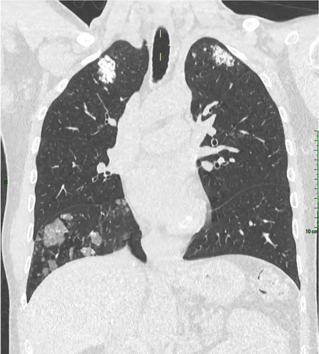 Figure 1
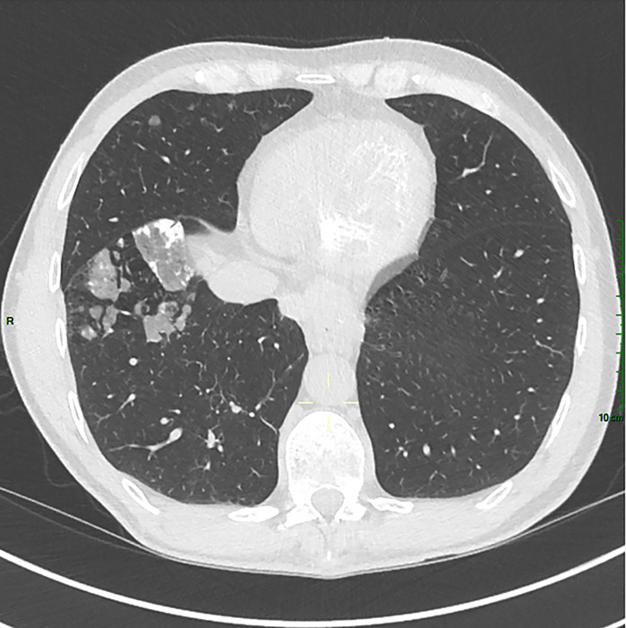 Figure 2
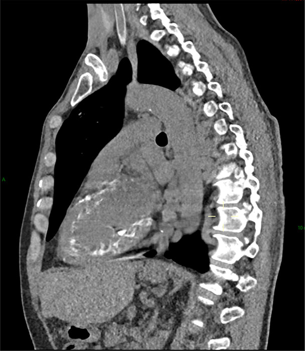 Figure 3
Peer Reviews
No public reviews on file for this paper yet. If you reviewed it on a platform where reviews are public (OpenReview, ICLR, NeurIPS, ICML), you can paste yours below so the community can read it here.
Videos
No videos yet. Explain this paper in a talk, walkthrough, or lecture? Add one.
Taxonomy
TopicsMedical Imaging and Pathology Studies · Pericarditis and Cardiac Tamponade · Inflammatory Myopathies and Dermatomyositis