Motion‐ and Field‐Robust Mesoscopic Whole‐Brain T2*‐Weighted Imaging at 7 and 11.7 T Using Servo Navigation
Matthias Serger, Rüdiger Stirnberg, Philipp Ehses, Malte Riedel, Thomas Ulrich, Caroline Le Ster, Franck Mauconduit, Vincent Gras, Alexis Amadon, Alexandre Vignaud, Son Chu, Shajan Gunamony, Maxim Zaitsev, Nicolas Boulant, Klaas P. Pruessmann, Tony Stoecker

TL;DR
This paper introduces a method to reduce motion and field artifacts in high-resolution brain imaging using servo navigation at ultra-high magnetic fields.
Contribution
The novel integration of servo navigation with segmented 3D-EPI improves motion and field correction in ultra-high field T2*-weighted imaging.
Findings
Servo navigation reduced blurring of small veins across all subjects and field strengths.
Prospective correction preserved image quality during large involuntary motion.
Field drift correction reduced shading and blurring in the frontal lobe due to microscopic motion.
Abstract
To mitigate artifacts related to motion and field changes in high‐resolution T2*‐weighted human brain imaging using servo navigation at ultra‐high fields up to 11.7 T. MR‐based servo navigators were integrated into a segmented 3D‐EPI sequence to allow for prospective correction of involuntary head motion and first‐order shim changes. Seven subjects were scanned with whole‐brain protocols at 0.3 mm isotropic resolution with and without correction at 7 and 11.7 T. Validation was performed on detailed brain vasculature in scans with involuntary motion. Blurring of small veins was reduced by servo navigation for all subjects and across field strengths. In case of involuntary large motion, the method preserved image quality, while uncorrected motion led to severe artifacts. In case of microscopic motion, reduced blurring and shading in the frontal lobe demonstrate the additional benefit of…
Genes, proteins, chemicals, diseases, species, mutations and cell lines named across the full text — each resolved to its canonical identifier and authoritative record.
Click any figure to enlarge with its caption.
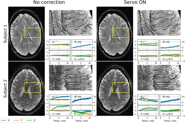 Figure 1
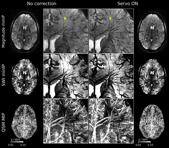 Figure 2
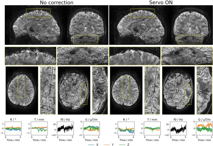 Figure 3
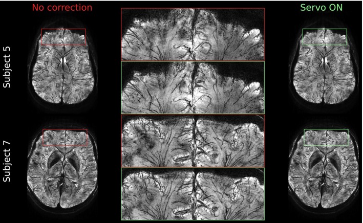 Figure 4
 Figure 5
 Figure 6
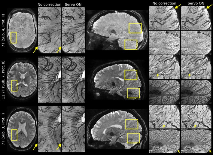 Figure 7
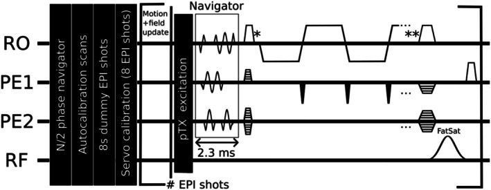 Figure 8
Peer Reviews
No public reviews on file for this paper yet. If you reviewed it on a platform where reviews are public (OpenReview, ICLR, NeurIPS, ICML), you can paste yours below so the community can read it here.
Videos
No videos yet. Explain this paper in a talk, walkthrough, or lecture? Add one.
Taxonomy
TopicsAdvanced MRI Techniques and Applications · Cerebrospinal fluid and hydrocephalus · Advanced Neuroimaging Techniques and Applications