Pole‐To‐Pole 3D Radial Trajectory Designs Improve Image Quality and Quantitative Parametric Mapping in the Brain and Heart
Eva S. Peper, Grzegorz Bauman, Matteo Tagliabue, Berk C. Açikgöz, Nils M. J. Plähn, Adèle L. C. Mackowiak, Yasaman Safarkhanlo, Joseph G. Woods, Davide Piccini, Li Feng, Christopher W. Roy, Oliver Bieri, Jessica A. M. Bastiaansen

TL;DR
New 3D radial MRI designs improve brain and heart image quality and parametric mapping accuracy by reducing phase inconsistencies.
Contribution
Pole-to-pole and continuous spiral phyllotaxis trajectories reduce phase inconsistencies in 3D radial MRI without additional corrections.
Findings
Pole-to-pole and continuous phyllotaxis improved image quality over original radial designs.
New trajectories reduced T1/T2 estimation errors compared to original and Cartesian methods.
Phase inconsistencies were mitigated without requiring extra correction steps.
Abstract
To design 3D radial spiral phyllotaxis trajectories aimed at removing phase inconsistencies, improving image quality, and enhancing parametric mapping accuracy by acquiring nearly opposing spokes starting from both hemispheres in 3D radial k‐space. Two 3D radial trajectories, pole‐to‐pole and continuous spiral phyllotaxis, were developed and implemented on a 3T MRI scanner in a phase‐cycled balanced steady‐state free precession (bSSFP) and a spoiled gradient‐echo (GRE) sequence. Image quality and k‐space center phase variations were evaluated in a spherical phantom using the original and new radial phyllotaxis designs. T1/T2 was quantified and compared using phase‐cycled bSSFP data acquired with the new radial trajectory designs, as well as the original phyllotaxis trajectory and a Cartesian trajectory as references, in both an MRI system phantom and the brains of three healthy…
Genes, proteins, chemicals, diseases, species, mutations and cell lines named across the full text — each resolved to its canonical identifier and authoritative record.
Click any figure to enlarge with its caption.
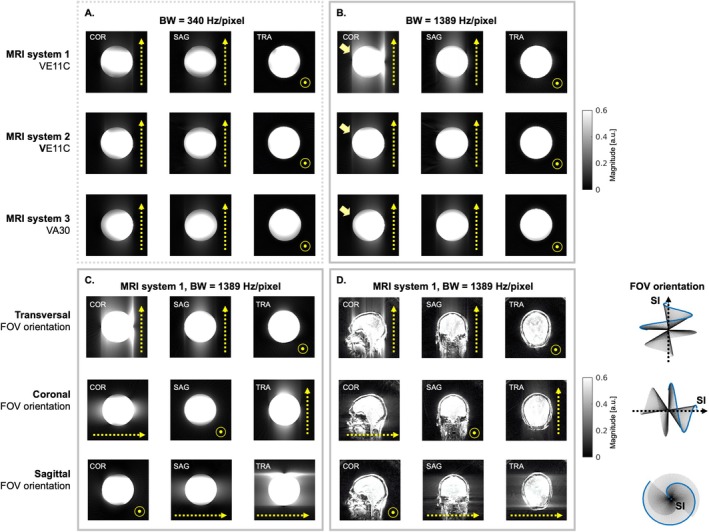 Figure 1
 Figure 2
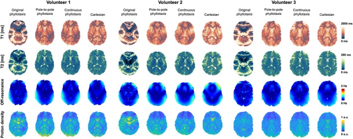 Figure 3
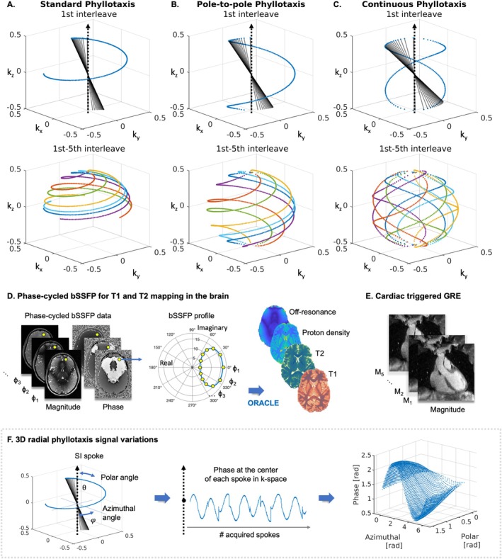 Figure 4
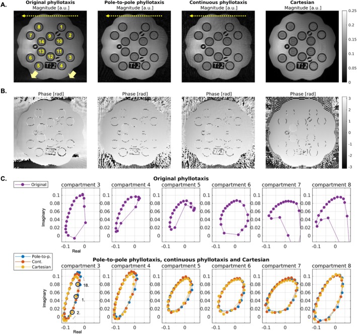 Figure 5
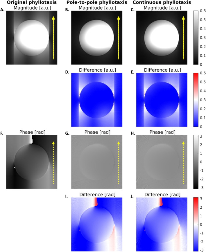 Figure 6
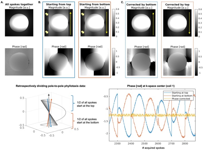 Figure 7
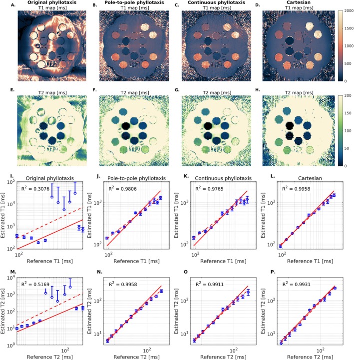 Figure 8
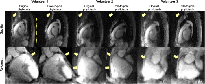 Figure 9
Peer Reviews
No public reviews on file for this paper yet. If you reviewed it on a platform where reviews are public (OpenReview, ICLR, NeurIPS, ICML), you can paste yours below so the community can read it here.
Videos
No videos yet. Explain this paper in a talk, walkthrough, or lecture? Add one.
Taxonomy
TopicsAdvanced MRI Techniques and Applications · Advanced Neuroimaging Techniques and Applications · Functional Brain Connectivity Studies