Jointly Learned 3D Non‐Cartesian Sampling With Wave Encoding and Reconstruction for Neurovascular Phase Contrast MRI
Chenwei Tang, Brock W. Jolicoeur, James Rice, Caroline A. Doctor, Zaynab S. Yardim, Leonardo A. Rivera‐Rivera, Laura B. Eisenmenger, Kevin M. Johnson

TL;DR
This paper introduces a new method for 3D phase contrast MRI that uses jointly learned wave encoding and reconstruction to improve image quality and flow measurements in neurovascular scans.
Contribution
The novelty is the joint learning of 3D non-Cartesian sampling with wave encoding and MoDL reconstruction for accelerated neurovascular phase contrast MRI.
Findings
Learned wave scans showed accurate flow rates in a phantom compared to reference measurements.
In vivo scans with learned wave sampling had reduced aliasing and better small vessel visibility than other methods.
Learned wave scans achieved flow measurements with variability comparable to longer reference scans.
Abstract
To develop accelerated 3D phase contrast (PC) MRI using jointly learned wave encoding and reconstruction. Pseudo‐fully sampled neurovascular 4D flow data (N = 40) and a simulation framework were used to learn phase encoding locations, wave readout parameters, and model‐based reconstruction network (MoDL) for a rapid 3D PC scan (2.25 min). Parameters were also learned for an otherwise identical scan without wave encoding. Prospective scans with and without wave sampling, time‐matched 3D radial, and reference 3D radial (5.65 min) were conducted in a flow phantom and 12 healthy participants. Flow rate, pixel‐wise velocity, and variability of maximum velocity (σvmax) were compared. In the phantom, learned wave scans provided accurate flow rates compared to flow probe values (0.170 ± 0.002 vs. 0.17, 0.152 ± 0.003 vs. 0.15, 1.838 ± 0.044 vs. 1.83 L/min) and showed high correlation with…
Genes, proteins, chemicals, diseases, species, mutations and cell lines named across the full text — each resolved to its canonical identifier and authoritative record.
Click any figure to enlarge with its caption.
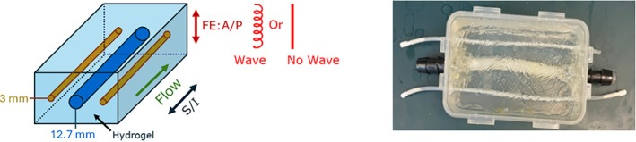 Figure 1
 Figure 2
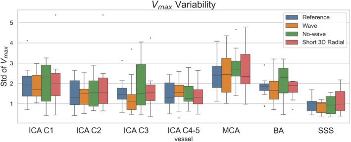 Figure 3
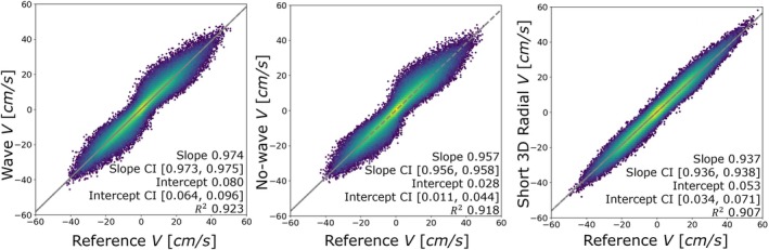 Figure 4
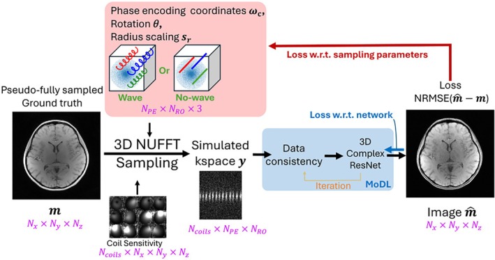 Figure 5
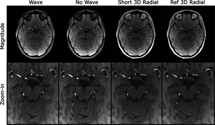 Figure 6
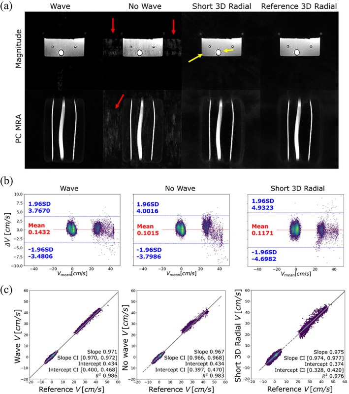 Figure 7
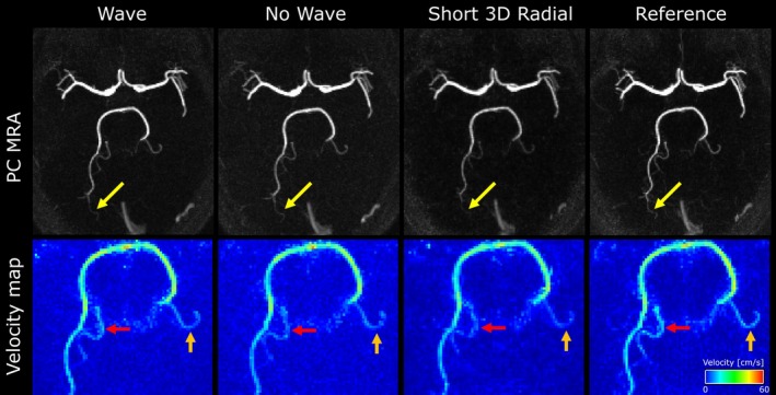 Figure 8
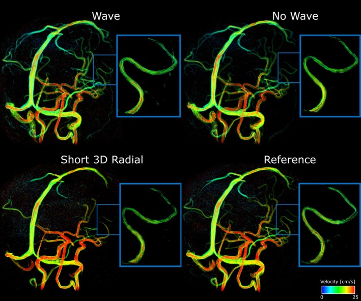 Figure 9
Peer Reviews
No public reviews on file for this paper yet. If you reviewed it on a platform where reviews are public (OpenReview, ICLR, NeurIPS, ICML), you can paste yours below so the community can read it here.
Videos
No videos yet. Explain this paper in a talk, walkthrough, or lecture? Add one.
Taxonomy
TopicsAdvanced MRI Techniques and Applications · Seismic Imaging and Inversion Techniques · Ultrasound Imaging and Elastography