Clinical feasibility of accelerated whole liver water T1 mapping with T2*-compensation
Elizabeth Huaroc Moquillaza, Lisa Steinhelfer, Kilian Weiss, Robert Walter, Jonathan Stelter, Mariya Doneva, Rickmer Braren, Dimitrios C. Karampinos

TL;DR
A new fast liver imaging method captures whole liver T1 values and compensates for fat and iron effects, potentially improving non-invasive fibrosis detection.
Contribution
A novel accelerated water T1 mapping method with T2*-compensation for liver fibrosis assessment is proposed and clinically evaluated.
Findings
The wT1 method provides broad liver coverage in an 11-second breath-hold and captures all liver segments.
T2*-compensation (wT1comp) outperforms wT1 in identifying fibrosis-related tissue changes in liver segments.
The wT1comp model effectively removes T1 bias from fat and iron, enabling contrast-free fibrosis assessment.
Abstract
Current liver T1 mapping methods present restricted liver coverage, take long acquisition times and mostly exclude the T1 bias induced by fat and iron effects. We evaluated the clinical feasibility of an accelerated water T1 (wT1) mapping method, including all liver segments and the potential of its T2*-compensation (wT1comp) for fibrosis tissue assessment. Forty-three patients were classified into three groups: benign without/with risk of developing fibrosis and hepatocellular carcinoma (HCC). A 9-slice accelerated single-shot spiral continuous inversion-recovery Look-Locker (CIR-LL) wT1 mapping acquisition, performed in an 11-s breath-hold, and clinical images (proton density fat fraction (PDFF), T2*, T1- and T2-weighted) were acquired for all patients. ROIs were defined on the PDFF, T2* and wT1 maps in all liver segments. wT1comp was estimated based on the wT1-T2* correlation of the…
Genes, proteins, chemicals, diseases, species, mutations and cell lines named across the full text — each resolved to its canonical identifier and authoritative record.
Click any figure to enlarge with its caption.
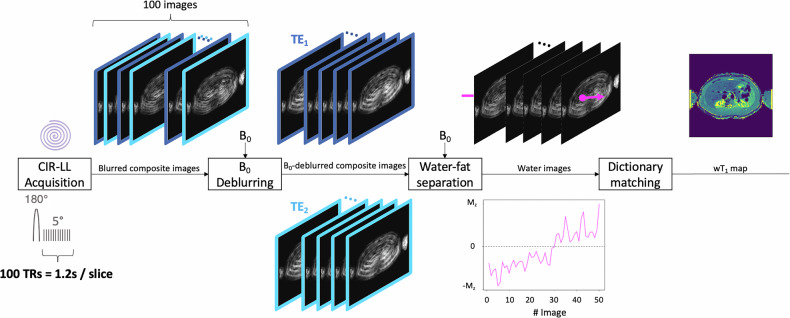 Figure 1
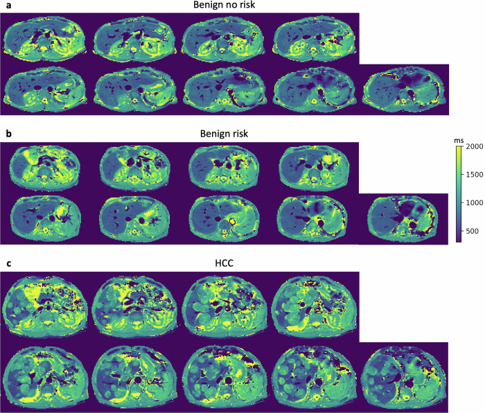 Figure 2
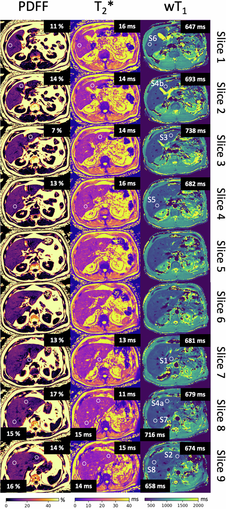 Figure 3
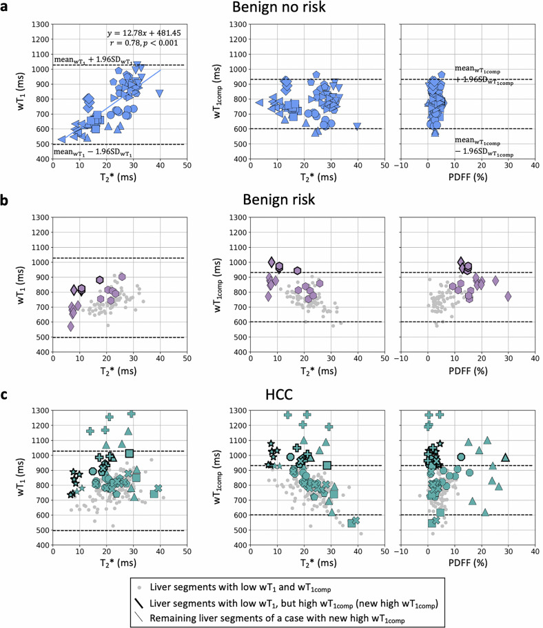 Figure 4
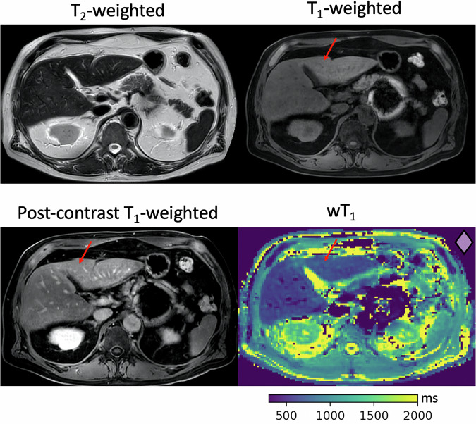 Figure 5
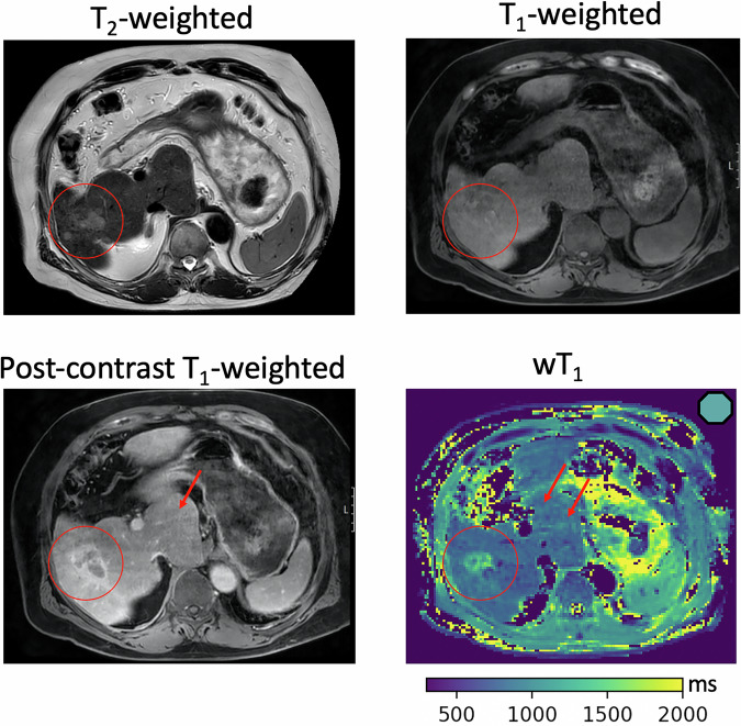 Figure 6
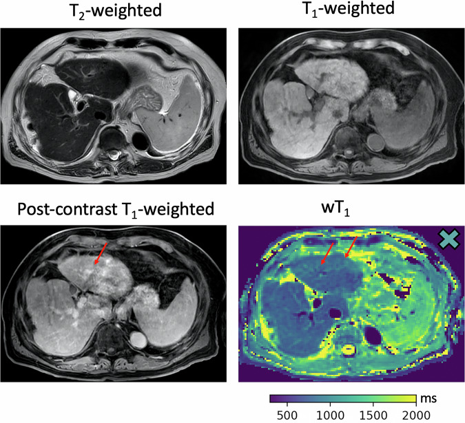 Figure 7
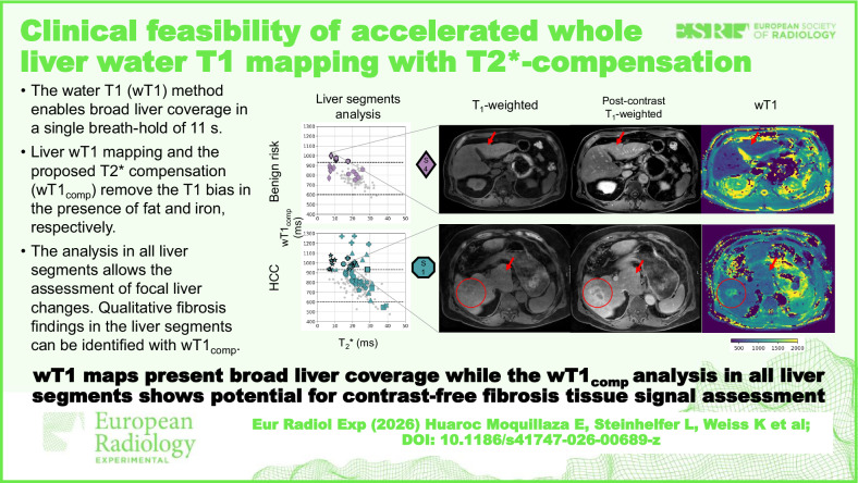 Figure 8
Peer Reviews
No public reviews on file for this paper yet. If you reviewed it on a platform where reviews are public (OpenReview, ICLR, NeurIPS, ICML), you can paste yours below so the community can read it here.
Videos
No videos yet. Explain this paper in a talk, walkthrough, or lecture? Add one.
Taxonomy
TopicsLiver Disease Diagnosis and Treatment · Hepatocellular Carcinoma Treatment and Prognosis · Advanced MRI Techniques and Applications