Differences in β-lactamase activity and carbapenem resistance among the Bacillus cereus group
Yuji Nishihara, Ryuichi Nakano, Akiyo Nakano, Yuki Suzuki, Miho Ogawa, Ryuji Sakata, Hisakazu Yano, Kei Kasahara

TL;DR
This study explores how different species in the Bacillus cereus group show varying levels of resistance to β-lactam antibiotics, including carbapenems, through differences in β-lactamase activity.
Contribution
The study introduces a method to measure β-lactamase enzyme activity and reveals interspecies variability in resistance mechanisms within the B. cereus group.
Findings
Bacillus luti isolates showed higher resistance to ampicillin and meropenem despite lacking carbapenemase genes.
β-lactamase activity in the B. cereus group was categorized as constitutive, inducible, or silent.
Inducible enzyme activity was linked to elevated penicillinase and carbapenemase expression.
Abstract
The Bacillus cereus group causes severe nosocomial infections. This group carries the chromosomal β-lactamases, including bla1 and BcII, which contribute to β-lactam resistance; however, the β-lactam resistance mechanisms are poorly understood. We performed genomic and phenotypic analyses of 48 clinical isolates from blood cultures and the reference strain ATCC14579 to clarify these mechanisms. Genomic analyses included species identification, multilocus sequence typing (MLST), and detection of β-lactamase genes using whole-genome sequencing. β-Lactam susceptibility testing, enzyme activity assays, and RT-qPCR of β-lactamases were performed. For this analysis, we developed a method to measure the enzyme activity of B. cereus group. The 48 isolates comprised three species (30 Bacillus mosaicus, 9 Bacillus cereus sensu stricto (s.s.), and 9 Bacillus luti) and 28 sequence types. Although…
Genes, proteins, chemicals, diseases, species, mutations and cell lines named across the full text — each resolved to its canonical identifier and authoritative record.
Click any figure to enlarge with its caption.
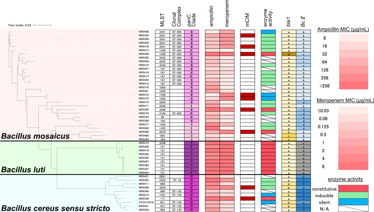 Fig 1
Fig 1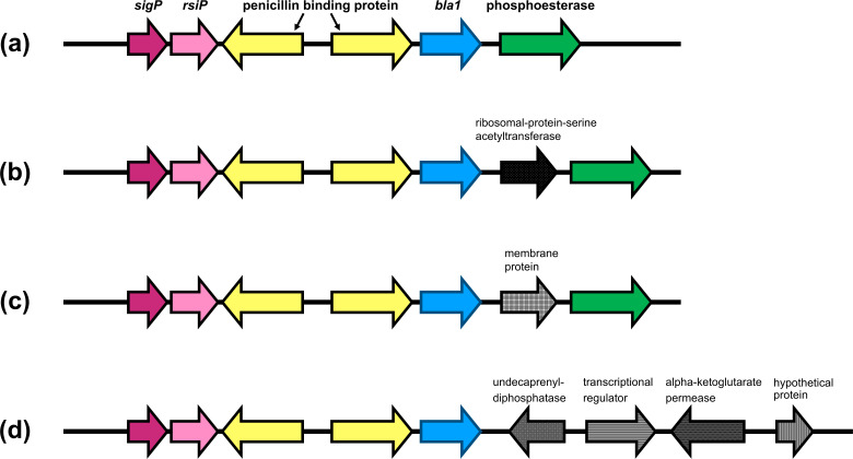 Fig 2
Fig 2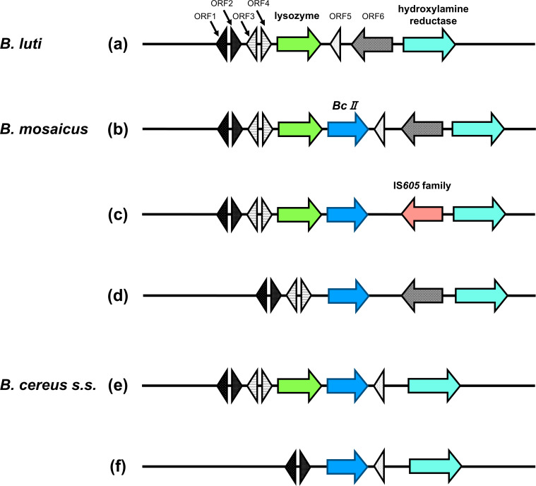 Fig 3
Fig 3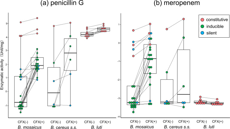 Fig 4
Fig 4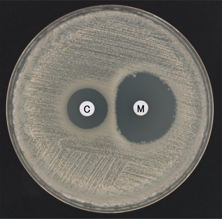 Fig 5
Fig 5| Enzyme category | NR isolate | Species | MIC (µg/mL) | mCIM | Enzyme activity (unit/mg) | mRNA | |||||||||||
|---|---|---|---|---|---|---|---|---|---|---|---|---|---|---|---|---|---|
| AMP | MEM | PCNG | PCNG with CFX | Fold | MEM | MEM with CFX | Fold | bla1 | Fold | BcII | Fold | ||||||
| Constitutive | 5368 |
| >256 | 8 | Positive | 60.29 | 72.76 | 1.21 | 1.97 | 2.80 | 1.42 | 0.62 | 0.86 | 1.39 | 8.22 | 9.08 | 1.11 |
| 5385 |
| >256 | 4 | Positive | 166.23 | 224.82 | 1.35 | 0.49 | 0.92 | 1.86 | 2.98 | 3.20 | 1.08 | 3.00 | 3.47 | 1.15 | |
| 5388 |
| 64 | 4 | Positive | 60.79 | 97.95 | 1.61 | 0.90 | 1.68 | 1.87 | 0.76 | 1.18 | 1.56 | 4.39 | 4.58 | 1.04 | |
| 5396 | >256 | 8 | Positive | 125.31 | 159.57 | 1.27 | 2.13 | 3.67 | 1.73 | 0.76 | 0.85 | 1.13 | 2.57 | 2.30 | 0.90 | ||
| 5372 |
| >256 | 8 | Negative | 113.46 | 148.25 | 1.31 | 0.04 | 0.03 | 0.95 | 1.82 | 1.55 | 0.85 | <0.01 | <0.01 | <0.01 | |
| 5390 |
| 256 | 8 | Negative | 67.47 | 88.17 | 1.31 | 0.04 | 0.04 | 0.94 | 1.91 | 1.58 | 0.83 | <0.01 | <0.01 | <0.01 | |
| 5395 |
| 256 | 8 | Negative | 61.70 | 101.64 | 1.65 | 0.04 | 0.04 | 1.04 | 1.88 | 1.68 | 0.89 | <0.01 | <0.01 | <0.01 | |
| 5400 |
| 256 | 8 | Negative | 50.84 | 110.93 | 2.18 | 0.05 | 0.04 | 0.76 | 1.40 | 1.70 | 1.22 | <0.01 | <0.01 | <0.01 | |
| 5404 |
| >256 | 8 | Negative | 73.01 | 85.42 | 1.17 | 0.04 | 0.04 | 0.98 | 1.82 | 1.68 | 0.92 | <0.01 | <0.01 | <0.01 | |
| Inducible | 5386 | 256 | 4 | Positive | 8.30 | 60.01 | 7.23 | 0.20 | 1.34 | 6.55 | 0.28 | 1.63 | 5.86 | <0.01 | 1.28 | >100 | |
| 5402 |
| 32 | ≤0.03 | Negative | 0.27 | 5.18 | 19.20 | 0.03 | 0.41 | 13.87 | 0.10 | 2.52 | 26.47 | <0.01 | 10.39 | >100 | |
| 5405 |
| 32 | 2 | Negative | 0.72 | 5.62 | 7.83 | 0.04 | 0.54 | 13.69 | 1.09 | 28.89 | 26.58 | <0.01 | 25.89 | >100 | |
| 5413 |
| 64 | ≤0.03 | Negative | 0.30 | 5.96 | 20.08 | 0.03 | 0.42 | 13.05 | 0.50 | 2.86 | 5.78 | 3.76 | 22.19 | 5.90 | |
| 5416 |
| 32 | 0.06 | Negative | 0.45 | 10.16 | 22.68 | 0.05 | 0.79 | 16.07 | 0.43 | 4.45 | 10.27 | <0.01 | 63.71 | >100 | |
| 5371 |
| 128 | 0.06 | Negative | 0.29 | 6.69 | 22.83 | 0.03 | 0.26 | 7.99 | 2.44 | 2.63 | 1.08 | 0.35 | 11.06 | 31.36 | |
| 5380 |
| >256 | ≤0.03 | Negative | 0.35 | 7.89 | 22.73 | 0.04 | 0.16 | 4.13 | 1.34 | 1.03 | 0.77 | <0.01 | 0.87 | >100 | |
| 5392 |
| 64 | 2 | Positive | 0.35 | 17.15 | 48.37 | 0.10 | 1.25 | 12.90 | 0.87 | 0.82 | 0.95 | 7.03 | 16.64 | 2.37 | |
| 5414 |
| 16 | ≤0.03 | Negative | 0.22 | 7.45 | 34.62 | 0.02 | 0.19 | 8.08 | 0.73 | 0.99 | 1.36 | 4.58 | 17.48 | 3.82 | |
| Silent | 5374 |
| 8 | 4 | Positive | 5.19 | 5.39 | 1.04 | 1.06 | 1.27 | 1.21 | 1.16 | 1.14 | 0.99 | 1.69 | 1.07 | 0.63 |
| 5375 |
| 16 | 4 | Positive | 2.43 | 6.49 | 2.68 | 0.71 | 1.70 | 2.39 | 0.91 | 1.10 | 1.21 | 0.91 | 0.49 | 0.54 | |
| 5408 |
| 8 | 1 | Negative | 2.81 | 2.35 | 0.84 | 0.04 | 0.04 | 1.12 | 2.37 | 2.14 | 0.90 | 0.93 | <0.01 | <0.01 | |
| 5415 | 64 | 0.06 | Negative | 0.38 | 0.36 | 0.93 | 0.04 | 0.04 | 0.93 | 1.40 | 1.21 | 0.86 | 1.38 | 0.93 | 0.67 | ||
| ATCC14579 | 64 | 0.06 | Negative | 2.15 | 3.61 | 1.68 | 0.04 | 0.04 | 1.01 | 1.00 | 0.89 | 0.89 | 1.00 | 0.51 | 0.51 | ||
| Gene | Orientation | Primer sequence (5' to 3') | Product size (bp) | Reference |
|---|---|---|---|---|
|
| F | 161 | This study | |
| R | ACRCCTGCACGAATAAGTTT | |||
|
| F |
| 99 | This study |
| R1 |
| |||
| R2 |
| |||
|
| F |
| 175 | ( |
| R |
| |||
|
| F |
| 101 | ( |
| R |
|
- —Japan Society for the Promotion of Sciencehttp://dx.doi.org/10.13039/501100001691
Peer Reviews
No public reviews on file for this paper yet. If you reviewed it on a platform where reviews are public (OpenReview, ICLR, NeurIPS, ICML), you can paste yours below so the community can read it here.
Videos
No videos yet. Explain this paper in a talk, walkthrough, or lecture? Add one.
Taxonomy
TopicsBacillus and Francisella bacterial research · Antibiotic Resistance in Bacteria · Antimicrobial agents and applications
INTRODUCTION
The Bacillus cereus group comprises gram-positive, spore-forming rod bacteria that are ubiquitously distributed in natural environments (1, 2). This group includes closely related species with high genetic similarity, such as Bacillus anthracis, Bacillus cereus, and Bacillus thuringiensis (3). B. anthracis, harboring virulence plasmids pXO1 and/or pXO2, causes anthrax, which is potentially fatal. B. cereus mainly causes gastrointestinal infections such as food poisoning in humans but can also cause severe, invasive infections, particularly in immunocompromised individuals and neonates. B. thuringiensis causes lethal infections in insects and is widely used as a biopesticide (4). To date, approximately 20 species have been proposed for inclusion in the B. cereus group (3, 5). Specific species within the B. cereus group cannot be reliably differentiated using the phenotypic identification method or matrix-assisted laser-desorption/ionization time-of-flight mass spectrometry (MALDI-TOF MS) with commercially available databases (3, 6). Whole-genome sequencing (WGS) is required for accurate identification of species within this group (3, 7). Because of the difficulties with identification, detailed species classification is not possible in routine clinical laboratory practice.
Except for B. anthracis, most species within the B. cereus group are resistant to penicillins and cephalosporins (2). Penicillin susceptibility is frequently used to differentiate B. anthracis from other species of the B. cereus group, such as B. cereus and B. thuringiensis (8), but sporadic penicillin resistance of B. anthracis has been reported (9). In the B. cereus group, including B. anthracis, the expression of chromosomal β-lactamase genes is thought to be partially responsible for β-lactam resistance (8, 9). Chromosomally encoded β-lactamases include serine-β-lactamase, bla1, and metallo-β-lactamase, bla2 (9, 10). In penicillin-sensitive B. anthracis strains, both bla1 and bla2 are almost silent transcriptionally, whereas semiquantitative RT-PCR analysis showed that these genes are expressed in penicillin-resistant strains (9, 11).
Previous studies have confirmed that bla1 contributes to penicillin resistance. The deletion of bla1 gene in penicillin-resistant B. anthracis strains results in a dramatic decrease in the minimum inhibitory concentration (MIC) of ampicillin (12), and B. thuringiensis, a species genetically similar to B. anthracis, also demonstrates a decreased MIC of ampicillin with the elimination of bla1 (13). However, the implications of bla2 for the susceptibility to carbapenems are not well understood. Discovered in 1966, bla2 was the first extracellular metallo-β-lactamase to be discovered (14). Despite the presence of chromosomal carbapenemase, members of the B. cereus group are generally sensitive to carbapenem antibiotics (5, 15), and the production of bla2 is insufficient to confer carbapenem resistance (9, 12).
The regulatory mechanisms underlying β-lactamase expression in the B. cereus group have been gradually elucidated, with recent studies demonstrating that extracytoplasmic function (ECF) sigma factors play a key role in this process (8, 13). However, the detailed regulatory mechanisms, especially those for carbapenemase expression, remain poorly understood.
In bacteria, the core RNA polymerase (RNAP) associates with a sigma factor to form the holoenzyme, which enables promoter recognition and transcription initiation. Sigma factors determine promoter specificity, thereby directing RNAP to transcribe distinct sets of genes (16). ECF sigma factors are a subfamily of σ^70^ sigma factors. They typically regulate cellular processes related to the cell envelope and are, therefore, termed “extracytoplasmic function” sigma factors (16). The general regulatory pathway of ECF sigma factors involves a membrane-bound anti-sigma factor that sequesters the sigma factor on the membrane, thereby suppressing its activity. Upon the detection of an extracytoplasmic signal, the anti-sigma factor is proteolytically degraded, releasing the sigma factor to activate transcription of the target genes (16, 17).
In the B. cereus group, SigP functions as an ECF sigma factor, and its activity is inhibited by its cognate anti-sigma factor, RsiP. These components are thought to participate in the regulation of β-lactamase expression. Penicillin-resistant B. anthracis strains constitutively express β-lactamase activity, whereas the deletion of the sigP–rsiP locus completely abolishes it (8). In B. thuringiensis, SigP regulates multiple β-lactamases and penicillin-binding proteins (PBPs), potentially contributing to β-lactam resistance (13). Additionally, ECF sigma factors upregulate β-lactamase activity in response to β-lactam antibiotic exposure (8, 13). However, few studies have investigated the levels of metallo-β-lactamase expression or the mechanisms of carbapenem resistance, and the mechanisms of metallo-β-lactamase and its contribution to carbapenem resistance remain unclear. Carbapenems are a crucial therapeutic option for treating infections caused by the B. cereus group (5); therefore, understanding carbapenem resistance mechanisms has important implications for treating clinical infectious diseases.
Measuring enzyme activity is crucial for clarifying the mechanisms of resistance to β-lactam antibiotics. However, in Bacillus species, β-lactamase activity has not been accurately quantified. Gram-positive bacteria secrete the β-lactamase enzymes that they produce into the extracellular environment (18, 19). Although previous studies have measured enzyme activity in liquid culture supernatants (11, 20), these measurements have not been normalized to protein content; therefore, enzyme activity assays have not been standardized.
In this study, we used 48 clinical isolates of the B. cereus group from patient blood cultures and conducted detailed species identification based on WGS analysis. We developed a novel method of quantifying enzyme activity toward β-lactam antibiotics and performed quantitative reverse transcription polymerase chain reaction (RT-qPCR) on a subset of the isolates to measure the levels of penicillinase and metallo-β-lactamase expression. This revealed that β-lactamase activity manifests in three distinct categories: constitutive, inducible, and silent and that carbapenem resistance levels and the expression of chromosomal β-lactamases vary considerably among species in the B. cereus group.
RESULTS
Species and MLST
Using the average nucleotide identity (ANI) method of the BTyper3 system (21), 48 isolates were taxonomically classified as three different species: 30 Bacillus mosaicus, 9 Bacillus cereus sensu stricto (s.s.), and 9 Bacillus luti (Fig. 1). All B. cereus s.s. isolates were classified as panC group IV, and all B. luti isolates were classified as panC group V. The B. mosaicus were classified as either panC group II or III.
Phylogenetic tree of 48 clinical isolates from the Bacillus cereus group and the reference strain B. cereus ATCC 14579, with associated antimicrobial susceptibility, mCIM results, enzyme activity, and β-lactamase gene profiles. A phylogenetic tree of 48 NR isolates of Bacillus cereus group combined with the reference strain B. cereus ATCC 14579 was constructed using Mashtree (22). All isolates were classified into three species: Bacillus mosaicus, Bacillus luti, and Bacillus cereus sensu stricto (s.s.). A total of 28 distinct sequence types (STs) were identified. B. cereus s.s. isolates belonged to panC group IV, B. luti belonged to group V, and B. mosaicus belonged to groups II or III. Minimum inhibitory concentrations (MICs) of ampicillin and meropenem were represented using a color gradient from white (low MICs) to red (high MICs). Positive results in the mCIM are indicated by red boxes. The enzyme activity categories are shown with colored boxes; red indicates constitutive activity, green indicates inducible activity, and blue indicates a silent phenotype. The genetic structures around bla1 and BcII are shown based on the classifications shown in Fig. 2 and 3, respectively. Abbreviation: mCIM, modified carbapenem inactivation method.
MLST analysis revealed that the 48 isolates were distributed across 28 distinct sequence types (STs). Of the nine B. luti isolates, eight were ST151. Of the 30 B. mosaicus isolates, 5 isolates each were ST167 and ST3341, and 2 isolates each were ST3343, ST1050, ST1420, and ST1425. Of the 30 B. mosaicus isolates analyzed, 15 were classified within clonal complex (CC) 365. The STs of B. cereus s.s. were different for all isolates, but four (44%) belonged to CC142. The species, MLST sequence type, CC, panC phylogenetic group, and other detailed genomic information are shown in Fig. 1 and Table S1.
Antimicrobial susceptibility and the modified carbapenem inactivation method
The MICs (µg/mL) for ampicillin and meropenem, and the modified carbapenem inactivation method (mCIM) results, are shown in Fig. 1. The MICs of both antibiotics against B. mosaicus and B. cereus s.s. displayed a broad distribution. In contrast, B. luti exhibited consistently elevated MIC values for both β-lactams.
Notably, despite higher MICs for meropenem, all B. luti isolates tested negative using mCIM. Among the other two species, 7 (23%) B. mosaicus and 2 (22%) B. cereus s.s. isolates showed positive mCIM results. For these two species, all isolates with meropenem MICs >4 µg/mL were mCIM positive. Among the isolates, 4 B. mosaicus isolates and 1 B. cereus s.s. isolate had a meropenem MIC of 2 µg/mL, and 2 of the B. mosaicus isolates were mCIM positive. Importantly, all isolates with an MIC of ≤1 µg/mL were mCIM negative.
Presence of β-lactamase genes among each species
The bla1 gene was found in all 48 isolates. The genetic structure surrounding bla1 was highly conserved, with the sigma factor genes sigP and rsiP arranged sequentially, followed downstream by two types of PBPs and bla1. In contrast, the downstream region of bla1 exhibited strain-specific variability, with diverse genetic elements, such as phosphoesterases, acetyltransferases, and membrane proteins, present in different isolates (Fig. 2).
Schematic representation of the genetic context surrounding bla1. The upstream region of bla1 was highly conserved across isolates and consistently contained sigP, rsiP, and two penicillin-binding protein genes. In contrast, the downstream region was classified into four distinct types based on the gene located immediately downstream of bla1. Forty of 48 isolates belonged to Group a; 5 isolates (NR5386, NR5388, NR5391, NR5411, and NR5415) and the reference strain, B. cereus ATCC 14579, belonged to Group b; two isolates (NR5384 and NR5406) belonged to Group c; and NR5368 belonged to Group d. Purple arrows represent sigP; pink, rsiP; yellow, penicillin-binding protein; blue, bla1; green, phosphoesterase; and black patterned arrows represent other genes. Detailed information on the isolates and their corresponding genetic types is provided in Table S2.
The homology levels of bla1 ranged from 85% to 99% at the nucleotide level and from 84% to 99% at the amino acid level. Relative to the ATCC reference strain, nucleotide homology varied among species. The homology ranged from 97% to 99% in B. cereus s.s., 91% to 92% in B. luti, and 85% to 99% in B. mosaicus.
The BcII (bla2) gene was detected in all the B. cereus s.s. and B. mosaicus isolates analyzed. In contrast with the relatively conserved region surrounding bla1, the genetic structure surrounding BcII displayed considerable variability. Notably, sigma factor genes such as sigP and rsiP were absent in the vicinity of BcII. Instead, the genetic region around BcII, and the corresponding genomic locus in B. luti, contained genes encoding lysozymes and hydroxylamine reductases, along with several genes of unknown function.
In B. mosaicus and B. cereus s.s., BcII appeared to be inserted between the forward-oriented lysozyme gene and the reverse-oriented hypothetical protein found in B. luti (Fig. 3). The genetic structure of B. luti is shown in Fig. 3a, and the genetic structures of 19 of the 30 B. mosaicus are shown in Fig. 3b. In four B. mosaicus isolates (NR5368, NR5382, NR5409, and NR5410), an IS605 element was present downstream of BcII (Fig. 3c). Additionally, in seven B. mosaicus isolates (NR5373, NR5377, NR5379, 5380, NR5385, NR5393, and NR5398), the lysozyme gene immediately upstream of BcII was absent (Fig. 3d).
Schematic representation of the genetic context surrounding BcII. The genetic structure surrounding BcII varied between species. All nine isolates of B. luti completely lacked the BcII gene (type a). B. mosaicus isolates were classified into three distinct structural types (19 type b, 4 type c, and 7 type d) based on the presence or absence of lysozyme and IS605 family elements. B. cereus s.s. was divided into two types (2 NR isolates and the reference strain B. cereus ATCC 14579 were type e, and 7 isolates were type f), depending on the presence of lysozyme. Green arrows represent lysozyme; blue, BcII; red, IS605 family; light blue, hydroxylamine reductase; gray, uncharacterized protein; black with pattern, hypothetical protein (ORF1–ORF6). Detailed information on the isolates and their corresponding genetic types is provided in Table S2. Abbreviation: ORF, open reading frame.
In B. cereus s.s., two isolates (NR5386 and NR5401) retained the lysozyme gene directly upstream of BcII (Fig. 3e). This lysozyme gene was absent in the remaining seven B. cereus s.s. isolates (NR5369, NR5383, NR5391, NR5394, NR5396, NR5411, and NR5415) (Fig. 3f). None of the bacterial isolates in this study showed evidence of genetic structures, such as homologous recombination or transposons, associated with the transmission of antimicrobial resistance genes.
The homology of BcII among the isolates ranged from 87% to 100% at the nucleotide level and from 86% to 100% at the amino acid level. The nucleotide homology of BcII was high in B. cereus s.s., ranging from 94% to 100%. In B. mosaicus, one isolate (NR5385) showed 99% homology, whereas the other isolates showed homology ranging from 87% to 89%. The detailed data on the homology and structure of each β-lactamase are summarized in Table S2.
Enzyme activity
In this study, we developed a novel method for measuring the enzyme activity of β-lactamases per unit mass of protein in gram-positive bacteria. This method is based on the release of β-lactamases produced by gram-positive bacteria into the extracellular environment (18, 19). The technique involves centrifuging the supernatant of liquid culture media to isolate and quantify enzyme activity against β-lactam antibiotics. In addition to ATCC14579, 35 of the 48 clinical isolates from blood culture were chosen for enzyme activity analysis. The enzyme activity against penicillin G and meropenem was assessed in all 23 isolates, with meropenem MICs ≥0.5 µg/mL. For the remaining 25 isolates with meropenem MICs <0.5 µg/mL, a subset of 12 isolates was selected to ensure a balanced distribution of ampicillin MIC values.
Fig. 1 shows the phylogenetic tree of 49 (48 NR isolates and ATCC14579) B. cereus group isolates with the results of susceptibility testing, mCIM, enzyme activity, and β-lactamase genes.
Penicillin G
The enzyme activity against penicillin G is shown in Fig. 4a. We identified three types of regulation system based on the relationship between enzyme activity levels and cefoxitin induction: constitutive, inducible, and silent. The constitutive group was defined as isolates with enzyme activity against penicillin G >50 U/mg without cefoxitin induction. The enzyme activity of the other two groups was <10 U/mg before induction. Isolates in which cefoxitin induction increased enzyme activity by more than threefold were classified as inducible, whereas isolates with a less than threefold increase were classified as silent.
Enzyme activity against penicillin G (a) and meropenem (b). Boxplots were generated using the statistical software R version 4.3.2 (23) and ggplot2 (24) to show the distribution of the enzyme activity. The box represents the interquartile range (IQR), the horizontal line indicates the median, and the whiskers extend to 1.5 × IQR. Outliers are plotted as individual points. The enzyme activity was plotted for each bacterial isolate. Data points obtained with (+) and without (–) cefoxitin (CFX) induction are connected by a line to illustrate inducibility. The color of each dot represents the mode of penicillin G enzyme activity: red indicates the constitutive type, green the inducible type, and blue the silent type. The enzyme activity values (U/mg) are shown on a logarithmic scale. The β-lactamase activity values are the mean of three measures, with standard deviations within 10%.
In addition to three B. mosaicus isolates (NR5368, NR5385, and NR5388) and one B. cereus s.s. isolate (NR5396), all B. luti isolates were included in the constitutive group. Most B. mosaicus and B. cereus s.s. isolates were included in the inducible group, and three B. mosaicus isolates (NR5374, NR5375, and NR5408) and two of B. cereus s.s. isolates (NR5415 and ATCC14579) were included in the silent group.
Meropenem
The enzyme activity against meropenem is shown in Fig. 4b. Notably, all B. luti isolates had low activity and were not induced by cefoxitin, consistent with their lack of metallo-β-lactamase. The B. mosaicus and B. cereus s.s. isolates in the constitutive group also showed high enzyme activity against meropenem and penicillin G.
In the inducible group, 13 of 18 isolates showed induced activity against meropenem and penicillin G. For the remaining five isolates (NR5373, NR5379, NR5383, NR5411, and NR5412) in the inducible group, meropenem enzyme activity was not induced by cefoxitin. Among the 13 isolates with induced β-lactamase activity against meropenem, isolate NR5416 was selected as a representative isolate and was used to assess whether the presence of cefoxitin affects meropenem resistance using the broth-microdilution and disk-diffusion methods. The MIC of meropenem was originally 0.06 µg/mL. In the presence of cefoxitin (1 µg/mL) (the same concentration used in the β-lactamase induction experiment), the MIC of meropenem increased to 1 µg/mL. Subsequently, a disk diffusion test was performed using 10-µg meropenem and 10-µg cefoxitin disks placed 20 mm apart on a Mueller-Hinton agar plate, followed by overnight incubation. After incubation, the inhibition zone around the meropenem disk adjacent to the cefoxitin disk was flattened, forming a characteristic D-shaped zone of inhibition (Fig. 5).
D-shaped zone of inhibition in inducible B. mosaicus. D-shaped zone of inhibition around the meropenem (M) disk adjacent to the cefoxitin (C) disk. Disks containing meropenem (10 µg) and cefoxitin (10 µg) were placed 20 mm apart on a Mueller-Hinton agar that had been inoculated overnight with B. mosaicus NR5416 in The induction group. The double-disk test was conducted in triplicate, and each replicate showed a similar D-shaped zone of inhibition.
Three silent isolates (NR5408, NR5415, and ATCC14579) showed minimal activity against meropenem, but NR5374 and NR5375 showed high levels of enzyme activity against meropenem, comparable with those of the constitutive group.
The detailed data on enzyme activity, antimicrobial susceptibility, and mCIM are summarized in Table S3. Meropenem hydrolytic activity exceeded 0.09 U/mg in all mCIM-positive isolates, suggesting that mCIM accurately reflected the carbapenemase activity in B. cereus group isolates.
RT-qPCR
In addition to the ATCC14579 strain, the expression levels of bla1 and BcII were quantified in 23 of the 35 isolates with data available on enzyme activity using RT-qPCR. Representative isolates from each of the three enzyme activity groups were selected for RT-qPCR analysis.
The RT-qPCR results, β-lactam susceptibility profiles, mCIM outcomes, and enzyme activity measurements are summarized in Table 1. In the constitutive group, all isolates of B. mosaicus and B. cereus s.s. exhibited constitutive expression of both bla1 and BcII, irrespective of cefoxitin induction. In contrast, all B. luti isolates demonstrated constitutive expression of bla1 but showed no detectable expression of BcII. The fold change in β-lactamase gene expression following cefoxitin induction ranged from 0.8 to 1.6, indicating no significant change.
The inducible isolates were categorized into two distinct groups based on the induction patterns of bla1 and BcII expression. The first group exhibited induced expression of both bla1 and BcII. This group included one B. cereus s.s. isolate (NR5386) and four B. mosaicus isolates (NR5402, NR5405, NR5413, and NR5416). In these isolates, cefoxitin induction led to a more than fivefold increase in bla1 expression. Although BcII was predominantly silent under baseline conditions, its expression surged dramatically on induction. In contrast, the second inducible group consisted of four B. mosaicus isolates (NR5371, NR5380, NR5392, and NR5414) that demonstrated elevated BcII expression levels in response to cefoxitin, whereas bla1 expression remained uninduced.
In the silent group, bla1 and BcII were consistently expressed despite lower enzyme activity against penicillin G and were not induced by cefoxitin. Although NR5374 and NR5375 showed high levels of meropenem activity, their BcII expression levels (NR5374: 1.69, and NR5375: 0.91) were comparable to those of the three silent isolates with minimal meropenem activity (NR5408:0.93, NR5415:1.38, and ATCC 14579:1.00).
DISCUSSION
This study provides a novel perspective on the distribution of species and genotypes within the B. cereus group isolated from clinical specimens. In addition, this is the first study to clarify the difference in β-lactam drug resistance among individual species within the B. cereus group and to identify the presence of three distinct types of regulatory mechanisms for β-lactamase activity: constitutive, inducible, and silent.
Genomic analysis
The 48 isolates used in this study were classified into 28 different STs. Previous studies have demonstrated genetic diversity within the B. cereus group. For example, in studies conducted in the United States, an analysis of 85 environmental isolates identified 62 unique STs (25), and 55 clinical isolates were classified into 38 distinct STs (26). However, the genomic accumulation of specific STs has also been observed. For example, ST1420 is a predominant ST among B. cereus group responsible for recent nosocomial infections in Japan (27). A comprehensive genomic analysis of 191 B. cereus s.s. isolates from the National Center for Biotechnology Information (NCBI) database further revealed that clonal complex (CC) 142 is the most widespread genetic cluster within the global B. cereus group (28).
In our study, eight new STs (ST3341, ST3342, ST3343, ST3344, ST3345, ST3346, ST3348, and ST3350) were submitted to the online MSLT database (29). ST151, belonging to B. luti, was the most frequently identified ST; however, detailed information regarding the source of isolation or geographic distribution is missing in most previous studies. CC142 was the most prevalent cluster among B. cereus s.s., consistent with previous reports (28), and CC365 was the most frequently detected genetic cluster among B. mosaicus. As this study is based on data from a single center in Japan, the findings cannot be directly generalized to other hospitals or regions. Nevertheless, these results suggest the involvement of specific species or STs in human infections.
The genomic analysis revealed a distinct distribution of metallo-β-lactamase among the species. All B. mosaicus and B. cereus s.s. isolates carried BcII, whereas all B. luti isolates lacked this gene. This complete segregation suggests species-specific differences in BcII distribution.
Fifteen whole-genome assemblies of B. luti have been entered into the NCBI database to date, of which none contain the BcII gene (30). A study conducted in Italy analyzed 17 blood culture isolates, all of which were closely related to either B. cereus s.s. or B. thuringiensis and carried the BcII gene (31). Another study found that 76 of 85 environmental isolates (89%) harbored the BcII gene. Among the nine isolates lacking BcII, five were identified as Bacillus pseudomycoides, three as B. cereus s.s., and one as B. mosaicus (25). Integrating our findings with those of previous reports suggests that B. luti does not inherently possess BcII, whereas the majority of other species within the B. cereus group probably carry BcII.
Considering the genetic characteristics identified in this study, specifically, the species-specific variations in the surrounding structure of BcII suggest that the development of a refined strategy for species differentiation may be possible using methods such as PCR-based assays. Currently, accurate identification of the B. cereus group requires WGS. The development of a simplified species identification method would greatly enhance the efficiency of rapid microbial classification. We plan to conduct further research focusing on expanding the strain collection, refining the data set, and advancing the development of a rapid detection method.
MIC distribution and mCIM
B. cereus group is inherently resistant to penicillins and cephalosporin β-lactam antibiotics due to the presence of chromosomally encoded β-lactamases (9, 32). Carbapenems are essential therapeutic agents for treating infections caused by this species (5); however, limited data are available on carbapenem susceptibility among species within the B. cereus group.
To our knowledge, this study is the first to demonstrate that the carbapenem susceptibility rate and mechanisms of carbapenem resistance vary among species within the B. cereus group and that the presence of BcII alone is not a reliable indicator of carbapenem resistance.
Notably, in this study, the B. luti isolates exhibited the highest meropenem MICs among the three species despite the absence of the BcII gene. All B. luti isolates tested mCIM negative, consistent with the absence of detectable carbapenemase activity. These findings suggest that carbapenem resistance in B. luti is mediated by mechanisms other than carbapenemase production.
To date, no comprehensive studies have been conducted focusing on non-β-lactamase-mediated mechanisms of carbapenem resistance in the B. cereus group. Generally, modified PBPs associated with β-lactam resistance are more common in gram-positive bacteria than in gram-negative bacteria (33). Gram-positive bacteria, including Streptococci pneumoniae, Streptococcus mitis/oralis, and Enterococcus faecium, are resistant to carbapenems because of the reduced affinity of PBPs for these antibiotics (34–36). Alterations in PBPs are responsible for penicillin resistance in Bacillus subtilis, a species closely related to B. cereus group (37). Efflux pumps have been implicated in the resistance of certain isolates within the B. cereus group to β-lactam antibiotics, including ampicillin and cefuroxime (38). Although this remains a subject for future research, it is possible that B. luti-specific mechanisms, such as mutations in PBPs or the involvement of active efflux pumps, may contribute to carbapenem resistance. Further studies, including a larger collection of clinical isolates, are needed to clarify the species-specific mechanisms of carbapenem resistance within the B. cereus group.
The other two species, B. mosaicus and B. cereus s.s., all had BcII and displayed a wide range of MICs for meropenem. The isolates with higher MICs for meropenem were mCIM positive, which suggests that carbapenem resistance might be caused by carbapenemase production in these two species. The mCIM was originally developed to detect carbapenemase production in Enterobacteriaceae and Pseudomonas aeruginosa (39, 40), and its applicability to other bacterial species remains unclear. However, our study suggests that in the B. cereus group, mCIM may be a useful tool for predicting carbapenemase production in isolates with elevated meropenem MICs. In carbapenem-resistant isolates, this approach could be used to differentiate B. luti, which lacks BcII, from other species that harbor metallo-β-lactamase.
Enzyme category and RT-qPCR
In this study, the β-lactamase activity in the B. cereus group was successfully measured using a newly developed method. The enzyme activity was able to be accurately assessed by concentrating the crude enzyme extract. Without concentration, the activity values could not be reliably determined, probably due to the low protein concentration (data not shown). In gram-negative bacilli, enzyme activity is typically assessed following sonication; however, in the B. cereus group, sonication did not yield a significant difference compared with that of non-sonicated samples (data not shown). This phenomenon may be attributable to the characteristic secretion of β-lactamase into the extracellular environment in gram-positive bacteria (18, 19). A positive correlation was observed between enzyme activity and β-lactamase gene expression levels in individual inducible isolates before and after cefoxitin induction, which suggests that the enzyme activity assay developed in this study is a reliable method for quantifying β-lactamase activity. Further research is warranted to evaluate the applicability of this assay to other gram-positive bacterial species.
This is the first study to identify three distinct types of β-lactamase activity in species within the B. cereus group: constitutive, inducible, and silent. In the constitutive group, all isolates of B. luti and several isolates of B. mosaicus and B. cereus s.s. showed the high enzyme activity for penicillin G without cefoxitin induction, which suggested this group constitutively produced β-lactamases. In accordance with the enzyme activity results, the β-lactamase gene was constitutively expressed in this group. In the silent group, all except two isolates of B. mosaicus and B. cereus s.s. demonstrated consistently low enzyme activity, and activity was not induced by cefoxitin exposure.
In the inducible group, baseline β-lactamase activity against penicillin G and meropenem was almost undetectable under baseline conditions, but cefoxitin dramatically induced this activity and upregulated transcription of bla1 and BcII. In NR5416, the meropenem MIC increased from 0.06 µg/mL to 1.0 µg/mL in the presence of cefoxitin, and the meropenem disk produced a D-shaped inhibition zone when placed adjacent to a cefoxitin disk. Similar phenomena have been reported in Enterobacterales, where double-disk synergy tests have shown that certain β-lactam agents can induce β-lactamase expression, resulting in flattening of the inhibition zone surrounding a nearby β-lactam disk (41, 42). The double-disk result observed with NR5416 provides visual evidence of the capacity for metallo-β-lactamase induction.
The tripartite pattern of enzyme activity observed in this study might have arisen from the involvement of an ECF sigma factor. The β-lactamase activity of the B. cereus group is regulated by ECF sigma factors, including SigP and its associated anti-sigma factor, RsiP. Degradation of RsiP triggers SigP activation, which subsequently drives the transcription of bla genes (8). β-Lactam antibiotics such as cefoxitin have been shown to promote RsiP degradation and SigP activation, resulting in increased β-lactamase expression (43). Previous studies have reported that mutations in rsiP can affect β-lactamase activity. In one study, silico substitution of Valine82 or Serine84 of rsiP with tryptophan eliminated the predicted cleavage site and prevented SigP activation, whereas substitution of Serine84 with alanine markedly increased the predicted probability of signal-peptidase cleavage, resulting in constitutive SigP activity (44). Another study found that in a penicillin-resistant B. anthracis isolate, RsiP was truncated due to a genomic mutation that prevented it from sequestering the cognate SigP. Consequently, SigP remained continuously activated, leading to constitutive expression of bla genes (8). These previous findings suggest that the SigP/RsiP regulatory system plays a pivotal role in modulating the enzyme activity of β-lactamases.
In our study, all isolates contained homologs corresponding to sigP/rsiP. However, no specific mutations were identified that could differentiate between constitutive, inducible, or silent β-lactamase activity phenotypes. Specifically, no isolate harbored mutations at Valine82 or Serine84, and none of the isolates in the constitutive group carried the rsiP mutation responsible for truncated RsiP. Genomic analysis of Bacillus anthracis has shown that mutations in the sigP-bla1 region cannot be exclusively used to predict penicillin resistance (11). A more comprehensive genomic analysis of sigma factors and other related elements in a larger sample of isolates could contribute to the elucidation of the regulatory mechanisms underlying β-lactamase production.
Another notable finding from the enzyme activity assay and RT-qPCR analysis is the lack of a consistent correlation between β-lactamase gene expression and enzyme activity. Despite the expression of BcII, isolates NR5392, NR5413, and NR5414 from the inducible group, and NR5408 and NR5415 from the silent group exhibited very low hydrolytic activity against meropenem. These findings indicate that BcII expression does not necessarily confer resistance to carbapenems. This observation is consistent with a previous report describing a penicillin-resistant B. anthracis strain, designated UT223, which expressed BcII but remained susceptible to meropenem, with a MIC of 0.06 µg/mL (12). Previous studies have demonstrated that specific amino acid substitutions within BcII can alter its enzyme activity toward β-lactam antibiotics. For example, individual substitutions such as G262S or N70S are associated with reduced catalytic efficiency against carbapenems, whereas their simultaneous presence results in enhanced catalytic activity against these agents (45). Although none of the isolates in this study harbored these specific mutations, the BcII gene sequences of our isolates showed a wide range of homology, with sequence similarities ranging from 87% to 100%, suggesting potential involvement of other as yet unidentified mutations that may influence catalytic efficacy and substrate specificity.
Limitations
This study has several limitations. First, the analysis was based on a relatively small number of isolates obtained from a single institution. Although B. luti isolates identified in this study demonstrated elevated MICs for carbapenems and lacked the BcII gene, further validation using a more extensive and geographically varied isolate set is necessary to ascertain whether these features are species-specific.
Second, the mechanisms responsible for carbapenem resistance in B. luti have not yet been elucidated. In addition, among non-B. luti species, some isolates, such as NR5405 and NR5408, showed high MICs for meropenem despite exhibiting very low metallo-β-lactamase activity. This phenotypic characteristic is similar to that observed in B. luti, suggesting that B. luti and certain isolates of non-B. luti species may possess an unknown resistance mechanism.
Third, the regulatory systems of β-lactamase gene expression and the consequent effects on both enzyme function and antimicrobial susceptibility have not yet been well characterized. The observation that certain isolates express BcII while displaying minimal enzyme activity raises the possibility that gene expression alone may not lead to phenotypic resistance. This highlights the need for further investigation into regulatory pathways, genomic mutation, and variety, as well as the structural determinants of substrate specificity.
To address these limitations, future studies should incorporate a broader range of clinical isolates and employ integrated genomic, transcriptomic, and functional approaches. Such efforts will be instrumental in advancing our understanding of the molecular basis of β-lactam resistance and may ultimately inform the development of more effective diagnostic tools and therapeutic strategies.
Conclusion
In this study, a comprehensive whole-genome analysis provides new insights into the distribution patterns of bacterial species and genotypes of B. cereus group isolated from blood cultures. Although the B. cereus group is generally recognized to harbor chromosomally encoded metallo-β-lactamases, B. luti lacked BcII yet exhibited high MICs for carbapenems. In addition, by developing a novel method for measuring enzyme activity, we identified three distinct types of activity: constitutive, inducible, and silent. Expanding this approach to additional bacterial species and isolates could lead to further facilitation of the understanding of β-lactam resistance.
MATERIALS AND METHODS
Bacterial isolates
A total of 48 isolates were collected from the blood cultures of different 48 patients between January 2009 and October 2021 at Nara Medical University Hospital, a tertiary teaching hospital in Japan with 992 beds. All isolates had already been identified as B. cereus group using MALDI-TOF MS of Vitek MS system (bioMérieux, France). Additionally, we included ATCC14579, a type strain of B. cereus, in our investigation.
WGS and genomic analysis
For WGS analysis, genomic DNA was extracted using a QIAGEN Genomic-tip 500/G (Qiagen, Germany) and sequenced using MiSeq (Illumina, United States). The assembled sequences were annotated using DFAST with default parameters (46). B. cereus isolates were taxonomically classified using BTyper3 based on pairwise genomic similarity, which was calculated using an integrated method, average nucleotide identity based on BLAST (ANIBlast) (21). BTyper3 was used to perform the analysis of the panC gene phylogenetic group. Mashtree was used to cluster the genomes into a phylogenetic tree (22), which was visualized using iTOL v7.0 (47, 48). The presence of β-lactamase genes was determined using ABRicate v1.0.1 (49) based on the NCBI database (50). For β-lactamase genes in the B. cereus group, we adopted the designation “bla1” for penicillinase, and “BcII” for metallo-β-lactamase, according to the terminology used in the NCBI database.
Antimicrobial susceptibility and modified carbapenem inactivation method
Based on the Clinical and Laboratory Standards Institute (CLSI) M45:ED3 guidelines, broth microdilution tests were conducted on every isolate to determine the MIC for ampicillin and meropenem (51). The modified carbapenem inactivation method (mCIM) was performed using a 10-µL loop for the detection of carbapenemase production (52). Each isolate was tested by mCIM in duplicate, and the interpretation of the results was concordant in both experiments.
Enzyme activity
β-Lactamase activity was measured using a colorimetric assay (53) based on a modification of a previously described method (54). Bacterial strains were inoculated into Mueller-Hinton broth and incubated for 5 h with shaking at 37℃. For the induction assay, bacterial strains were cultured in Mueller-Hinton broth to mid-logarithmic phase and subjected to cefoxitin 1 μg/mL as the inducer for 3 h. After incubation for 5 h, the culture broth was centrifuged at 3,500 rpm for 20 min at 4℃. The supernatant was ultrafiltered using a Vivaspin Turbo 15 (Sartorius, Germany) with a 10,000 Da molecular weight cutoff filter. Filtrated supernatants (50 µL) were suspended in 2 mL of 50 mM phosphate buffers (pH 7.0) with 50 µM MgSO_4_. Enzyme activity was determined at 30℃ using a spectrophotometer (UV-1900i Plus; Shimadzu, Japan) with penicillin G (233 nm) and meropenem (298 nm) as the substrate. The final concentration of penicillin G and meropenem was adjusted to 100 µM. The enzyme activity was measured three times, and the mean value was used. Protein concentrations were measured using the Bradford assay (55). One unit of β-lactamase activity is equivalent to the amount of β-lactamase that hydrolyzes 1 μmol of β-lactams in 1 min at 30℃.
RT-qPCR
The expression levels of bla1 and BcII were measured using RT-qPCR. After incubation for 5 h, RNA was extracted from the cell pellet using an RNeasy Mini Kit (Qiagen, Germany) following the manufacturer’s protocol. Real-time PCR amplification was performed using the Power SYBR Green RNA-to-C_T_ 1-Step kit on a QuantStudio 5 Real-Time PCR system (Thermo Fisher Scientific, the United States). One-step real-time PCR was performed under the following conditions: 48°C for 30 min for cDNA synthesis, 95°C for 10 min for transcriptase inactivation, followed by 40 cycles of 95°C for 15 s and 60°C for 1 min. Based on a previous study, gatB_Yqey and udp were used as internal reference genes (56). The primers for bla1 and BcII were newly designed in this study. To accommodate sequence variations, two reverse primers targeting BcII were combined in equal proportions. The relative expression levels of bla1 and BcII were determined by 2^−ΔΔCt^ method, using the ATCC14579 strain without cefoxitin induction as a control sample. The primers used in this study are shown in Table 2. Real-time PCR assays were performed in triplicate for each sample, and the mean Ct value was used in the analysis.
The reference list from the paper itself. Each links out to its DOI / PubMed record.
- 1Drobniewski FA. 1993. Bacillus cereus and related species. Clin Microbiol Rev 6:324–338. doi:10.1128/CMR.6.4.3248269390 PMC 358292 · doi ↗ · pubmed ↗
- 2Bottone EJ. 2010. Bacillus cereus, a volatile human pathogen. Clin Microbiol Rev 23:382–398. doi:10.1128/CMR.00073-0920375358 PMC 2863360 · doi ↗ · pubmed ↗
- 3Carroll LM, Cheng RA, Wiedmann M, Kovac J. 2022. Keeping up with the Bacillus cereus group: taxonomy through the genomics era and beyond. Crit Rev Food Sci Nutr 62:7677–7702. doi:10.1080/10408398.2021.191673533939559 · doi ↗ · pubmed ↗
- 4Ehling-Schulz M, Lereclus D, Koehler TM. 2019. The Bacillus cereus group: Bacillus species with pathogenic potential. Microbiol Spectr 7. doi:10.1128/microbiolspec.gpp 3-0032-2018 PMC 653059231111815 · doi ↗ · pubmed ↗
- 5Lotte R, Chevalier A, Boyer L, Ruimy R. 2022. Bacillus cereus invasive infections in preterm neonates: an up-to-date review of the literature. Clin Microbiol Rev 35:e 0008821. doi:10.1128/cmr.00088-2135138121 PMC 8826972 · doi ↗ · pubmed ↗
- 6Pauker VI, Thoma BR, Grass G, Bleichert P, Hanczaruk M, Zöller L, Zange S. 2018. Improved discrimination of Bacillus anthracis from closely related species in the Bacillus cereus sensu lato Group based on matrix-assisted laser desorption ionization–time of flight mass spectrometry. J Clin Microbiol 56:e 01900-17. doi:10.1128/JCM.01900-1729514939 PMC 5925723 · doi ↗ · pubmed ↗
- 7Carroll LM, Wiedmann M, Kovac J. 2020. Proposal of a taxonomic nomenclature for the Bacillus cereus Group which reconciles genomic definitions of bacterial species with clinical and industrial phenotypes. m Bio 11:e 00034-20. doi:10.1128/m Bio.00034-2032098810 PMC 7042689 · doi ↗ · pubmed ↗
- 8Ross CL, Thomason KS, Koehler TM. 2009. An extracytoplasmic function sigma factor controls beta-lactamase gene expression in Bacillus anthracis and other Bacillus cereus group species. J Bacteriol 191:6683–6693. doi:10.1128/JB.00691-0919717606 PMC 2795285 · doi ↗ · pubmed ↗