Telomere-to-telomere reference genome of the common five-lined skink, Plestiodon fasciatus (Squamata: Scincidae)
Jon J Hoffman, Frank T Burbrink, R Alexander Pyron, Christopher J Raxworthy

TL;DR
This paper presents the first complete genome for the common five-lined skink, a lizard species in the eastern U.S., offering insights into its genetic makeup and evolutionary biology.
Contribution
The study provides the first telomere-to-telomere chromosome-level genome for a skink species, Plestiodon fasciatus, with high completeness and detailed annotations.
Findings
The genome includes 6 macrochromosome pairs and 7 microchromosome pairs with 98% of BUSCO genes represented.
Transposable elements make up 46.7% of the genome and show conservation across the Scincidae family.
Functional annotation identified 32,520 protein-coding genes with an average length of 9,372 base pairs.
Abstract
Although the publication of high-quality reference genomes is steadily increasing, many clades remain chronically neglected. Skinks (order, Squamata; family, Scincidae) are one of the most diverse lizard families (1,785 species), yet there are currently just six published chromosome-level skink genomes. Here, we present the first telomere-to-telomere, chromosome-level reference genome for one of the most abundant lizards in the eastern United States, the common five-lined skink (Plestiodon fasciatus). Through the sequencing of RNA, long-read DNA, and Hi-C chromatin interactions, we produced an annotated reference genome (N50 = 227MB, L50 = 3) consisting of 6 macrochromosome pairs and 7 microchromosome pairs with 98% of BUSCO genes represented (lineage, Sauropsida; 7480 BUSCO markers), providing one of the most complete skink genomes to date: rPleFas1.1. Functional annotation predicts…
Genes, proteins, chemicals, diseases, species, mutations and cell lines named across the full text — each resolved to its canonical identifier and authoritative record.
Click any figure to enlarge with its caption.
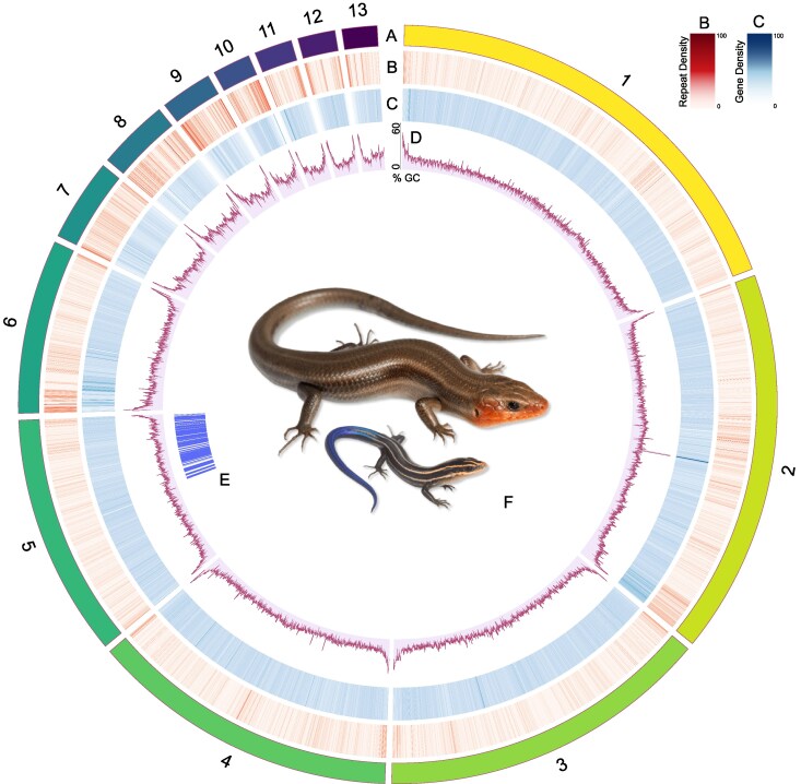 Fig. 1
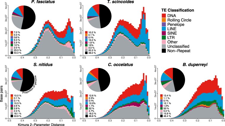 Fig. 2
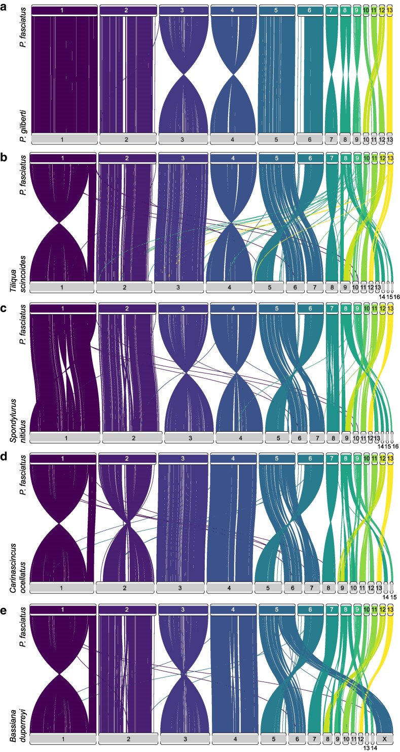 Fig. 3
| Metric | Draft genome | Hi-C informed genome | |
|---|---|---|---|
| Basic Stats | Total assembly size (bp) | 1,537,706,373 | 1,537,706,373 |
| Number of contigs | 34 | 34 | |
| Number of scaffolds | 34 | 31 | |
| GC content (%) | 45.6 | 45.6 | |
| Contiguity | Contigs N50 (bp) | 166,278,287 | 166,278,287 |
| Contigs L50 (bp) | 3 | 3 | |
| Largest contig (bp) | 244,253,184 | 244,253,184 | |
| Scaffolds N50 (bp) | 166,278,287 | 227,050,938 | |
| Scaffolds L50 | 3 | 2 | |
| Largest scaffold (bp) | 244,253,184 | 304,269,845 | |
| Completeness | BUSCO complete (%) | 98.17 | 98.17 |
| Missing BUSCOs (%) | 1.24 | 1.24 | |
| Fragmented BUSCOs (%) | 0.19 | 0.19 | |
| Sequencing coverage (X) | 53 | 116 | |
| Telomere-to-telomere chromosomes | 6 | 13 |
| Category | Metric | Value |
|---|---|---|
| Gene content | Number of protein-coding genes | 32,520 |
| Unique genes with common names | 16,100 | |
| Gene structure | Average gene length (bp) | 9,372.34 |
| Total number of exons | 252,300 | |
| Average exon length (bp) | 255.27 | |
| Multiple-exon transcripts | 25,572 | |
| Single-exon transcripts | 7,372 | |
| Functional annotation | Genes with GO term annotations | 21,578 |
| Genes with InterProScan hits | 24,316 | |
| Genes with eggNOG annotations | 26,007 | |
| Genes with Pfam domains | 19,449 | |
| CAZyme-annotated genes | 319 | |
| MEROPS-annotated genes | 1,143 |
- —American Museum of Natural History’s Richard Gilder Graduate School10.13039/100005835
- —Theodore Roosevelt Memorial Fund10.13039/501100007435
- —Society for the Study of Amphibians and Reptiles’ Roger Conant Grants-in-Herpetology10.13039/100007119
- —U.S. National Science Foundation10.13039/100000001
Peer Reviews
No public reviews on file for this paper yet. If you reviewed it on a platform where reviews are public (OpenReview, ICLR, NeurIPS, ICML), you can paste yours below so the community can read it here.
Videos
No videos yet. Explain this paper in a talk, walkthrough, or lecture? Add one.
Taxonomy
TopicsGenomics and Phylogenetic Studies · Chromosomal and Genetic Variations · Telomeres, Telomerase, and Senescence
Introduction
With the advent of whole-genome sequencing, numerous genomic resources are now available for many clades across the vertebrate tree of life, with birds and mammals comprising most of these genomes. Despite the abundance of ecological and evolutionary research on squamate reptiles, the availability of high-quality genomes has, until recently, lagged far behind other amniote groups. There has been a recent drastic push to increase the production of long-read genomes for many squamate groups (Gable et al. 2023). As a result, the number of reference-level genomes of NCBI has risen from 115 in 2023 to 292 in Spring of 2025, a ∼150% increase over the last year and a half. Still, there is an underrepresentation of diverse families such as skinks, geckos, chameleons, and amphisbaenids (Pinto et al. 2023). Generating high-quality reference genomes across squamates is paramount, as broad sampling is an important aspect of understanding and preserving a genetic record of biodiversity, hypothesizing species relationships, and conservation of endangered taxa (Worley et al. 2017; Brandies et al. 2019).
For example, there is a large variation of chromosomal architecture within squamates, with karyotypes ranging from 2n = 16 in the gecko Gonatodes taniae to 2n = 62 in the microteiid Notobachia ablephara and the chameleon Rieppeleon brevicaudatus (Schmid et al. 1994; Pellegrino et al. 1999; Rovatsos et al. 2017; Mezzasalma et al. 2024). In skinks, the number of chromosomes ranges from 2n = 22 to 2n = 32, and genomic architecture is relatively conserved (Deweese and Wright 1970; Giovannotti et al. 2009). Most Plestiodon have 13 chromosome pairs, where 2n = 26 with 6 macrochromosome pairs and 7 microchromosome pairs (Xu and Zhu 2024), though Plestiodon anthracinus has 12 chromosome pairs (Hardy et al. 2017).
There is also great diversity of sex-determination systems in squamates (Janzen and Phillips 2006; Ezaz et al. 2009; Alam et al. 2018), ranging from temperature-dependent sex determination to genotypic sex determination with XY/XX (male heterogamy) and ZZ/ZW (female heterogamy). Sex-determining systems are extremely labile in squamates (Ezaz et al. 2009; Mezzasalma et al. 2021). For example, it is estimated that there have been 17 to 25 in sex-determination transitions in geckos, with multiple sex-determining systems in some genera (Gamble et al. 2015). Unlike geckos but similar to iguanas, skinks have a conserved XY sex-determining system (male heterogamy) with homomorphic sex chromosomes that are difficult to distinguish (Kostmann et al. 2021; Xu et al. 2024). There is little apparent variation in the sex-determining system of skinks, but this may be an artifact of limited genomic resources (Dodge et al. 2025). It has been hypothesized that the XY sex-determination system may have evolved independently from that of other squamates, such as Podarcis mularis (Lacertidae) and Anolis carolinensis (Dactyloidae), which are commonly used in comparative studies (Kostmann et al. 2021).
Skinks represent 4% of amniote diversity, yet there are currently only 6 chromosome-level skink genomes, 4 of which are Australian. To increase both the phylogenetic and biogeographic diversity of genomic resources for skinks, we present a telomere-to-telomere annotated reference genome for Plestiodon fasciatus. The common five-lined skink, P. fasciatus (Linnaeus 1758), is one of the most abundant lizards of the eastern United States and southeastern Canada (Powell et al. 2016). These generalist skinks are small (total length, 12.5 to 22.2 cm; maximum snout–vent length, 8.6 cm), are found in mesic wooded areas, and reside in cover materials, such as rock crags, logs, and tree bark but will emerge to thermoregulate or search for invertebrate prey (Fitch and Fitch 1954; Brazeau et al. 2015; Powell et al. 2016). This genome is the seventh chromosome-level in the family Scincidae, which currently includes Bassiana duperreyi (Hanrahan et al. 2025), Spondylurus nitidus (Rivera et al. 2024), Carinascincus ocellatus, Tiliqua scincoides, Cryptoblepharus egeriae (Dodge et al. 2025), and the congener Plestiodon gilberti (Richmond et al. 2026). Barring P. gilberti, all of the other species represented which shares a common ancestor with P. fasciatus ∼115 Ma (Title et al. 2024), while P. gilberti and P. fasciatus diverged ∼17 Ma (Brandley et al. 2011).
Materials and methods
Sample acquisition
We collected an individual female Plestiodon fasciatus in Allegan County, Michigan (Lat, 42.5394; Long, −85.9949), and euthanized with MS-222 prior to tissue collection following Michigan DNR permits and approved IACUC protocols at the American Museum of Natural History (AMNH). We sampled liver, lung, heart, skeletal muscle, kidney, and skin from the individual and stored them in NAP buffer (Camacho-Sanchez et al. 2013) to preserve the RNA. Due to a suboptimal PacBio Revio whole-genome sequencing effort of the NAP-preserved liver tissue from this individual, we collected a female P. fasciatus in McCracken County, Kentucky (Lat, 37.1501; Long, −88.7953), with Kentucky Department of Fish and Wildlife permits to provide a blood sample (stored in EDTA) for genomic sequencing. Despite the 650 km between these localities, there are no major biogeographical barriers (Soltis et al. 2006). Furthermore, there is little mitochondrial phylogeographic structure within the population (Howes and Lougheed 2008) and therefore likely little genomic variation, though this has not been tested. Both individuals are vouchered specimens, cataloged in the Herpetology Collections at the AMNH as AMNH-179334 (Allegan County, MI) and AMNH-179327 (McCracken County, KY).
RNA extraction and sequencing
For RNA sequencing, we sent 6 tissues (liver, lung, heart, skeletal muscle, kidney, and skin) to Azenta/Genewiz for extraction, library preparation with poly(A) selection to target eukaryotic strand-specific mRNA, and sequencing on an Illumina NovaSeq 2 × 150 bp, generating ∼100 M paired-end reads. All RNA sequences passed the initial quality check run with FastQC (Brown et al. 2017). Sequencing adapters in the resulting sequences were filtered and trimmed with trimmomatic using the default settings (Bolger et al. 2014).
Genomic DNA extraction and sequencing
Genomic DNA was extracted from blood stored in EDTA from AMNH-179327 using the Qiagen MagAttract High Molecular Weight DNA Kit following their “Manual Purification of High-Molecular Weight Genomic DNA from Whole Blood” protocol from the MagAttract HMW DNA Handbook in the AMNH ICG. The extracted DNA was then sent to Azenta/Genewiz, where it was sequenced with PacBio Revio HiFi sequencing on 1 SMRT cell, which typically results in ∼15 million reads and ∼100 GB of data, depending on the quality of the input sample. We expected a coverage of ∼66× based on an estimated 1.5 GB genome size. Sequencing generated ∼6.3 million read-pairs and ∼79 GB of data, with an average read length of ∼12,000 bp, resulting in ∼53× coverage.
Finally, we generated Hi-C data with Phase Genomics using the Proximo Hi-C animal genome scaffolding platform from a collected blood sample. Proximity-ligated fragments were sequenced on an Illumina NovaSeq to produce 2 × 150 bp paired-end reads. The sequencing generated 300 million read-pairs, of which 56% were high quality, yielding 1,721,863 read-pairs per contig that were “usable,” indicating that they mapped to different >5 kb contigs.
Draft genome assembly
A draft genome assembly was made from the HiFi long-read sequencing using Hifiasm v0.25.0, a haplotype-resolved de novo assembly tool for PacBio HiFi reads (Cheng et al. 2021). Hifiasm was run without the Hi-C sequences as its inclusion led to a drastic increase of contigs, from 34 without Hi-C to ∼6,000 with Hi-C. Due to the high content of low-divergence repeats, we soft-masked the draft genome with the Earlgrey v4.1.1pipeline (Baril et al. 2024) prior to Hi-C mapping, which also provide transposable element (TE) annotations (see below).
Hi-C incorporation
To incorporate the Hi-C sequencing with the draft genome assembly, we initially aligned the Hi-C sequences to the HiFi draft genome with a Burrow-Wheeler Alignment (BWA v 0.7.19) (Li, 2013). Then, we processed the resulting alignments with SAMtools v1.22.1 (Danecek et al. 2021) to remove duplicate sequences. We then scaffolded the assembly using the de novo YAHS v1.2.2 assembly pipeline (Zhou et al. 2023). From there, we used Juicer Tools to generate a Hi-C contact map (Durand et al. 2016). Finally, we visualized the scaffolding of the chromosome-level assembly with Juicebox Assembly Tools v2.20.0 (Robinson et al. 2018).
TE and gene annotation
We annotated the final assembly by first modeling and quantifying TEs by soft-masking the genome with EarlGrey v4.1.1, a fully automated TE annotation pipeline (Baril et al. 2024) with the default settings, which includes a 100 bp minimum length and 10 iterations of the BLAST, extract, and extend process. Next, we functionally annotated the predicted gene regions using the train, predict, update, fix, and annotate steps of the funannotate pipeline (Palmer and Stajich 2020). The “train” step aligns RNA-seq data, assembles it with Trinity (Grabherr et al. 2011), and runs PASA, which models gene structures based on alignments of expressed transcripts (Haas et al. 2003). The “predict” step uses PASA gene models to train Augustus, a de novo gene finder (Stanke et al. 2008), prior to running EvidenceModeler (Haas et al. 2008). We included RNA-seq data from the liver, lung, heart, skeletal muscle, kidney, and skin as evidence for EvidenceModeler. We used the Tetrapoda BUSCO database with Taeniopygia guttata as the seed species for the “predict” step and kept the default options for each step. The “update” step fixes gene models that disagree with RNA-seq data, which are corrected in the “fix” step. Prior to running the “annotate” step, we ran InterProScan v5.74-105.0 (Jones et al. 2014) to run predicted genes against the InterPro database for gene families and downloaded Eggnog-mapper v2.1.12 (Cantalapiedra et al. 2021) locally to be run during the “annotate” step. The “annotate” step incorporates the generated data into an annotated genome. We used the default functional annotation databases in the annotation step. To compare across existing chromosome-level skink genomes, we ran the above pipeline on the assemblies of T. scincoides, S. nitidus, B. duperreyi, and C. ocellatus.
Synteny analysis
To assess genomic synteny of the P. fasciatus genome and other chromosome-level squamate assemblies, we created a custom pipeline called Synk (https://github.com/jomhoff/Synk) that uses the output files from compleasm (Huang and Li 2023) and isolates the BUSCO genes to create comparative text files and uses RIdeogram (Hao et al. 2020) to plot the syntenic chromosomal regions from BUSCO genes between species in one script. Compared to methods for calculating and visualizing whole-genome synteny, Synk runs much faster. In addition, limiting the dataset to BUSCO genes minimizes paralogy issues, as BUSCO genes are highly conserved, single-copy orthologs. This is especially effective in ensuring appropriate estimates of synteny in cross-species and cross-genera comparisons. Here, we show synteny between P. fasciatus, B. duperreyi, S. nitidus, C. ocellatus, T. scincoides, and P. gilberti.
Results and discussion
Assembly of the P. fasciatus genome
After completing Hifiasm with the long-read PacBio sequences, the draft assembly of the genome was close to complete, with an L50 of 3 and a total of 34 contigs, 9 of which represented near telomere-to-telomere chromosomes (Table 1). With the Hi-C data incorporated, we successfully generated a chromosome-level assembly of Plestiodon fasciatus with 18 unplaced scaffolds that we refer to as rPleFas1.1 after Vertebrate Genome Project naming rules (Rhie et al. 2021) (Table 1; Fig. 1). One of the 18 unplaced scaffolds includes the complete mitochondrial genome. The other 17 unplaced scaffolds range in size from 8,262 bp to 1,241,752 bp and consist of unplaced TEs and mRNA sequences. For the complete autosomal genome, the presence of telomeres was estimated with the characteristic rise in GC content at the ends of chromosomes (Fig. 1) and confirmed with tidk v0.2.65, a toolkit for identifying telomeres that outputs a count of telomeric repeats in windows across the genome (Brown et al. 2025). There is also a large spike in GC content in the middle of the second chromosome, which corresponds to a repeat-dense region consisting of Long Interspersed Nuclear Elements (LINEs), simple, and unclassified repeats.
Ideogram for Plestiodon fasciatus. Row A represents chromosome number and relative size. Row B is a heatmap of gene density for 50 kb windows, where darker colors represent a higher density of genes. Row C is a heatmap of repeat elements for 50 kb windows, where darker colors represent a higher density of repeat elements. Row D is a line chart of GC content across the genome. Row E is the region of the genome that is syntenic with the X chromosome of Bassiana duperreyi. Row F is a photograph of an adult male and juvenile P. fasciatus (photo via Herps of Arkansas). Plot made with Circos (Krzywinski et al. 2009).
Annotation of genes and TEs
The resulting annotations from the funannotate pipeline consist of an estimated 32,520 genes. Filtering for named genes and removing isoforms resulted in 16,100 unique genes with common names (Table 2). Like other squamates, the genome of P. fasciatus has a high proportion of repetitive regions which account for 46.7% of the genome (Pasquesi et al. 2018). Unclassified repeats account for 23.3% of the genome, while DNA repeats and LINEs, respectively, make up 7.0% and 9.3% of the genome (Fig. 2). The Kimura 2-parameter distance between the repeat sequences produces an approximately bimodal distribution, with a peak indicating a large degree of moderately diverged repeats and a peak indicating recently diverged repeats (Fig. 2). The more diverged peak is mainly driven by unclassified repeats. Interestingly, this a pattern that is seen in all 5 skinks tested. Furthermore, the more recently diverged peak is driven by a large proportion of both DNA and LINE repeats and is also conserved across the species analyzed (Fig. 2).
Divergence estimates of TEs shown with Kimura 2-parameter distance for 5 chromosome-level skink genomes. Higher Kimura 2-parameter distance indicates more diverged sequences. The proportion of each TE subdivision in the genome for each species is displayed with a pie chart.
Although the impact of TEs on the diversification and adaptation of squamates has yet to be studied in detail, it has been hypothesized that variation in TEs can impact phenotypic adaptation through TE domestication, exaptation, host-gene regulation, formation of retrogenes, and genomic plasticity (reviewed in Schrader and Schmitz 2019) and that TEs contribute significantly to genomic variation (Catlin and Josephs 2022). Since TEs are highly mobile across the genome, are abundant and labile, and have been major players in the evolution of eukaryotic genomes (Bowen and Jordan 2002; Oliver and Greene 2009), we expect variation of TEs among distantly diverged clades. Despite originating around ∼115 mya, the skinks in this study display remarkable conservation of TE composition, especially when considering the variation seen in younger clades, such as extant mammals (Platt et al. 2018) and plethodontid salamanders (Sun et al. 2012).
Synteny of skink genomes
The genomes of P. gilberti and P. fasciatus appear highly syntenic, with the only rearrangement being an inversion in the eighth chromosome. Despite ∼150 ma divergence (Title et al. 2024), the macrochromosomes are largely conserved across the skink species analyzed here, aside from a large inversion on chromosome 1 between T. scincoides and all other species (Fig. 3). Compared to the other scincids, P. fasciatus and P. gilberti have fewer microchromosomes, where chromosomes 14, 15, and 16 in S. nitidus and T. scincoides are syntenic with chromosome 8 in P. fasciatus and P. gilberti. There is also a rearrangement creating syntenic blocks relating chromosomes 6 and 7 in S. nitidus and T. scincoides to chromosome 5 in P. fasciatus and P. gilberti.
Synteny of BUSCO genes between Plestiodon fasciatus and other skinks. a) Plestiodon gilberti; b) Tiliqua scincoides; c) Spondylurus nitidus; d) Carascincus ocellatus; e) Bassiana duperreyi.
Due to the cryptic, homogametic XY sex-determining system in many skinks, little is known about the position of sex-determining regions on the chromosomes; however, hypothetical sex chromosomes have been identified in B. duperreyi (Dissanayake et al. 2020; Hanrahan et al. 2025). Here, we show that a block of chromosome 5 is syntenic with the X chromosome in B. duperreyi, indicating a potential location for sex-determining regions in P. fasciatus (Fig. 3). The same block of the fifth chromosome of P. gilberti is syntenic with the X chromosome of B. duperreyi (Richmond et al. 2026). In an attempt to further identify sex-linked regions, we used the FindZX pipeline (Sigeman et al. 2022) with 2 male and 2 female P. fasciatus; however, the results were inconclusive. Despite some preliminary work, more research is required to further classify sex determination in skinks, ideally with population-level sampling of populations with numerous representatives from both sexes across Scincidae.
Conclusion
We present a high-quality, telomere-to-telomere, chromosome-level annotated reference assembly of the North American common five-lined skink Plestiodon fasciatus (Linnaeus 1758), representing one of the most complete reference genomes (rPleFas1.1) to date of any species of Scincidae. We find that macrochromosome structure is conserved across the family, but there are common rearrangements of the microchromosomes, including a likely fusion in P. fasciatus, which has fewer microchromosomes than many other skink species. We also present insight into the content and evolution of TEs in skinks, which show remarkable conservation between species over the last ∼115 Ma.
The reference list from the paper itself. Each links out to its DOI / PubMed record.
- 1Alam SMI, Sarre SD, Gleeson D, Georges A, Ezaz T. 2018. Did lizards follow unique pathways in sex chromosome evolution? Genes (Basel). 9:239. 10.3390/genes 9050239.29751579 PMC 5977179 · doi ↗ · pubmed ↗
- 2Baril T, Galbraith J, Hayward A. 2024. Earl grey: a fully automated user-friendly transposable element annotation and analysis pipeline. Mol Biol Evol. 41:msae 068. 10.1093/molbev/msae 068.38577785 PMC 11003543 · doi ↗ · pubmed ↗
- 3Bolger AM, Lohse M, Usadel B. 2014. Trimmomatic: a flexible trimmer for Illumina sequence data. Bioinformatics. 30:2114–2120. 10.1093/bioinformatics/btu 170.24695404 PMC 4103590 · doi ↗ · pubmed ↗
- 4Bowen NJ, Jordan IK. 2002. Transposable elements and the evolution of eukaryotic complexity. Curr Issues Mol Biol. 4:65–76. 10.21775/cimb.004.065.12074196 · doi ↗ · pubmed ↗
- 5Brandies P, Peel E, Hogg CJ, Belov K. 2019. The value of reference genomes in the conservation of threatened species. Genes (Basel). 10:846. 10.3390/genes 10110846.31717707 PMC 6895880 · doi ↗ · pubmed ↗
- 6Brandley MC et al 2011. Accommodating heterogenous rates of evolution in molecular divergence dating methods: an example using intercontinental dispersal of plestiodon (Eumeces) lizards. Syst Biol. 60:3–15. 10.1093/sysbio/syq 045.20952756 · doi ↗ · pubmed ↗
- 7Brazeau D, Freitag R, Hecnar SJ, Hecnar DR. 2015. Comparing Common Five-lined Skink (Plestiodon fasciatus) Diet Among Populations and Time. EBSC Ohost [WWW Document]. https://openurl.ebsco.com/contentitem/gcd:115178012?sid=ebsco:plink:crawler&id=ebsco:gcd:115178012 (accessed 6.11.25).
- 8Brown MR, Manuel Gonzalez de La Rosa P, Blaxter M. 2025. Tidk: a toolkit to rapidly identify telomeric repeats from genomic datasets. Bioinformatics. 41:btaf 049. 10.1093/bioinformatics/btaf 049.39891350 PMC 11814493 · doi ↗ · pubmed ↗