Magnetism and 3D Electron Diffraction Solution of Hydrated Rubidium–Ruthenium Oxide Rb2Ru2O7·H2O
Krystof Chrappova, Jeremiah P. Tidey, Christopher Bell, Simon R. Hall

TL;DR
This paper reports the crystal structure and magnetic properties of a hydrated rubidium-ruthenium oxide compound using 3D electron diffraction.
Contribution
The study provides a novel crystal structure determination and magnetic analysis of Rb2Ru2O7·H2O using 3D electron diffraction.
Findings
The compound crystallizes in space group C2/c with specific lattice parameters.
Magnetic measurements show a diamagnetic baseline with a small Curie tail below 55 K.
Bond-valence-sum analysis suggests high-valent Ru and a local singlet state.
Abstract
The crystal structure of Rb2Ru2O7·H2O was determined by three-dimensional electron diffraction from the individual crystallites of a solid-state powder product. Rb2Ru2O7·H2O crystallizes in space group C2/c (a = 7.841(3) Å, b = 12.500(3) Å, c = 8.392(2) Å, β = 93.57(4)°, Z = 4). The structure contains infinite chains that run normal to the (101) plane and consist of alternating RuO6 octahedra and square-pyramidal RuO5 units connected via shared O–O edges. Magnetic properties were measured on the bulk powder, showing a diamagnetic baseline from 300 K to 60 K with a small Curie tail below 55 K. The magnetic moment, calculated from the 1.8 K isotherm, saturates at M = 4.4 × 10⁻³ μB Ru–1, much less than would be expected for S = 1 ruthenium. Bond-valence-sum analysis indicates high-valent Ru, and the near-diamagnetic response is consistent with the edge-sharing Ru–Ru motif, where weak…
Genes, proteins, chemicals, diseases, species, mutations and cell lines named across the full text — each resolved to its canonical identifier and authoritative record.
Click any figure to enlarge with its caption.
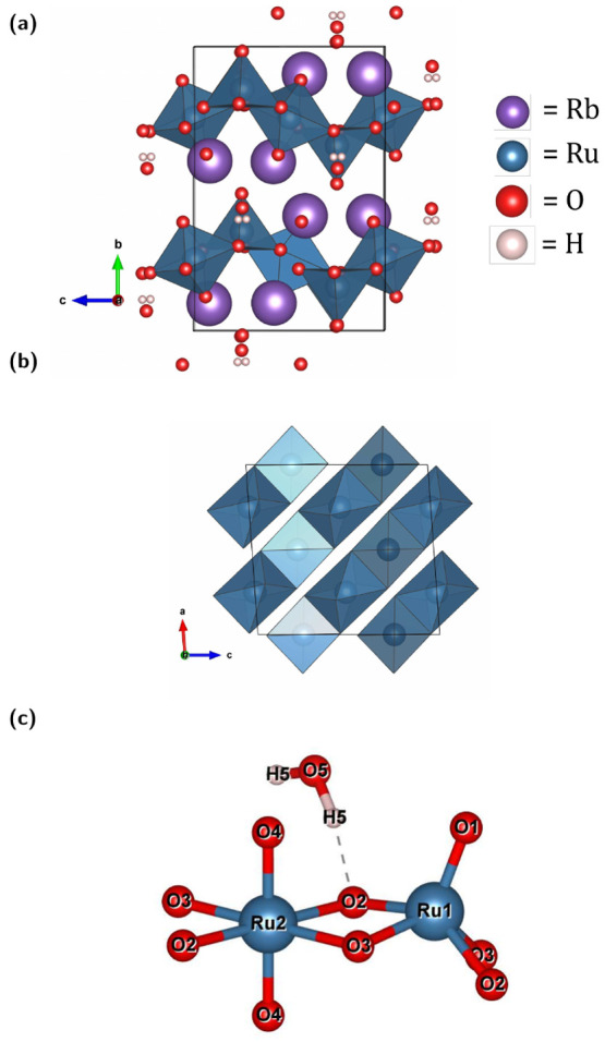 Figure 1
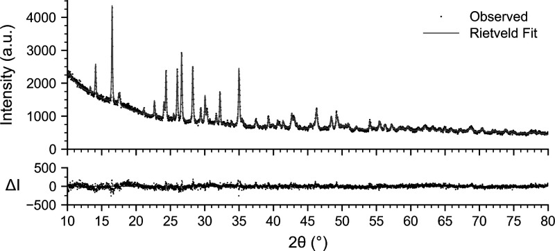 Figure 2
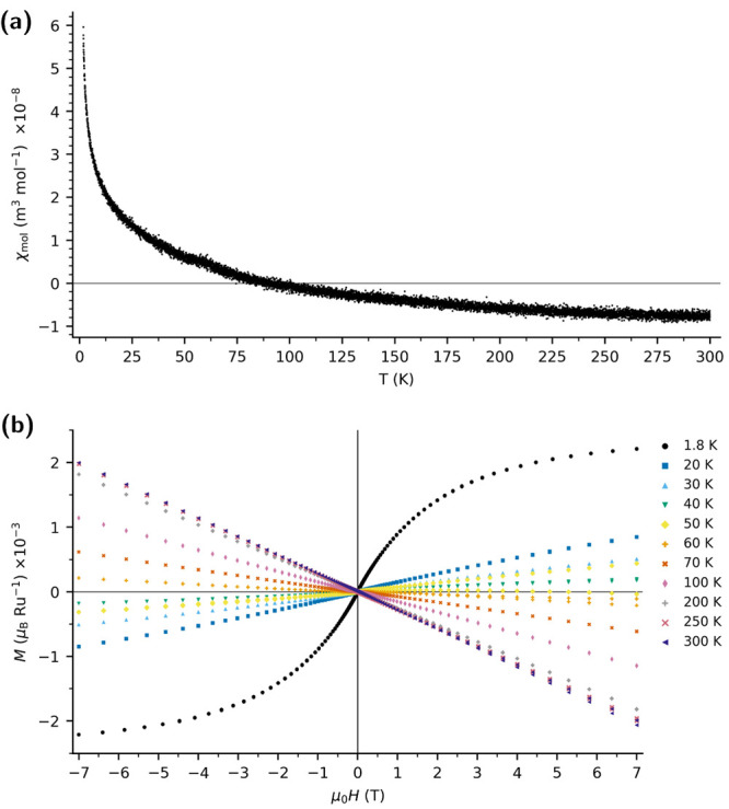 Figure 3
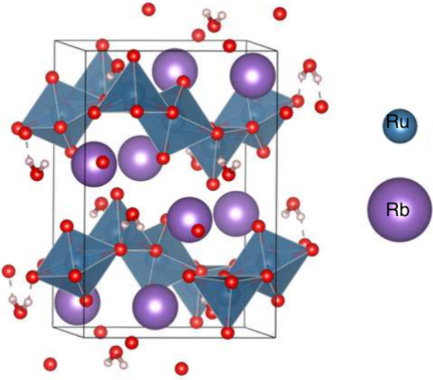 Figure 4
- —Henry Royce Institute10.13039/100016128
- —EPSRC Centre for Doctoral Training in Technology Enhanced Chemical Synthesis10.13039/501100018959
Peer Reviews
No public reviews on file for this paper yet. If you reviewed it on a platform where reviews are public (OpenReview, ICLR, NeurIPS, ICML), you can paste yours below so the community can read it here.
Videos
No videos yet. Explain this paper in a talk, walkthrough, or lecture? Add one.
Taxonomy
TopicsMagnetism in coordination complexes · Layered Double Hydroxides Synthesis and Applications · Advanced Condensed Matter Physics
Ruthenium oxides display a wide range of unusual electronic ground states. Among alkaline metal ruthenates, SrRuO_3_ is an itinerant ferromagnet (T Curie = 150 K), whereas its iso-structural analogue CaRuO_3_ remains paramagnetic when undoped. ?,? Sr_2_RuO_4_ exhibits unconventional superconductivity (superconducting T c = 1.5 K).?
In the alkali-ruthenate family, intermediate valence Ru^4+/5+^ allow diverse extended frameworks. Two-dimensional honeycomb layers have been reported for Li_2_RuO_3_ and Na_2_RuO_3_, and isolated octahedral chains are present in Li_3_RuO_4_. ?−? ?
Here, we present the previously unknown hydrated oxide Rb_2_Ru_2_O_7_·H_2_O, whose structure comprises isolated zigzag chains of alternating edge-sharing RuO_6_ octahedra and RuO_5_ square pyramids. The compound was solved by three-dimensional electron diffraction (3D ED) and its magnetic properties were determined on the bulk polycrystalline solid-state product.
A polycrystalline sample of Rb_2_Ru_2_O_7_·H_2_O was obtained by reacting ground Rb_2_CO_3_ (1.5 mmol) with RuO_2_ (0.75 mmol) in an alumina crucible at 1000 °C (4 h; 5 °C min^–1^ ramp). The structure (Figure) was solved from micrometer-sized crystals (Figure S1) using continuous rotation, selected-area 3D ED performed at 100(5) K. Rietveld refinement using the 3D ED model fits the laboratory powder X-ray diffraction (PXRD) pattern of the bulk sample well (Figure). Scanning electron microscopy with energy-dispersive X-ray spectroscopy (SEM/EDS) shows colocalization of both metals used in the synthesis (Figures S3 and S4). Transmission electron microscopy reveals the powder consists of polycrystalline, needle-like particles up to 6 μm in length, and the corresponding selected area electron diffraction shows rings that can be indexed to the lattice planes in the 3D ED structure (Figure S5, Table S3).
The results from 3D ED show two crystallographically independent Ru sites are present in the crystal structure (Figurec). Ru1 is five-coordinate, forming a distorted square pyramid with an apical Ru1–O1 bond of 1.656(8) Å and basal bonds Ru1–O3 = 1.896(6) Å (×2) and Ru1–O2 = 1.910(6) Å (×2). Ru2 is six-coordinate, with two short axial contacts Ru2–O4 = 1.741(6) Å and four equatorial contacts Ru2–O3 = 1.994(6) Å (×2) and Ru2–O2 = 2.032(5) Å (×2). The polyhedra share the O2–O3 edge, giving Ru1**···**Ru2 = 3.045(4) Å. Edge-sharing continues normal to the (101) plane (Figure).
Figure (a) shows the temperature dependence of the molar susceptibility. A temperature-independent baseline χ_0_ = −1.06 × 10^–8^ m^3^ mol^–1^ persists from 300 to 60 K, followed by a Curie upturn below ∼55 K. A linearization of [χ(T) – χ_0_]^−1^ against temperature over 5–55 K yields a Curie constant C = 1.32 × 10^–6^ m^3^ mol^–1^ K and an effective moment μ_eff_ = 0.65 μ_B_ Ru^–1^. The 1.8 K, 7 T isotherm saturates at M = 4.4 × 10^–3^ μ_B_ Ru^–1^, implying that only a small fraction of Ru sites carry S = ^1^/2 (or S = 1) moments, or that the moments form some kind of antiferromagnetic-like order whose antiparallel moments are not significantly canted at an applied field of 7 T. Rietveld refinement with an Al_2_O_3_ internal standard shows that 27% of the bulk mass is crystalline Rb_2_Ru_2_O_7_·H_2_O (Figure S2). All χ and M data are normalized to the crystalline Rb_2_Ru_2_O_7_·H_2_O mass fraction within the sample. Thus, trivial dilution by amorphous material has already been removed. Therefore, the vanishing net moment is intrinsic to Rb_2_Ru_2_O_7_·H_2_O.
Bond-valence-sum (BVS) analysis was carried out for both Ru coordination polyhedra. Using Brese–O’Keeffe parameters for Ru^4+^ (R 0 = 1.834 Å, B = 0.37 Å) gives V Ru1 = 4.94 and V Ru2 = 5.04 v.u.? Parameters for higher oxidation states are not available using this reference. Using the Gagné–Hawthorne Ru^4+^ parameters (R 0 = 1.833 Å, B = 0.366 Å) produces similar results V Ru1 = 4.93 and V Ru2 = 5.02 v.u., whereas their Ru^5+^ set (R 0 = 1.894 Å, B = 0.346 Å) increases the sum to V Ru1 = 5.89 and V Ru2 = 5.95 v.u.? Parameters for Ru^6+^ are not available. Because the Gagné–Hawthorne parameter sets were calibrated predominantly for regular RuO_6_ octahedra, the distortion of the Ru2 octahedron and the short apical bond in Ru1 inflate the valence sum and the BVS is in this case semiquantitative allowing for both +5 and +6 formal charge.?
Both BVS-permitted oxidation states can plausibly produce Ru–Ru bonding combinations that yield a local singlet consistent with the observed near-diamagnetism. For Ru^5+^ (d^3^ + d^3^), edge-sharing Ru pairs permit three t _2g –t 2g _ overlaps that form bonding combinations denoted σ, π, δ, as has been previously shown for the Li_2_RuO_3 system.? Six electrons can then fill this bonding set as σ^2^π^2^δ^2^, giving a closed-shell S = 0. For the Ru^6+^ (d^2^ + d^2^) analogue, either a molecular orbital configuration that results in S = 1 (σ^2^π^1^δ^1^) or closed shell configuration (σ^2^π^2^δ^0^) is possible. However, only the latter is consistent with the observed magnetism. The Ru1–Ru2 separation of 3.045(4) Å lies within the range where weak direct overlap has been invoked (viz., 2.76–3.06 Å in Y_5_Ru_2_O_12). ?,?
We conclude, therefore, that the magnetism here is likely governed through overlap of Ru–Ru neighbors. The open-shell Ru^6+^ molecular orbital configuration or Ru^4+^/Ru^6+^ disproportionation would contradict either the Curie constant or the single set of Ru–O distances and are thus ruled out.
Supplementary Material
The reference list from the paper itself. Each links out to its DOI / PubMed record.
- 1Jeong D. W.Choi H. C.Kim C. H.Chang S. H.Sohn C. H.Park H. J.Kang T. D.Cho D.-Y.Baek S. H.Eom C. B.Shim J. H.Yu J.Kim K. W.Moon S. J.Noh T. W.Temperature Evolution of Itinerant Ferromagnetism in Sr Ru O 3 Probed by Optical Spectroscopy Phys. Rev. Lett.201311024720210.1103/Phys Rev Lett.110.24720225165956 · doi ↗ · pubmed ↗
- 2Shepard M.Cao G.Mc Call S.Freibert F.Crow J. E.Magnetic and transport properties of Na doped Sr Ru O 3 and Ca Ru O 3 J. Appl. Phys.1996794821482310.1063/1.361619 · doi ↗
- 3Maeno Y.Hashimoto H.Yoshida K.Nishizaki S.Fujita T.Bednorz J. G.Lichtenberg F.Superconductivity in a layered perovskite without copper Nature 199437253253410.1038/372532 a 0 · doi ↗
- 4Miura Y.Yasui Y.Sato M.Igawa N.Kakurai K.New-Type Phase Transition of Li 2Ru O 3 with Honeycomb Structure J. Phys. Soc. Jpn.20077603370510.1143/JPSJ.76.033705 · doi ↗
- 5Wang J. C.Terzic J.Qi T. F.Ye F.Yuan S. J.Aswartham S.Streltsov S. V.Khomskii D. I.Kaul R. K.Cao G.Lattice-tuned magnetism of Ru 4+(4d 4) ions in single crystals of the layered honeycomb ruthenates Li 2Ru O 3 and Na 2Ru O 3 Phys. Rev. B 20149016111010.1103/Phys Rev B.90.161110 · doi ↗
- 6Alexander A.Battle P. D.Burley J. C.Gallon D. J.Grey C. P.Kim S. H.Structural and magnetic properties of Li 3Ru O 4 J. Mater. Chem.200313261210.1039/b 305220 f · doi ↗
- 7Brese N. E.O’Keeffe M.Bond-valence parameters for solids Acta Crystallogr., Sect. B: Struct. Sci.19914719219710.1107/S 0108768190011041 · doi ↗
- 8GagnéO. C.Hawthorne F. C.Comprehensive derivation of bond-valence parameters for ion pairs involving oxygen Acta Crystallogr., Sect. B: Struct. Sci., Cryst. Eng. Mater.20157156257810.1107/S 2052520615016297 PMC 459155626428406 · doi ↗ · pubmed ↗