Shenqi Dihuang Decoction Attenuates ALOX5‐Mediated Ferroptosis in Diabetic Nephropathy via AMPK/mTOR and TGF‐β/Smads Pathways
Li Zhao, Danna Zheng, Wenjuan Gu, Yanna Liu, Jinlong Lyu

TL;DR
This study shows that Shenqi Dihuang Decoction reduces kidney damage in diabetic mice by targeting ALOX5 and related pathways.
Contribution
The study identifies ALOX5-mediated ferroptosis as a novel mechanism in diabetic nephropathy and demonstrates how SDD modulates AMPK/mTOR and TGF-β/Smads pathways.
Findings
SDD improves kidney function and reduces fibrosis and inflammation in diabetic nephropathy.
SDD inhibits ALOX5 and ferroptosis markers while modulating AMPK/mTOR and TGF-β/Smads pathways.
Upregulation of ALOX5 reverses the protective effects of SDD, confirming its role in the mechanism.
Abstract
To explore the underlying mechanisms about that Shenqi Dihuang decoction (SDD) attenuated diabetic nephropathy (DN), mice were fed on high‐sugar and high‐fat diet and treated with streptozotocin (STZ) to induce DN model, as well as HK‐2 cells treated with D‐glucose to establish a DN cell model. High‐performance liquid chromatography (HPLC) analysis was carried out to excavate the chemical compositions existed in SDD. The network pharmacological analysis was performed to screen the key genes involved in SDD treating DN. Subsequently, the effects of SDD on ALOX5, ferroptosis‐ and AMPK/mTOR pathway‐associated indices were examined. Finally, whether SDD attenuated ALOX5‐mediated ferroptosis in DN via AMPK/mTOR and TGF‐β/Smads pathways were validated using gain‐of‐function experiment. SDD exerted a therapeutic effect on DN mice by improving kidney function, kidney fibrosis and reducing…
Genes, proteins, chemicals, diseases, species, mutations and cell lines named across the full text — each resolved to its canonical identifier and authoritative record.
Click any figure to enlarge with its caption.
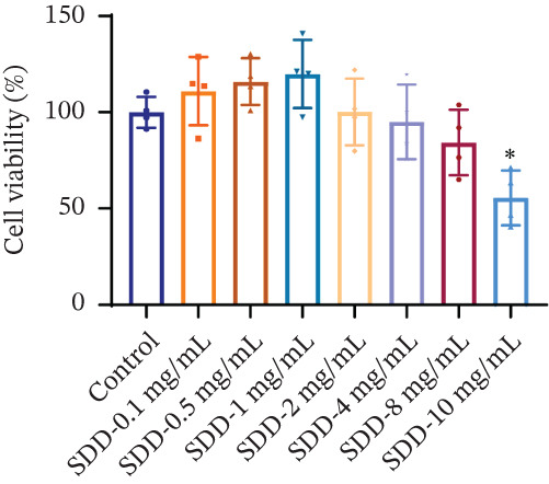 Figure 1
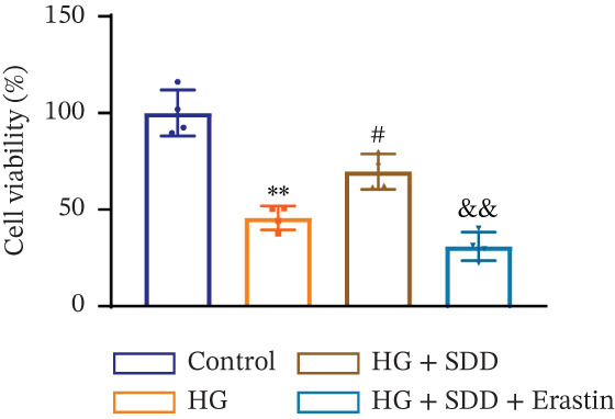 Figure 2
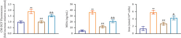 Figure 3
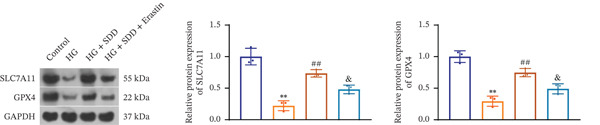 Figure 4
 Figure 5
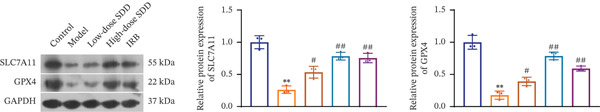 Figure 6
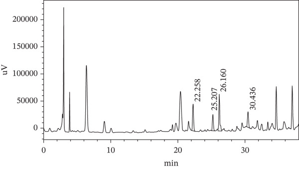 Figure 7
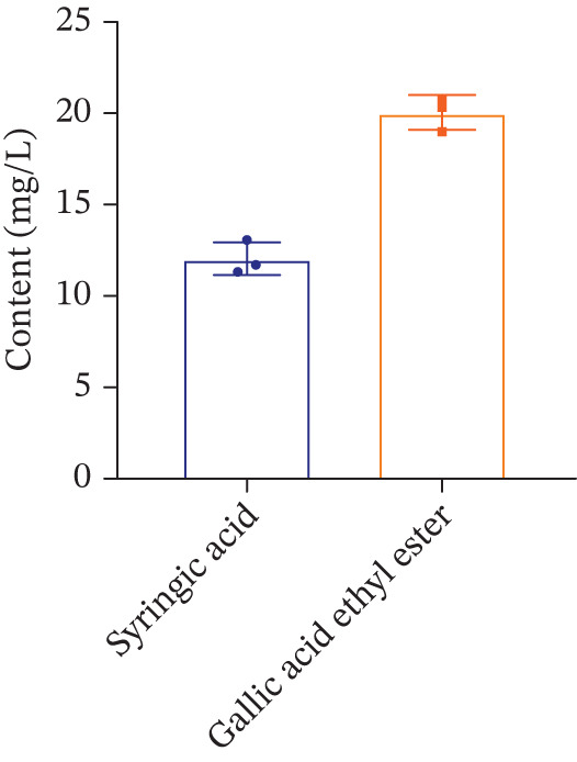 Figure 8
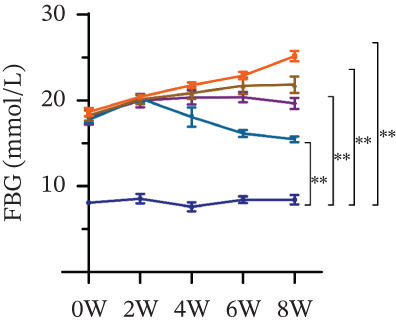 Figure 9
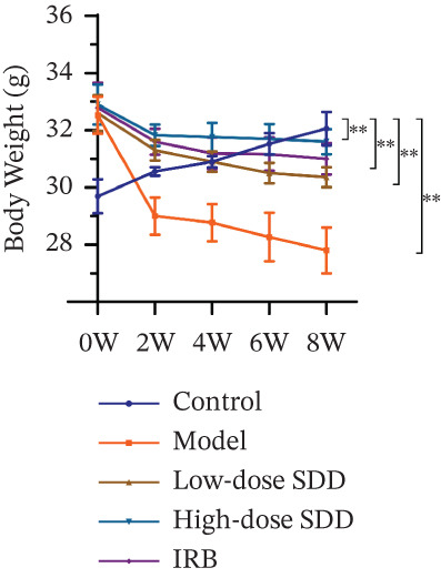 Figure 10
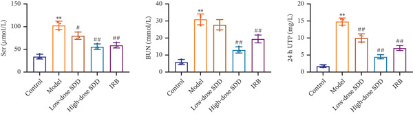 Figure 11
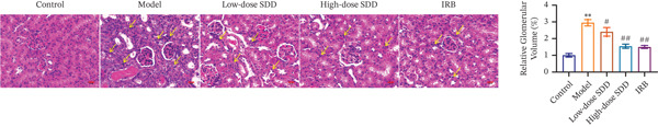 Figure 12
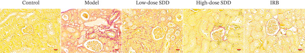 Figure 13
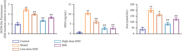 Figure 14
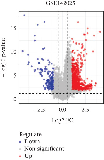 Figure 15
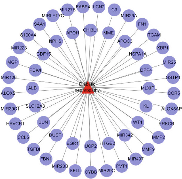 Figure 16
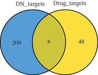 Figure 17
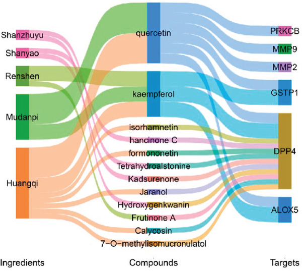 Figure 18
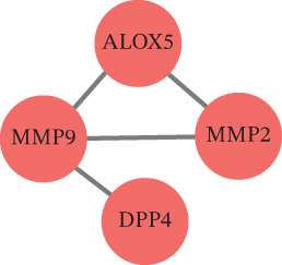 Figure 19
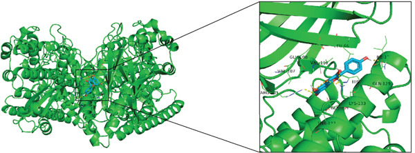 Figure 20
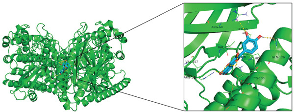 Figure 21
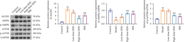 Figure 22
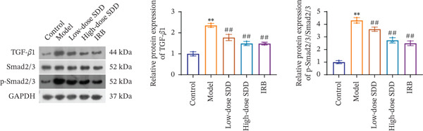 Figure 23
 Figure 24
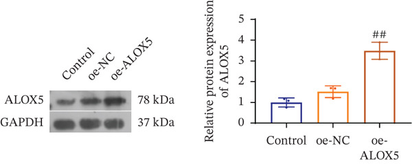 Figure 25
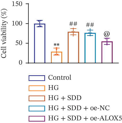 Figure 26
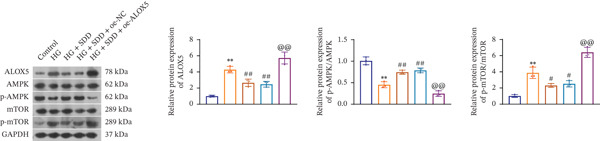 Figure 27
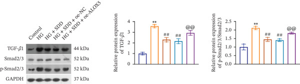 Figure 28
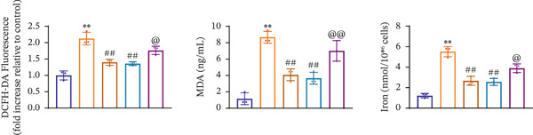 Figure 29
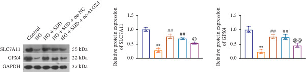 Figure 30
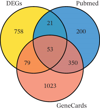 Figure 31
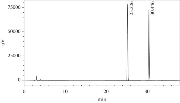 Figure 32
| Time | A: acetonitrile (%) | B: 0.2% phosphoric acid aqueous solution (%) |
|---|---|---|
| 0 | 5 | 95 |
| 5 | 5 | 95 |
| 20 | 15 | 85 |
| 30 | 20 | 80 |
| 30.1 | 80 | 20 |
| 35 | 80 | 20 |
| 35.1 | 5 | 95 |
| 38 | 5 | 95 |
| CurPocket ID | Vina score | Cavity volume ( | Center( | Docking size (x, y, z) | Contact residues | Download |
|---|---|---|---|---|---|---|
|
| −8.4 | 2859 | 16, 15, −23 | 27, 21, 33 |
| Ligand [ |
|
| −8.2 | 3516 | −11, 77, 31 | 21, 27, 35 |
| Ligand [ |
|
| −7.9 | 1950 | 14, 57, 0 | 21, 21, 21 |
| Ligand [ |
|
| −7.9 | 1938 | 11, 38, 13 | 21, 27, 21 |
| Ligand [ |
|
| −7.5 | 1915 | 2, 46, 3 | 21, 21, 33 |
| Ligand [ |
| CurPocket ID | Vinascore | Cavity volume ( | Center( | Docking size ( | Contact residues | Download |
|---|---|---|---|---|---|---|
|
| −8.5 | 3516 | −11, 77, 31 | 21, 27, 35 |
| Ligand [ |
|
| −8.4 | 2859 | 16, 15, −23 | 27, 21, 33 |
| Ligand [ |
|
| −8.3 | 1950 | 14, 57, 0 | 21, 21, 21 |
| Ligand [ |
|
| −8.2 | 1938 | 11, 38, 13 | 21, 27, 21 |
| Ligand [ |
|
| −7.2 | 1915 | 2, 46, 3 | 21, 21, 33 |
| Ligand [ |
- —Department of Health of Zhejiang Province10.13039/501100008856
- —Zhejiang Traditional Chinese Medicine Administration10.13039/501100012175
Peer Reviews
No public reviews on file for this paper yet. If you reviewed it on a platform where reviews are public (OpenReview, ICLR, NeurIPS, ICML), you can paste yours below so the community can read it here.
Videos
No videos yet. Explain this paper in a talk, walkthrough, or lecture? Add one.
Taxonomy
TopicsFerroptosis and cancer prognosis · Immune cells in cancer · Chronic Kidney Disease and Diabetes
Summary
- 1.Shenqi Dihuang decoction (SDD) is a promising drug for diabetic nephropathy (DN) treatment.
- 2.SDD inhibits ALOX5‐mediated ferroptosis in DN via AMPK/mTOR and TGF‐β/Smads pathways.
- 3.This study provides new treatments and insights for DN treatment.
1. Introduction
Diabetes mellitus (DM) is a metabolic disease characterized by high blood sugar levels, which is caused by insufficient insulin secretion or insulin action impairment [1, 2]. The renal functional and structural changes induced by DM are regarded as the most common microvascular complications of DM, seriously affecting people′s lives and health [3]. It is estimated that the number of DM patients will rise to 700 million by 2045 [4], and 30%–40% of DM patients will eventually develop DN [5]. DN seriously endangers people′s life and health, and it is not only a common complicating disease of Type 1 and Type 2 diabetes mellitus (T2DM), but also one of the most significant kidney disorders leading to end‐stage renal disease [6–9]. However, due to the current lack of effective drug treatments for DN, it is of utmost importance to search for drugs that can effectively treat DN.
Traditional Chinese medicine (TCM) has been widely used in the treatment of kidney diseases with satisfactory therapeutic effects [10–12]. Among them, SDD has been reported to have significant effects in reducing proteinuria, improving inflammatory conditions, and protecting renal function [13]. Interestingly, Zhang et al. have found that SDD can alleviate the clinical symptoms of patients and can also effectively control the renal function and blood sugar level of patients with early DN (Z. [14]). Li et al. have highlighted that SDD can effectively improve blood glucose and kidney function with good therapeutic effect [15]. These data have shown that SDD exerts satisfactory therapeutic effects on DN. However, the underlying mechanisms involved in SDD treating DN are still unclear.
Ferroptosis is a type of programmed cell death caused by iron‐dependent lipid peroxidation (S. [16, 17]). Numerous evidence have shown that ferroptosis is concerned with the development of DN, and DN can be effectively alleviated by inhibiting ferroptosis. For instance, Li et al. uncovered that fenofibrate upregulates Nrf2 to postpone the DN progression by suppressing ferroptosis (S. [18]). Jin et al. discovered a novel circular RNA that regulates ferroptosis by targeting the miR‐188‐3p/GPX4 axis, thereby improving germacrone‐mediated DN [19]. However, whether SDD plays a therapeutic function in DN by suppressing ferroptosis is unknown. Thus, to investigate the protective effect of SDD on DN, as well as the involved potential mechanisms, in vivo and in vitro experiments were performed. This study proposes a novel perspective for SDD in DN treatment.
2. Methods
2.1. High‐Performance Liquid Chromatography (HPLC)
HPLC analysis was applied to analyze the chemical composition of SDD using LC‐20A Prominence liquid chromatograph. The 6 g Panax ginseng C. A. Mey, 15 g Astragali Radix, 15 g Rehmanniae Radix, 15 g Dioscoreae Rhizoma, 9 g Poria, 9 g Moutan Cortex, and Corni Fructus powders purchased from Xiumingtang pharmacy flagship store were precisely weighed and mixed, and 10 times and 8 times the amount of water were added, respectively, for reflux extraction for 30 and 20 min. After the mixture concentrated under reduced pressure in a water bath at 75°C, the supernatant was centrifuged and filtered through a 0.45 μm microporous membrane. Then the test sample stock solution of the SDD was obtained. For the control substance treatment, the syringic acid (#S817528, MACKLIN) and gallic acid ethyl ester (#E809094, MACKLIN) were accurately weighed, and methanol was added to prepare mixed control solution working solutions with concentrations of 30.031 μg/mL and 50.045 μg/mL, respectively, followed by filtration. Chromatographic separation was performed utilizing NanoChrom ChromCore C18‐AC column (5 μm, 4.6 × 250 mm; #S009‐050018‐04625S, NanoChrom). The column temperature was set as 30°C, and the automatic sampler injection volume was 10 μL. The mobile phases were acetonitrile and 0.2% phosphoric acid aqueous solution. Table 1 showed the gradient elution procedure.
2.2. Cell Culture and Treatment
Human kidney proximal tubule epithelial cells HK‐2 (#CL‐0109, Procell) were exposed in DMEM medium (#C11885500BT, Gibco) maintained with 10% FBS and 1% penicillin‐streptomycin (#C0222, Beyotime) and subcultured at 37°C with 5% CO_2_. Then the cells were administrated with 0.1, 0.5, 1, 2, 4, 8, 10 mg/mL SDD for 48 h. CCK‐8 assay was applied to assess the safety and safe concentration of SDD for HK‐2 cells by CCK‐8 kit (#C0037, Beyotime). In addition, the cells were treated in DMEM medium containing 30 mM D‐glucose (#S11022, Shanghai yuanye Bio.) for 48 h to construct a DN model (H. [20]). Then 2 mg/mL SDD was used to treat the cells for 24 h, and 35 μM erastin (#E793566, MACKLIN) was utilized to administrate cells for 1 h. Besides, for ALOX5 overexpression, the ALOX5 overexpression plasmids were established; then the ALOX5 coding sequences were cloned into the vectors to produce the overexpression construct. Lentiviral particles were produced by cotransfecting 293 T cells in 96‐well plates with ALOX5 overexpression constructs. Viral supernatants were harvested and used to infect HK‐2 cells for 48 h. Then the transfection efficiency was evaluated by western blot and qRT‐PCR.
2.3. Animal Study
A total of 25 C57BL/6 J male mice (6–8 weeks, 10–25 g) were stochastically categorized into control, model and treatment groups [low‐dose SDD, high‐dose SDD and Irbesartan (IRB)] after 1 week of adaptation (n = 5). After feeding the mice in model and treatment groups with high‐sugar and high‐fat diet (HFD) for 8 weeks, they were fasted for 12 h; then the mice were intraperitoneally injected with 120 mg/kg streptozotocin (STZ; #S17049‐100 mg, Shanghai yuanye Bio.) [21]. Mice in the control group received a normal diet and were intraperitoneally injected with an equal volume of 1% sodium citrate solution. After injection for 72 h, the blood glucose levels were detected. If the blood glucose level is above 16.7 mmol/L, it is considered that the T2DM model is successfully constructed. The content of 24 h urinary total protein (UTP) was measured weekly. The positive results for glucose and protein in the urine and the blood glucose level above 16.7 mmol/L were judged as the modeling criteria for DN [21]. After successful modeling, the mice in low‐ and high‐dose SDD groups were administrated with 5.4 and 21.3 g/kg of SDD daily via gavage for 8 consecutive weeks, respectively. IRB group mice were administered irbesartan (IRB; #Y259351‐5 g, Beyotime) at doses of 30 mg/kg by gavage once a week for 8 consecutive weeks. The control and model groups received an equal volume of 0.9% sodium chloride solution by gavage once a day for 8 consecutive weeks. After 8 weeks, fasting blood glucose (FBG) level and body weight were determined once a week. After administration, 0.5/100 g telazol was administered to the mice for anesthesia. Blood was taken from the abdominal aorta and serum was separated, as well as the kidney collection. Then serum creatinine (Scr; #E‐BC‐K188‐M, Elabscience), blood urea nitrogen (BUN; #E‐BC‐K183‐M, Elabscience), and UTP (#E‐BC‐K252‐M, Elabscience) levels were detected using corresponding assay kits. The animal experiments were conformed to ARRIVE guidelines and approved by the laboratory animal management and ethics committee of Zhejiang Provincial People′s Hospital (No. 20250122404494).
2.4. Pathological Examination
The kidney tissues were obtained for pathological examination. After kidney tissues collection, 4% paraformaldehyde was employed to fix the kidney tissues for 24 h, then the tissues were cut into 4‐7 μm sections for HE staining and sirius red staining after embedding in paraffin. Finally, an Olympus microscope (CKX53, Tokyo, Japan) was employed to randomly capture images.
2.5. Malondialdehyde (MDA), ROS, and Iron Detection
The levels of MDA (#E‐EL‐0060, Elabscience), ROS (#E004‐1‐1, Nanjing Jiancheng Bio.), and iron (#E‐BC‐K880‐M/E‐BC‐K772‐M, Elabscience) were measured using MDA, ROS, and iron assay kits, respectively.
2.6. qRT‐PCR
Total RNA was collected in cells utilizing Trizol (#DP424, TIANGEN) and reversely transcribed into cDNA by FastKing‐RT SuperMix (#KR118‐02, TIANGEN). SYBR Green PCR Master mix (#Q712‐02, Vazyme) was utilized to perform the qRT‐PCR. The utilized primers were ALOX5 (human) forward 5 ^′^‐TGCCAACAAAACAGACCCCT‐3 ^′^ and reverse 5 ^′^‐GAAGGTGGGTGATGGTCTGG‐3 ^′^; GAPDH (human) forward 5 ^′^‐CCTGCACCACCAACTGCTTA‐3 ^′^ and reverse 5 ^′^‐TGAGTCCTTCCACGATACCA‐3 ^′^. GAPDH was applied as an internal reference.
2.7. Western Blot
RIPA (#P0013B, Beyotime) was added in tissues and cells to acquire the total protein, then the proteins were separated to SDS‐PAGE and transferred onto PVDF membranes (#FFP24, Beyotime). The blocked membranes were incubated with antibodies anti‐SLC7A11 (1:1000; #DF12509), anti‐GPX4 (1:2000; #DF6701), anti‐ALOX5 (1:2000; #AF4699), anti‐AMPK (1:2000; #DF6361), anti‐p‐AMPK (1:2000; #AF3423), anti‐mTOR (1:1000; #AF6308), anti‐p‐mTOR (1:1000; #AF3308), anti‐TGF‐β1 (1:1000; #AF1027), anti‐Smad2/3 (1:1000; #AF6367), anti‐p‐Smad2/3 (1:1000; #AF3367), and anti‐GAPDH (1:2000; #AF7021, Abcam) obtained from Affinity, then incubated with secondary antibody Goat Anti‐Rabbit IgG H&L (HRP) (1:2000, #ab6721, Abcam) for 1 h. Membranes were developed by ECL (#P1000, Applygen).
2.8. Network Pharmacological Analysis
2.8.1. Screening of Active Compounds of SDD and Potential Targets
The candidate active compounds of P. ginseng C. A. Mey, Astragali Radix, Rehmanniae Radix, Dioscoreae Rhizoma, Poria, Moutan Cortex, and Corni Fructus were predicted in the TCMSP database with the oral bioavailability (OB) ≥ 30% and drug‐likeness (DL) ≥0.18. After removing duplicates, the potential targets of the active compounds of SDD were identified in the TCMSP database. Then the targets were imported into the Unitprot database for gene name standardization processing, and the active compounds‐target network was constructed using the Cytoscape software (Version 3.7.1).
2.8.2. Collection of Target Genes of DN
The DN‐related candidate target genes were searched in Pubmed, GeneCards (GeneCards Score > 6) databases with the keyword of “diabetic nephropathy”. Also, the GSE96804 dataset was obtained from the GEO database, then the differentially expressed genes (DEGs) were screened using “limma” package with the cutoff value of P.adj < 0.05 and |log2FC| > 1. The p value was corrected using the BH method. Then the DEGs were intersected with the DN‐related candidate target genes searched in Pubmed, GeneCards databases, and the overlapped genes were considered as the DN‐related target genes, followed by the DN‐target network construction using Cytoscape software.
2.8.3. Collection of Target Genes of SDD in Treating DN
The “draw. triple. venn” function in VennDiagram package was used to acquire overlapped genes after intersecting the DN‐related target genes, target genes of active compounds of SDD, and the overlapped genes were considered as the target genes of SDD in treating DN.
2.8.4. PPI Network Construction
“STRING” database was utilized to predict the protein–protein interactions of target genes of SDD in treating DN with the following considerations: species—Homo sapiens; minimum required interaction score—0.4; network type—full STRING network; active interaction sources—All sources enabled (experiments, databases, text mining, coexpression, neighborhood, gene fusion, co‐occurrence). Then the PPI network was visualized using Cytoscape software.
2.8.5. Molecular Docking
The “PDB” format protein structures of key genes were acquired from the PDB database. The “SDF” format of the 3D structure of active ingredients was obtained from the PubChem database. Blind docking was performed using CB‐Dock2. This tool integrates the AutoDock Vina algorithm and pocket prediction function, and supports the automatic identification of potential binding sites. Then the docking result file was imported into the PyMOL software.
2.9. Statistical Analysis
Data are expressed as mean ± SD. One‐way analysis of variance was employed to determine the differences between groups. p < 0.05 was deemed to suggest statistical differences.
3. Results
3.1. Identification of Chemical Compositions of SDD
To confirm the chemical compositions existed in SDD, HPLC analysis was performed. Figure 1 illustrated the chromatograms of reference substance and SDD. The detection wavelengths were detected at 25.207 and 30.436 min, respectively, and chemical compounds corresponding to these peaks were then identified as syringic acid and gallic acid ethyl ester (Figures 1a,b). The content of syringic acid and gallic acid ethyl ester was 11.746 and 19.023 mg/L, respectively (Figure 1c).
Figure 1. Chemical compositions of Shenqi Dihuang decoction (SDD) detected by high performance liquid chromatography (HPLC) analysis. Chromatograms of (a) reference substance and (b) SDD. (c) The content of syringic acid and gallic acid ethyl ester.(a)(b)(c)
3.2. Therapeutic Effect of SDD on DN Mice
To assess the effect of SDD on DN, FBG level and body weight were determined. As demonstrated in Figure 2a, the mice in the model group showed an obvious increase in FBG level during the 8 weeks, whereas SDD and IRB administration could reverse this trend. In addition, SDD and IRB implement both prominently elevated the body weight to varying degrees in DN mice (all p < 0.01; Figure 2b). The renal function test results uncovered that Scr, BUN, and 24 h UTP levels were remarkedly elevated in the model group relative to the control group, whereas the opposite results were observed after SDD and IRB treatment (all p < 0.05; Figure 2c). The HE staining uncovered that the renal structure in the control group was clearly visible and intact, and glomerular mesangial hyperplasia, thickening of the basement membrane, atrophy of the renal tubules, and infiltration of inflammatory cells in the tubulointerstitium were observed in the model group. However, SDD and IRB treatment significantly reduced the glomerular and tubular lesions in DN mice (Figure 2d). Moreover, sirius red staining revealed that obvious fibrosis occurred in the kidneys of mice in the model group, whereas the fibrosis was significantly alleviated after SDD and IRB administration (Figure 2e). Collectively, these findings emphasize that SDD is a promising therapeutic method for DN.
Figure 2. Therapeutic effect of Shenqi Dihuang decoction (SDD) in diabetic nephropathy (DN) mice. The changes in (a) fasting blood glucose levels and (b) body weight in DN mice after SDD and irbesartan administration. (c) The changes in serum creatinine (Scr), blood urea nitrogen (BUN), and 24 h urinary total protein (UTP). (d) Hematoxylin‐eosin (HE) staining. Arrows indicated inflammatory cell infiltration into the interstitium. Scale = 20 μm. (e) Sirius red staining. Scale = 20 μm. ^∗∗^ p < 0.01, compared with control group; ^#^ p < 0.05, ^##^ p < 0.01, compared with model group.(a)(b)(c)(d)(e)
3.3. SDD Alleviates DN by Inhibiting Ferroptosis
To explore whether ferroptosis is involved in the effect of SDD on DN, ferroptosis‐related marks were detected. As shown in Figure 3a, ROS, MDA, and iron levels were obviously increased in the model group relative to the control group, whereas SDD and IRB treatment could reverse these trends. Additionally, the protein expression levels of SLC7A11 and GPX4 were dramatically reduced in DN mice, whereas opposite results were observed after SDD and IRB administration (Figure 3b). Besides, the ferroptosis‐related marks were further detected in HG‐induced HK‐2 cells after erastin treatment. Firstly, CCK‐8 assay was employed to assess the safety and safe concentration of SDD for HK‐2 cells, and 2 mg/mL SDD was selected to treat the cells (Figure 3c). Also, HG treatment could obviously decrease the HK‐2 cell viability, whereas SDD treatment could reverse this trend. Notably, erastin administration could eliminate the effect of SDD on the cell viability of HG‐induced HK‐2 cells (Figure 3d). Similarly, obviously reduced ROS, MDA, and iron levels were found in HG‐induced HK‐2 cells after SDD treatment, whereas opposite results were observed after erastin administration (Figure 3e). Also, the effects of SDD on the SLC7A11 and GPX4 protein expression levels in HG‐induced HK‐2 cells were also eliminated by erastin (Figure 3f). Thus, these results fully suggest that SDD alleviates DN by inhibiting ferroptosis.
Figure 3. Shenqi Dihuang decoction (SDD) alleviates diabetic nephropathy (DN) by inhibiting ferroptosis. (a) Reactive oxygen species (ROS), malondialdehyde (MDA), and iron levels in DN mice. (b) Protein expression levels of SLC7A11 and GPX4 in DN mice. (c) Cell viability of HK‐2 cells. (d) Cell viability of HK‐2 cells after erastin administration. (e) ROS, MDA, and iron levels in HK‐2 cells after erastin administration. (f) Protein expression levels of SLC7A11 and GPX4 in HK‐2 cells after erastin administration. ^∗^ p < 0.05, ^∗∗^ p < 0.01, compared with control group; ^#^ p < 0.05, ^##^ p < 0.01, compared with model/HG group;^&^ p < 0.05, ^&&^ p < 0.01, compared with HG + SDD group.(a)(b)(c)(d)(e)(f)
3.4. SDD Might Inhibit DN by Targeting ALOX5
To excavate the molecular mechanism involved in the SDD treated in DN, the network pharmacological analysis was performed. Based on the TCMSP database, a total of 22, 20, 2, 16, 15, 11, and 20 active compounds of P. ginseng C. A. Mey, Astragali Radix, Rehmanniae Radix, Dioscoreae Rhizoma, Poria, Moutan Cortex, and Corni Fructus were predicted, respectively. Then 93 active compounds of SDD were screened after removing duplicates, followed by 215 target genes of active ingredients of SDD acquisition. Then the active compounds‐target network was constructed (Figure 4a). Besides, a total of 910 DEGs were identified (Figure 4b), and a total 624, 911 DN‐related candidate target genes were searched in Pubmed and GeneCards, respectively. Then 53 overlapped genes were obtained (Figure 4c), which were considered as the DN‐related target genes, followed by the DN‐target network construction (Figure 4d). After intersecting the DN‐related target genes, target genes of active compounds of SDD, a total of six common genes were screened (Figure 4e), namely ALOX5, MMP9, MMP2, PRKCB, GSTP1, and DPP4, which were considered as the target genes of SDD in treating DN. Then the ingredient–compound‐target network was visualized (Figure 4f) and the PPI network was constructed (Figure 4g) with four nodes, including ALOX5, MMP9, MMP2, and DPP4.
Figure 4SDD might inhibit DN by targeting ALOX5. (a) The active compounds‐target network. (b) Volcano plot of DEGs. (c) Venn diagram of DN‐related target genes. (d) DN‐target network. (e) Venn diagram of the target genes of SDD in treating DN. (f) The ingredient–compound‐target network. (g) PPI network construction. Docking poses of (h) kaempferol and (i) quercetin with protein ALOX5, respectively.(a)(b)(c)(d)(e)(f)(g)(h)(i)
Due to numerous studies reported MMP9, MMP2, and DPP4 in DN [22–26] and few studies on ALOX5, thus, the subsequent analysis focuses on the role of ALOX5 in DN. Molecular docking was applied to evaluate the binding interactions between active compounds and ALOX5. Kaempferol and quercetin demonstrated affinity towards ALOX5 with respective binding energies of −8.4 and −8.5 kcal/mol, respectively (Figures 4h,i and Tables 2 and 3). The binding site of kaempferol was located within the active pocket of ALOX5, with the specific coordinates being (27, 21, 33). At this binding site, kaempferol formed multiple hydrogen bonds with the amino acid residues of ALOX5 (LYS‐133, GLN‐129, and ARG‐101). Also, the binding site of quercetin was located within the active pocket of ALOX5, with the specific coordinates being (21, 27, 35). At this binding site, quercetin formed multiple hydrogen bonds with the amino acid residues of ALOX5 (LYS‐133, ARG‐68, GLN‐129, and ARG‐101).
3.5. SDD Inhibits ALOX5‐Mediated Ferroptosis in DN via AMPK/mTOR and TGF‐β/Smads Pathways
Wang et al. have unveiled that ALOX5 facilitates autophagy‐dependent ferroptosis in melanoma through the AMPK/mTOR pathway [27]. Also, the TGF‐β/Smads pathway is a classic signaling pathway for DN [28]. Thus, we suspected that SDD might attenuate ALOX5‐mediated ferroptosis in DN via the AMPK/mTOR and TGF‐β/Smads pathways. To verify our hypothesis, the ALOX5 and AMPK/mTOR and TGF‐β/Smads pathways‐associated indices were detected. The ALOX5 protein expression level was prominently elevated in DN mice, whereas SDD and IRB treatment could reverse this trend, as well as the p‐mTOR/mTOR and TGF‐β1, p‐Smad2/3/Smad2/3 levels (all p < 0.05; Figures 5a,b). However, opposite results were observed on the p‐AMPK/AMPK level. To explore the role of ALOX5 in DN, the oe‐ALOX5 was transfected into HK‐2 cells to upregulate the ALOX5 expression. Then the transfection efficiency was determined by qRT‐PCR and western blot (Figures 5c,d). Of note, ALOX5 overexpression could remarkably plummet the cell viability of HG‐induced HK‐2 cells after SDD treatment (Figure 5e). Also, the protein expression levels of ALOX5, p‐AMPK/AMPK, and p‐mTOR/mTOR levels, as well as TGF‐β1, p‐Smad2/3/Smad2/3, were both prominently changed in SDD treated HG‐induced HK‐2 cells after ALOX5 upregulation (all p < 0.05; Figures 5f,g). Also, ALOX5 overexpression could eliminate the SDD effects on the ROS, MDA, and iron levels in HG‐induced HK‐2 cells (Figure 5h), as well as the levels of SLC7A11 and GPX4 (Figure 5i). Overall, these data strongly indicate that SDD inhibits ALOX5‐mediated ferroptosis in DN via the AMPK/mTOR and TGF‐β/Smads pathways.
Figure 5. Shenqi Dihuang decoction (SDD) inhibits ALOX5‐mediated ferroptosis in diabetic nephropathy (DN) via AMPK/mTOR and TGF‐β/Smads pathways. (a) Protein expression levels of ALOX5 and AMPK/mTOR pathway‐associated marks detected in DN mice. (b) Protein expression levels of TGF‐β/Smads pathway‐associated marks detected in DN mice. Transfection efficiency measured by (c) qRT‐PCR and (d) western blot. (e) Cell viability of HK‐2 cells. (f) Protein expression levels of ALOX5 and AMPK/mTOR pathway‐associated marks in HK‐2 cells after ALOX5 upregulation. (g) Protein expression levels of TGF‐β/Smads pathway‐associated marks in HK‐2 cells after ALOX5 upregulation. (h) Reactive oxygen species (ROS), malondialdehyde (MDA), and iron levels in HK‐2 cells after ALOX5 upregulation. (i) Protein expression levels of SLC7A11 and GPX4 in HK‐2 cells after ALOX5 upregulation. ^∗∗^ p < 0.01, compared with control group; ^#^ p < 0.05, ^##^ p < 0.01, compared with model/oe‐NC/HG group; ^@^ p < 0.05, ^@@^ p < 0.01, compared with HG + SDD + oe − NC group.(a)(b)(c)(d)(e)(f)(g)(h)(i)
4. Discussion
DN can gradually damage the renal function, ultimately leading to end‐stage renal disease, and significantly increase the risk of cardiovascular diseases, seriously threatening the life, health and life quality of patients [29, 30]. Thus, it is extremely urgent to find the therapeutic methods for DM. In this study, we found that SDD administration could obviously decrease the FBG, Scr, BUN, and 24 h UTP levels in DN mice, while prominently elevating the body weight to varying degrees. Also, SDD treatment could effectively alleviate pathological changes in the kidneys of DN mice. These findings emphasize that SDD is a promising therapeutic method for DN. Ferroptosis, as an iron‐dependent lipid peroxidization‐driven cell death mode, exerts a primary function in the occurrence and development of DN [31]. The kidney cells undergoing ferroptosis will release damage‐related molecules, which will further activate the local inflammatory response in the kidneys and promote the infiltration of macrophages and the release of inflammatory factors (such as TNF‐α and IL‐6), thereby exacerbating glomerular sclerosis and tubulointerstitial fibrosis, and accelerating the progression of DN [13, 32]. In this study, ROS, MDA, and iron levels were obviously increased in the model group when compared with the control group, whereas SDD treatment could reverse these trends. Moreover, the protein expression levels of SLC7A11 and GPX4 were remarkably reduced in DN mice, whereas opposite results were observed after SDD administration. Interestingly, similar results were observed in HG‐induced HK‐2 cells. Thus, these results elucidate that SDD exerts therapeutic effects in DN by targeting ferroptosis.
ALOX5 is a member of the ALOX family and is involved in the metabolism of arachidonic acid, which is a metabolic pathway that initiates the biosynthesis of cysteine leukotrienes (F. [33]). ALOX5 has been reported to regulate the activation of immune cells and inflammatory responses, and its dysfunction is closely related to inflammatory diseases; it may also participate in the progression of some cancers by influencing the tumor microenvironment ([15, 34]; L. [35]). Interestingly, Cilenšek et al. have uncovered that ALOXA5AP is strongly related to DN in Slovenian patients with T2DM [36]. Of note, Chen et al. have found that ALOX5 is upregulated in HG‐induced renal mesangial cells, and interference of ALOX5 attenuates the fibrosis and inflammation (X. [37]). Consistently, network pharmacological analysis found that SDD might inhibit DN by targeting ALOX5. We also found that the ALOX5 protein expression was prominently raised in DN mice, whereas SDD could reverse this trend. Besides, accumulating studies imply that ALOX5 participates in the progression of many diseases by mediating ferroptosis [38, 39]. Herein, ALOX5 overexpression could eliminate the SDD effects on the ROS, MDA, and iron levels in HG‐induced HK‐2 cells, as well as the protein expression levels of SLC7A11 and GPX4. Overall, these data strongly indicate that SDD can inhibit ALOX5‐mediated ferroptosis in DN.
JAK is in an abnormally activated state in DN, and the abnormal activation of JAK can lead to renal fibrosis in DN. The mTOR, as the downstream component of the JAK signaling pathway, can regulate renal interstitial fibrosis [40, 41]. Also, inflammation plays essential roles in the development and progression of DN [42, 43]. IL‐6, as an inflammatory cytokine, participates in the progression of DN by regulating inflammasomes, and it can exacerbate renal tubular damage by promoting the activation of the mTOR signaling pathway in DN ([44]; L. [45]). It is reported that the AMPK/mTOR pathway is concerned with the occurrence of various diseases related to metabolic and proliferative abnormalities, such as DM, cancer, and neurodegenerative disorders ([46, 47]; H. [48]). Interestingly, Zhang et al. have elucidated that compound XueShuanTong accelerates podocyte mitochondrial autophagy in DN through the AMPK/mTOR pathway (C. [49]). In addition, a previous study found that kirenol alleviates DN via targeting the TGF‐β/Smads pathway [28]. Li et al. have revealed that HDAC6 is involved in DN by regulating the TGFβ/Smads and NF‐κB signaling pathways (J. [50]). Consistently, we also found that the AMPK/mTOR and TGF‐β/Smads pathways are concerned with the SDD treating DN. Also, the p‐AMPK/AMPK, p‐mTOR/mTOR, TGF‐β1, and p‐Smad2/3/Smad2/3 levels were both changed in SDD treated HG‐induced HK‐2 cells after ALOX5 upregulation. Notably, a previous study has found that ALOX5 facilitates autophagy‐dependent ferroptosis in melanoma through the AMPK/mTOR pathway [27]. These findings clearly demonstrate that SDD inhibits ALOX5‐mediated ferroptosis in DN via the AMPK/mTOR and TGF‐β/Smads pathways.
However, this research also has numerous limitations. First, the STZ‐induced DN mouse model cannot completely reconstruct human DN status. Second, an inhibitor and activator of the AMPK/mTOR pathway or TGF‐β/Smads pathway should be utilized to further analyze the underlying mechanisms involved in SDD treating DN. In addition, the findings obtained in this study should be confirmed at the clinical level.
5. Conclusion
In summary, this study revealed that SDD inhibits ALOX5‐mediated ferroptosis in DN via AMPK/mTOR and TGF‐β/Smads pathways. Our findings offer a novel therapeutic strategy for DN treatment and new insight into the underlying mechanisms involved in SDD treating DN.
Author Contributions
Li Zhao: conceptualization, data curation, formal analysis, funding acquisition, writing—original draft, writing—review and editing. Danna Zheng: data curation, formal analysis, investigation, methodology. Wenjuan Gu: data curation, resources, visualization. Yanna Liu: formal analysis, visualization. Jinlong Lyu: conceptualization, project administration, resources, writing—original draft, writing—review and editing.
Funding
This study was supported by Zhejiang Provincial Administration of Traditional Chinese Medicine (2024ZL247); the Project of the Zhejiang Provincial Health Department (2023KY482).
Disclosure
All authors have read and approved the final manuscript.
Ethics Statement
The animal experiments were conformed to ARRIVE guidelines and approved by the laboratory animal management and ethics committee of Zhejiang Provincial People′s Hospital (No. 20250122404494).
Conflicts of Interest
The authors declare no conflicts of interest.
The reference list from the paper itself. Each links out to its DOI / PubMed record.
- 1Harreiter J. and Roden M. , Diabetes Mellitus: Definition, Classification, Diagnosis, Screening and Prevention (Update 2023), Wiener Klinische Wochenschrift. (2023) 135, no. supplement 1, 7–17, 10.1007/s 00508-022-02122-y, 37101021.37101021 PMC 10133036 · doi ↗ · pubmed ↗
- 2Zeyfang A. , Wernecke J. , and Bahrmann A. , Diabetes Mellitus at an Elderly Age, Experimental and Clinical Endocrinology & Diabetes. (2023) 131, no. 1-02, 24–32, 10.1055/a-1946-3728, 36638808.36638808 · doi ↗ · pubmed ↗
- 3Thipsawat S. , Early Detection of Diabetic Nephropathy in Patient With Type 2 Diabetes Mellitus: A Review of the Literature, Diabetes & Vascular Disease Research. (2021) 18, no. 6, 14791641211058856, 10.1177/14791641211058856, 34791910.34791910 PMC 8606936 · doi ↗ · pubmed ↗
- 4Li X. , Lu L. , Hou W. , Huang T. , Chen X. , Qi J. , Zhao Y. , and Zhu M. , Epigenetics in the Pathogenesis of Diabetic Nephropathy, Acta Biochimica et Biophysica Sinica. (2022) 54, no. 2, 163–172, 10.3724/abbs.2021016.35130617 PMC 9909329 · doi ↗ · pubmed ↗
- 5Gupta S. , Dominguez M. , and Golestaneh L. , Diabetic Kidney Disease: An Update, Medical Clinics of North America. (2023) 107, no. 4, 689–705, 10.1016/j.mcna.2023.03.004.37258007 · doi ↗ · pubmed ↗
- 6Gaddy A. , Elrggal M. , Madariaga H. , Kelly A. , Lerma E. , and Colbert G. B. , Diabetic Kidney Disease, Disease-a-Month. (2025) 71, no. 4, 101848, 10.1016/j.disamonth.2024.101848.39753456 · doi ↗ · pubmed ↗
- 7Kushwaha K. , Kabra U. , Dubey R. , and Gupta J. , Diabetic Nephropathy: Pathogenesis to Cure, Current Drug Targets. (2022) 23, no. 15, 1418–1429, 10.2174/1389450123666220820110801.35993461 · doi ↗ · pubmed ↗
- 8Mima A. , Mitochondria-Targeted Drugs for Diabetic Kidney Disease, Heliyon. (2022) 8, no. 2, e 08878, 10.1016/j.heliyon.2022.e 08878, 35265754.35265754 PMC 8899696 · doi ↗ · pubmed ↗