Beyond the Prostate: Incidental Detection of Male Breast Carcinoma on [18F]DCFPyl
Farzana Z. Ali, Pawan K. Gupta, Martin S. Allen-Auerbach

Abstract
Genes, proteins, chemicals, diseases, species, mutations and cell lines named across the full text — each resolved to its canonical identifier and authoritative record.
Click any figure to enlarge with its caption.
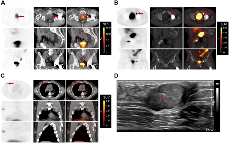 Figure 1
Peer Reviews
No public reviews on file for this paper yet. If you reviewed it on a platform where reviews are public (OpenReview, ICLR, NeurIPS, ICML), you can paste yours below so the community can read it here.
Videos
No videos yet. Explain this paper in a talk, walkthrough, or lecture? Add one.
Taxonomy
TopicsMale Breast Health Studies · Prostate Cancer Treatment and Research · Prostate Cancer Diagnosis and Treatment
Prostate-specific membrane antigen (PSMA) is consistently expressed in tumor neovasculature but variably in tumor cells, with prior reports demonstrating PSMA uptake in invasive ductal carcinoma in men (1). This case reinforces the value of PSMA in characterizing breast cancer in men, particularly for tumors with low [^18^F]FDG avidity.
A 72-y-old man with prostate cancer (Gleason score, 4 + 3) who underwent radical prostatectomy and chemoradiation presented with an elevated prostate-specific antigen level (149.93 ng/mL). A follow-up PET/CT scan with ^18^F-piflufolastat ([^18^F]DCFPyl) demonstrated intense tracer uptake (SUV_max_, 16.8) at the vesicourethral anastomosis extending along the bladder wall and neck (Fig. 1A), consistent with residual or recurrent disease. [^18^F]DCFPyl uptake (SUV_max_, 7.7) was also noted in his known osseous metastasis in the left superior pubic ramus (Fig. 1B).
Incidentally, moderate [^18^F]DCFPyl uptake (SUV_max_, 2.8) was seen in the retroareolar region of the right breast, corresponding to a subcutaneous ovoid mass on CT, measuring 17 mm (transverse) × 27 mm (anteroposterior) × 17 mm (craniocaudal) (Fig. 1C). Ultrasound-guided core needle biopsy of the mass (Fig. 1D) confirmed a grade 2 invasive ductal carcinoma that was estrogen receptor– and progesterone receptor–positive and human epidermal growth factor receptor 2–negative, with a Ki-67 index of 10%.
This case illustrates atypical PSMA expression in estrogen receptor–positive, low-grade invasive ductal carcinoma. It highlights the potential role of PSMA PET/CT in the evaluation of breast cancer in men, especially those with low [^18^F]FDG-avid tumors. PSMA is expressed in 84% of breast cancer lesions (2) and outperforms [^18^F]FDG PET/CT for detecting distant metastases (3), highlighting the need for broader PSMA imaging in nonprostatic malignancies (4) and validation in larger cohorts.
DISCLOSURE
No potential conflict of interest relevant to this article was reported.
The reference list from the paper itself. Each links out to its DOI / PubMed record.
- 1Kumar R Mittal BR Bhattacharya A Singh H Singh SK. Synchronous detection of male breast cancer and prostatic cancer in a patient with suspected prostatic carcinoma on 68Ga-PSMA PET/CT imaging. Clin Nucl Med. 2018;43:431–432.29538032 10.1097/RLU.0000000000002063 · doi ↗ · pubmed ↗
- 2Sathekge M Lengana T Modiselle M. 68Ga-PSMA-HBED-CC PET imaging in breast carcinoma patients. Eur J Nucl Med Mol Imaging. 2017;44:689–694.27822700 10.1007/s 00259-016-3563-6PMC 5323468 · doi ↗ · pubmed ↗
- 3Andryszak NŚwiniuch DWójcik E Ramlau R Ruchała M Czepczyński R. Head-to-head comparison of [18F]PSMA-1007 and [18F]FDG PET/CT in patients with triple-negative breast cancer. Cancers (Basel). 2024;16:667.38339419 10.3390/cancers 16030667 PMC 10854516 · doi ↗ · pubmed ↗
- 4Unger C Bronsert P Michalski K Bicker A Juhasz-Böss I. Expression of prostate specific membrane antigen (PSMA) in breast cancer. Geburtshilfe Frauenheilkd. 2022;82:50–58.35027860 10.1055/a-1638-9429 PMC 8747897 · doi ↗ · pubmed ↗