Erratum to “NUFIP1-Mediated Ribophagy Alleviates PANoptosis of CD4+ T Lymphocytes in Sepsis via the cGAS-STING Pathway”
Pengyue Zhao, Jingyan Li, Pengyi He, Yao Wu, Liyu Zheng, Xingpeng Yang, Jiaqi Yang, Ze Fu, Yun Xia, Ning Chen, Ning Dong, Zhiwen Luo, Renqi Yao, Xiaohui Du, Yongming Yao

Abstract
Genes, proteins, chemicals, diseases, species, mutations and cell lines named across the full text — each resolved to its canonical identifier and authoritative record.
Click any figure to enlarge with its caption.
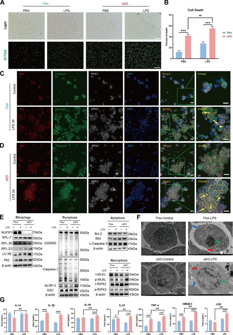 Figure 4
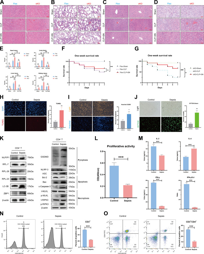 Figure 8
Peer Reviews
No public reviews on file for this paper yet. If you reviewed it on a platform where reviews are public (OpenReview, ICLR, NeurIPS, ICML), you can paste yours below so the community can read it here.
Videos
No videos yet. Explain this paper in a talk, walkthrough, or lecture? Add one.
Taxonomy
Topicsinterferon and immune responses · Ubiquitin and proteasome pathways · Inflammasome and immune disorders
The authors have identified several errors in the Research Article entitled “NUFIP1-mediated Ribophagy Alleviates PANoptosis of CD4^+^ T Lymphocytes in Sepsis via the cGAS-STING Pathway” [1].
Specifically, the Western blot bands of β-actin and Bax in the Fig. 4E panel and the SYTOX green images for the CLP group (Fig. S3A) were incorrectly presented. After carefully re-examining the original data, we determined that these discrepancies resulted from operator error during the figure assembly process in Adobe Illustrator.
In Fig. 4F, the TEM images for the Flox groups were inadvertently duplicated from the Control group shown in Fig. 2I. This occurred during the selection of images from different folders while preparing the figures. A similar duplication error is present in the HE staining images of Fig. 8C.
To facilitate verification, all original data corresponding to the affected figures have been provided for review. The authors would like to emphasize that these inadvertent errors do not affect the data analysis, results, or overall conclusions of the study. Corrected versions of the Figures 4 and 8 are below, and the online HTML and PDF versions of the article have been updated accordingly. Additionally, the Supplementary Materials package has been updated to reflect the revised Figure S3.
The reference list from the paper itself. Each links out to its DOI / PubMed record.