Effect of advanced pregnancy on intraocular pressure, tear secretion and hemogram values in Holstein cows
Tuba Özge Yaşar, Kudret Yenilmez

TL;DR
This study examines how advanced pregnancy affects eye pressure, tear production, and blood parameters in Holstein cows.
Contribution
The study identifies pregnancy-related asymmetry in tear production and hematological changes in cows.
Findings
Pregnant cows showed significant asymmetry in tear production between eyes.
Pregnancy caused significant changes in mean platelet volume and atypical lymphocyte counts.
No significant differences were found in intraocular pressure or average tear production between pregnant and non-pregnant cows.
Abstract
This study investigates how pregnancy influences ocular physiology and hematological parameters in Holstein cows as a topic with limited representation in veterinary research. Thirty clinically healthy cows, aged 2–6 years, were examined and categorized into pregnant (n = 15) and non-pregnant (n = 15) groups. Intraocular pressure (IOP) and tear production (Schirmer Tear Test, STT) were measured bilaterally, and comprehensive blood profiles were evaluated. No statistically significant differences were observed between groups in IOP and mean STT values (p > 0.05); however, a notable asymmetry in tear production was found between the right and left eyes of pregnant cows (p < 0.05), suggesting a localized physiological modulation during gestation. Hematological results revealed significant changes in mean platelet volume (MPV) and atypical lymphocyte percentages and counts (%ALY, ALY) (p <…
Genes, proteins, chemicals, diseases, species, mutations and cell lines named across the full text — each resolved to its canonical identifier and authoritative record.
Click any figure to enlarge with its caption.
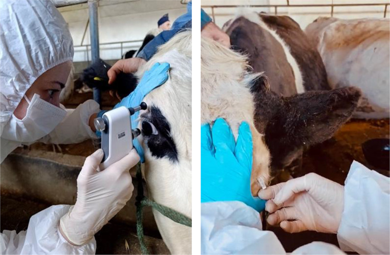 Figure 1
- —Tekirdag Namık Kemal University
Peer Reviews
No public reviews on file for this paper yet. If you reviewed it on a platform where reviews are public (OpenReview, ICLR, NeurIPS, ICML), you can paste yours below so the community can read it here.
Videos
No videos yet. Explain this paper in a talk, walkthrough, or lecture? Add one.
Taxonomy
TopicsReproductive Physiology in Livestock · Ocular Surface and Contact Lens · Corneal surgery and disorders
Introduction
Intraocular pressure (IOP) values can be influenced by various factors, including the type of measurement device used, the experience of the examiner, the species and breed of the animal, stress levels, the time of day the measurement is taken, and environmental conditions (Cahacaltana et al. 2016; Ghaffari et al. 2011). During gestation, multiple organ systems undergo adaptive physiological modifications (Soma-Pillay et al. 2016). The eye is among the organs affected by these physiological changes during pregnancy. Human studies indicate that gestation is associated with widespread ocular alterations, suggesting that pregnancy-related systemic changes extend to visual structurespregnancy, including the eyelids, cornea, intraocular lens, vitreous, macula, and optic nerve (Kalogeropoulos et al. 2019; Naderan 2018). Previous reports indicate that intraocular pressure tends to decline during pregnancy, particularly as gestation progresses into later stages (Artunay et al. 2010).
At the molecular level, pregnancy-associated systemic adaptations are tightly regulated by epigenetic mechanisms. Recent evidence highlights the role of DNA methyltransferases in regulating trophoblast fusion and placental development, which are critical for maintaining maternal–fetal homeostasis. Altered DNA methylation patterns have been shown to influence vascular remodeling, immune tolerance, and metabolic signaling during pregnancy, thereby contributing to systemic physiological adaptations observed in gestation (Yang et al. 2024). These molecular regulatory processes provide a biological framework for understanding pregnancy-related changes in hematological and ocular physiology.
The assessment of hematological parameters provides valuable insight into overall animal health, as blood reflects systemic physiological status. The evaluation of blood parameters can assist in diagnosing disorders related to the hematopoietic system, general metabolic imbalances, and diseases of various systems and organs (Bezerra et al. 2017). Achieving sustainable livestock production in cattle farming requires enhancing reproductive potential without compromising animal welfare. Hematological evaluation is widely employed in dairy practice as a practical indicator of systemic health status (Hasan et al. 2021; Mekroud et al. 2021). By examining the hematological profile, it is possible to detect reproductive disorders in cows and to identify factors that influence biological markers such as disease, pregnancy or stress (Abramowicz et al. 2019; Loi et al. 2021; Noya et al. 2019; Purwar et al. 2019).
Despite the extensive body of research on bovine reproductive physiology, pregnancy-associated ocular changes remain poorly characterized. Considering the profound hormonal, vascular and immunological alterations that occur during gestation, the lack of bovine-specific data on ocular parameters constitutes a significant knowledge gap.
Pregnancy is also characterized by profound metabolic reprogramming aimed at supporting fetal growth and maternal adaptation. Recent studies focusing on reproductive physiology have demonstrated that altered lipid, glucose and amino acid metabolism is closely linked to hormonal fluctuations and immune regulation (Ruan et al. 2024). Such systemic metabolic shifts may indirectly influence hematological parameters and tissue-specific physiological responses, including ocular structures, emphasizing the importance of evaluating pregnancy-related changes within a holistic physiological context.
A review of the existing literature indicates that only a limited number of studies have assessed intraocular pressure and Schirmer tear test values in cows and to date, no studies conducted in Turkey have examined the impact of pregnancy on ocular parameters in Holstein cattle.
In this context, accurate ocular screening during pregnancy is clinically important to prevent misdiagnosis, safeguard animal welfare, and optimize herd health management. Therefore, the present study aims to evaluate the effects of advanced pregnancy on intraocular pressure, tear secretion and selected hemogram parameters in Holstein cows.
Materials and methods
The present study was conducted on 30 clinically healthy Holstein cows, aged between 2 and 6 years (n = 30), housed on a private farm in the Tekirdağ province. Ethical approval was obtained from the Local Animal Ethics Committee of Tekirdağ Namık Kemal University. All animals underwent ultrasonographic examination (Hasvet WED-3100 V) and were allocated into two groups: cows in advanced pregnancy (n = 15) and non-pregnant cows (n = 15). The study was carried out during the spring season under natural photoperiod conditions. All animals were maintained under standardized housing, feeding, and reproductive management practices, and were kept in semi-open barns with natural daylight exposure.
Immediately following pregnancy diagnosis, intraocular pressure (IOP) measurements were performed in both eyes of each animal using a Tonovet Icare Plus device. All ocular examinations were conducted in the morning hours (between 09:00 and 11:00) to minimize diurnal variations in IOP and tear secretion. Ambient temperature during measurements ranged between 18 and 22 °C. Tear secretion was subsequently assessed using Schirmer tear test strips, and the obtained values were recorded (Fig. 1a, and b). Following ocular examinations, blood samples were collected into EDTA-containing tubes for hematological analysis and promptly transported to the Veterinary Clinical Practice and Research Center Laboratory of Tekirdağ Namık Kemal University, where hemogram analyses were performed using an automated analyzer.
To minimize stress-related variability and operator bias, all ocular measurements were carried out by the same investigator within the animals’ housing environment. During the procedure, animals were gently restrained by a trained assistant using minimal manual head fixation for a short duration.
Fig. 1a, b. Measurement of IOP using the tonovet icare plus and measurement of STT
Statistical analysis
Normality tests were first applied to the data obtained from the groups. For comparisons of intraocular pressure (IOP), Schirmer Tear Test (STT), and blood parameter test results between groups, the Independent Samples t-test was used for normally distributed paired groups, while the Mann–Whitney U test was applied for non-normally distributed data. A significance level of 5% (p = 0.05) was adopted for all statistical analyses.
Results
According to the descriptive statistical results, the mean intraocular pressure (IOP) in pregnant cows was calculated as 33.07 mmHg, with a minimum value of 23 mmHg and a maximum of 50 mmHg. The mean value for the Schirmer Tear Test (STT) was 27.73 mm/min, with a minimum of 14 mm/min and a maximum of 35 mm/min (Table 1).
Table 1. Descriptive statistics of intraocular pressure (IOP) (mmHg) and Schirmer Tear Test (STT) (mm/min) results in pregnant and non-pregnant groupsGroupTestEyes n
\documentclass[12pt]{minimal} \usepackage{amsmath} \usepackage{wasysym} \usepackage{amsfonts} \usepackage{amssymb} \usepackage{amsbsy} \usepackage{mathrsfs} \usepackage{upgreek} \setlength{\oddsidemargin}{-69pt} \begin{document}$$\:\stackrel{-}{x}$$\end{document} SS \documentclass[12pt]{minimal} \usepackage{amsmath} \usepackage{wasysym} \usepackage{amsfonts} \usepackage{amssymb} \usepackage{amsbsy} \usepackage{mathrsfs} \usepackage{upgreek} \setlength{\oddsidemargin}{-69pt} \begin{document}$$\:\stackrel{-}{x}$$\end{document} MinMaxPregnantIOPRight1532.336.6511.7172548Left1533.806.4051.6542350Mean33.076.4581.1792350STTRight1525.077.2651.8761435Left1530.404.3721.1292335Mean27.736.4861.1841435Non-pregnantIOPRight1528.674.4511.1492336Left1528.007.7001.9881335Mean29.804.7590.8692341STTRight1530.934.9351.2742441Left1527.007.9642.0561535Mean27.507.7141.4081335
In non-pregnant cows, the mean intraocular pressure was 29.80 mmHg, ranging from a minimum of 23 mmHg to a maximum of 41 mmHg. The mean STT value was 27.50 mm/min, with a minimum of 13 mm/min and a maximum of 35 mm/min.
Although a slight numerical difference was observed in intraocular pressure (IOP) values between the pregnant and non-pregnant groups, this difference was not statistically significant (p > 0.05; Table 2).
Table 2. Comparison of intraocular pressure (IOP) (mmHg) results between pregnant and non-pregnant groupsGroup n Mean rankSum of ranksU P Pregnant1534.731042.0323.00.060Non-pregnant1526.27788.0p < 0.05: p=Mann Whitney U Test
Regarding STT values, no statistically significant difference was observed between the pregnant and non-pregnant groups (p > 0.05: Table 3).
Table 3. Comparison of Schirmer Tear Test (STT) (mm/min) results between pregnant and non-pregnant groupsGroup n Mean rankSum of ranksU P Pregnant1529.83895.00430.00.764Non-pregnant1531.17935.00p < 0.05: p=Mann Whitney U Test
No statistically significant difference was found between the intraocular pressure values of the right and left eyes in pregnant cows (p > 0.05: Table 4).
Table 4. Comparison of intraocular pressure (IOP) (mmHg) values between the right and left eyes of pregnant cowsGroup n
\documentclass[12pt]{minimal} \usepackage{amsmath} \usepackage{wasysym} \usepackage{amsfonts} \usepackage{amssymb} \usepackage{amsbsy} \usepackage{mathrsfs} \usepackage{upgreek} \setlength{\oddsidemargin}{-69pt} \begin{document}$$\:\stackrel{-}{x}$$\end{document} S \documentclass[12pt]{minimal} \usepackage{amsmath} \usepackage{wasysym} \usepackage{amsfonts} \usepackage{amssymb} \usepackage{amsbsy} \usepackage{mathrsfs} \usepackage{upgreek} \setlength{\oddsidemargin}{-69pt} \begin{document}$$\:\stackrel{-}{x}$$\end{document} P Right eye1532.331.7170.543Left eye1533.801.654p < 0.05: p = Independent Samples t-Test
A statistically significant difference was observed between the Schirmer Tear Test results of the right and left eyes in pregnant cows (p < 0.05; Table 5).
Table 5. Comparison of Schirmer Tear Test (STT) (mm/min) results between the right and left eyes of pregnant cowsGroup n
\documentclass[12pt]{minimal} \usepackage{amsmath} \usepackage{wasysym} \usepackage{amsfonts} \usepackage{amssymb} \usepackage{amsbsy} \usepackage{mathrsfs} \usepackage{upgreek} \setlength{\oddsidemargin}{-69pt} \begin{document}$$\:\stackrel{-}{x}$$\end{document} S \documentclass[12pt]{minimal} \usepackage{amsmath} \usepackage{wasysym} \usepackage{amsfonts} \usepackage{amssymb} \usepackage{amsbsy} \usepackage{mathrsfs} \usepackage{upgreek} \setlength{\oddsidemargin}{-69pt} \begin{document}$$\:\stackrel{-}{x}$$\end{document} P Right eye1525.071.8760.021Left eye1530.401.129p < 0.05: p = Independent Samples t-Test
No statistically significant difference was observed between the intraocular pressure values of the right and left eyes in non-pregnant cows (p > 0.05; Table 6).
Table 6. Comparison of Intraocular Pressure (IOP) (mmHg) results between the right and left eyes of non-pregnant cowsGroup n Mean rankSum of ranksU P Right eye1513.53203.0083.000.233Left eye1517.47262.00p < 0.05: p=Mann Whitney U Testi
No statistically significant difference was observed between the intraocular pressure values of the right and left eyes in non-pregnant cows (p > 0.05; Table 7).
Table 7. Comparison of Schirmer Tear Test (STT) (mm/min) results between the right and left eyes of non-pregnant animalsGroup n Mean rankSum of ranksU P Right eye1515.83237.50107.500.838Left eye1515.17227.50p < 0.05: p=Mann Whitney U Testi
Hemogram Results
A statistically significant difference was observed only in MPV, %ALY and ALY values between pregnant and non-pregnant cows according to hemogram results (p < 0.05; Tables 8 and 9).
Table 8. Comparison of blood parameter results between pregnant and non-pregnant cowsGroup n Parameters \documentclass[12pt]{minimal} \usepackage{amsmath} \usepackage{wasysym} \usepackage{amsfonts} \usepackage{amssymb} \usepackage{amsbsy} \usepackage{mathrsfs} \usepackage{upgreek} \setlength{\oddsidemargin}{-69pt} \begin{document}$$\:\stackrel{-}{x}$$\end{document} S \documentclass[12pt]{minimal} \usepackage{amsmath} \usepackage{wasysym} \usepackage{amsfonts} \usepackage{amssymb} \usepackage{amsbsy} \usepackage{mathrsfs} \usepackage{upgreek} \setlength{\oddsidemargin}{-69pt} \begin{document}$$\:\stackrel{-}{x}$$\end{document} P Pregnant15WBC7.060.330.673Non-pregnant156.850.35Pregnant15%LYM49.101.690.475Non-pregnant1551.8813.38Pregnant15%NEU38.501.890.471Non-pregnant1535.303.96Pregnant15LYM3.460.200.786Non-pregnant153.570.32Pregnant15MON0.670.040.391Non-pregnant150.750.07Pregnant15NEU2.740.220.334Non-pregnant152.390.28Pregnant15RBC5.960.120.059Non-pregnant156.540.27Pregnant15HGB10.070.160.102Non-pregnant1510.730.40Pregnant15HCT29.670.490.077Non-pregnant1532.201.30Pregnant15MCV50.091.030.687Non-pregnant1549.491.02Pregnant15MCH16.830.360.538Non-pregnant1516.470.45p < 0.05: p = Independent Samples t-Test
Table 9. Comparison of blood parameter results between pregnant and non-pregnant cowsGroup n ParametersMean rankSum of ranksU P Pregnant15%MON13.93209.0089.000.330Non-pregnant1517.07256.00Pregnant15EOS18.10271.5073.500.106Non-pregnant1512.90193.50Pregnant15BASO14.57218.5098.500.557Non-pregnant1516.43246.50Pregnant15%EOS17.80267.0078.000.152Non-pregnant1513.20198.00Pregnant15%BASO14.47217.0097.000.515Non-pregnant1516.53248.00Pregnant15MCHC16.80252.0093.000.418Non-pregnant1514.20213.00Pregnant15RDW_CV15.13227.00107.000.819Non-pregnant1515.87238.00Pregnant15PLT17.87268.0062.000.061Non-pregnant1511.93167.00Pregnant15MPV19.93299.0031.000.001Non-pregnant159.71136.00Pregnant15%ALY11.17167.5047.500.007Non-pregnant1519.83297.50Pregnant15ALY10.93164.0044.000.004Non-pregnant1520.07301.00Pregnant15%LIC13.67205.0085.000.254Non-pregnant1517.33260.00Pregnant15LIC13.70205.5085.500.262Non-pregnant1517.30259.50Pregnant15SMEAN(PLT)18.60279.0066.000.054Non-pregnant1512.40186.00Pregnant15SMEAN(MPV)20.93314.0031.000.001Non-pregnant1510.07151.00p < 0.05: p=Mann Whitney U Test
Discussion
In this study, intraocular pressure (IOP), Schirmer Tear Test (STT) and various hemogram parameters were compared between pregnant and non-pregnant cows. Overall, gestation appeared to exert minimal influence on selected ocular measurements, whereas distinct alterations were evident in specific hematological indices.
Numerical differences were detected in IOP values between pregnant and non-pregnant cows; however, these differences were not statistically significant. This result is consistent with some studies in the literature. For example, Gebru et al. (2011) reported that pregnancy does not cause significant changes in intraocular pressure. In contrast, some human studies indicate that hormonal changes during pregnancy may reduce intraocular pressure (Qureshi et al. 1996). However, these effects may vary among species, and there is limited data on cattle.
Regarding the Schirmer Tear Test results, no significant difference was found between the pregnant and non-pregnant groups, suggesting that pregnancy does not affect lacrimation levels. However, a statistically significant difference was observed between the right and left eyes of pregnant cows. This finding is presented as a hypothesis and may reflect local physiological or anatomical variability rather than a definitive biological effect. Although bovine-specific data on interocular STT differences are limited, previous studies in cattle have reported wide ranges of normal STT values without significant interocular differences, while highlighting considerable variability in tear production among individuals and under different conditions (Tofflemire et al. 2015). Comparable interocular variability has also been described as part of normal physiological variation in human ophthalmologic studies (Gayton 2009).
When examining the hemogram parameters, significant differences were observed in MPV (Mean Platelet Volume), %ALY (Percentage of Atypical Lymphocytes) and ALY (Atypical Lymphocyte Count), between pregnant and non-pregnant cows. Elevated MPV may reflect physiological platelet activation during pregnancy. Indeed, various studies have documented increased platelet activity throughout pregnancy as a physiological adaptation to maintain hemostatic balance (Rath et al. 2015; Bozkurt et al. 2013).
Recent clinical evidence further supports the concept of immune modulation during reproductive processes. A prospective cohort study evaluating immune responses in couples undergoing assisted reproductive technologies demonstrated that controlled immune activation and lymphocyte profile modulation are essential components of reproductive success (Yang et al. 2025). These findings reinforce the interpretation that reduced %ALY and ALY values observed in pregnant cows may reflect physiological immune adaptation rather than pathological immune suppression.
Pregnancy is accompanied by controlled oxidative stress and adaptive antioxidant responses that support immune balance and tissue integrity. Experimental studies in animal models have shown that amino acids such as glutamate and aspartate play a protective role by enhancing antioxidant enzyme activity and supporting immune defense mechanisms (Tang et al. 2020). Although conducted in male reproductive physiology, these findings provide relevant insights into how systemic antioxidant and immune modulation may contribute to the physiological hematological adaptations observed during pregnancy.
Furthermore, lower %ALY and ALY values may indicate modulation of the immune system during pregnancy. Pregnancy induces immune tolerance by regulating immune cell populations. The literature describes changes in lymphocyte subgroups during pregnancy, including increased regulatory T cells and suppression of certain pro-inflammatory cells (Aluvihare et al. 2004).
Changes in iron metabolism during pregnancy may affect MCHC, but this effect may be less pronounced in cattle and requires further research in larger populations. Indeed, the current study did not find a statistically significant difference (Gül et al. 2010).
Limitations of this study include a relatively small sample size and lack of evaluation of hormonal variables. Additionally, ophthalmological parameters such as intraocular pressure and tear production may be influenced by seasonal and environmental factors.
Conclusion
This study aimed to compare intraocular pressure (IOP), Schirmer Tear Test (STT), and various hematological parameters between pregnant and non-pregnant cows. The results showed no statistically significant differences between the groups regarding IOP and STT, except for a significant difference between the right and left eye Schirmer Test results in pregnant cows. This suggests that pregnancy may asymmetrically affect tear production between eyes, possibly due to local factors.
Hematological data indicated that pregnancy has statistically significant effects on certain blood parameters. Changes in MPV, %ALY, ALY, and MCHC values demonstrate that pregnancy modulates hematological responses. These differences can be associated with physiological immune adaptations and hematological dynamics during pregnancy.
The findings emphasize that pregnancy is a physiological state that should be considered during ophthalmological and hematological evaluations. Clinical examinations and laboratory assessments of pregnant animals should take these physiological changes into account, and pregnancy-related variations should not be mistaken for pathological conditions.
Recognizing physiological pregnancy-related variations in hematological and ocular parameters is clinically important, as it may help veterinarians avoid misinterpretation of normal gestational changes as pathological conditions, thereby reducing the risk of misdiagnosis in pregnant cows.
Future studies with larger sample sizes and longitudinal designs could better elucidate the temporal course of these physiological changes. Additionally, assessing the relationship between hormonal levels and these parameters would provide a more comprehensive understanding.
Supplementary Information
Below is the link to the electronic supplementary material.
Supplementary Material 1
The reference list from the paper itself. Each links out to its DOI / PubMed record.
- 1Tang W, Wu J, Jin S, He L, Lin Q, Luo F, Yin Y (2020) Glutamate and aspartate alleviate testicular/epididymal oxidative stress by supporting antioxidant enzymes and immune defense systems in boars. Sci China Life Sci 63(1):116–124. https://link.springer.com/article/10.1007/s 11427-018-9492-8