Pulmonary Artery Sarcoma With Lung Metastasis
Ryoju Sato, Tomoki Teramoto, Masamitsu Hamakawa, Toshihide Yokoyama, Tadashi Ishida

TL;DR
A rare case of pulmonary artery sarcoma with lung metastasis was diagnosed in a 77-year-old woman using contrast-enhanced CT.
Contribution
This case highlights the diagnostic value of contrast-enhanced CT in identifying pulmonary artery sarcoma.
Findings
Contrast-enhanced CT revealed a contrast defect in the main pulmonary artery.
The patient was diagnosed with pulmonary artery sarcoma based on imaging findings.
Abstract
Pulmonary artery sarcoma is a rare disease that has been frequently misdiagnosed. A 77‐year‐old woman presented with palpitations and was found to have pulmonary hypertension. Contrast‐enhanced computed tomography showed a characteristic contrast defect occupying the entire lumen of the main pulmonary artery, leading to a diagnosis of pulmonary artery sarcoma. A 77‐year‐old woman presented with palpitations and was found to have pulmonary hypertension. Contrast‐enhanced computed tomography showed a characteristic contrast defect occupying the entire lumen of the main pulmonary artery, leading to a diagnosis of pulmonary artery sarcoma.
Genes, proteins, chemicals, diseases, species, mutations and cell lines named across the full text — each resolved to its canonical identifier and authoritative record.
Click any figure to enlarge with its caption.
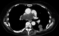 Figure 1
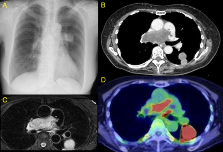 Figure 2
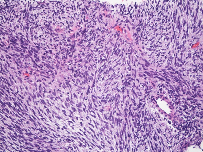 Figure 3
Peer Reviews
No public reviews on file for this paper yet. If you reviewed it on a platform where reviews are public (OpenReview, ICLR, NeurIPS, ICML), you can paste yours below so the community can read it here.
Videos
No videos yet. Explain this paper in a talk, walkthrough, or lecture? Add one.
Taxonomy
TopicsCardiac tumors and thrombi · Lung Cancer Diagnosis and Treatment · Vascular Anomalies and Treatments
A 77‐year‐old woman presented with a one‐month history of palpitations. The electrocardiogram showed sinus tachycardia with a heart rate of 109 beats/min. Echocardiography demonstrated pulmonary hypertension with a tricuspid regurgitation pressure gradient (TRPG) of 67 mmHg. A chest radiograph showed a mass shadow in the left lung field (Figure 1A). Contrast‐enhanced computed tomography (CT) showed a contrast defect occupying the entire lumen of the main pulmonary artery and a mass shadow in the left lower lobe (Figure 1B). Contrast‐enhanced magnetic resonance imaging (MRI) showed a mass lesion with contrast enhancement in the main pulmonary artery (Figure 1C). Positron emission tomography (PET) exhibited increased uptake in the main pulmonary artery (Figure 1D). A CT‐guided biopsy of the left lung mass confirmed metastasis from pulmonary artery sarcoma (Figure 2).
Pulmonary artery sarcoma was frequently misdiagnosed as pulmonary vascular diseases such as chronic thromboembolic pulmonary hypertension (CTEPH) or acute pulmonary embolism. Diagnosis requires chest contrast‐enhanced CT and cardiac ultrasound. Pulmonary artery sarcoma shows higher contrast enhancement than thrombi on contrast‐enhanced MRI and exhibits higher uptake than thrombi on PET. Clinicians should be familiar with the characteristic imaging found on contrast‐enhanced CT: a heterogeneous, low‐density filling the pulmonary artery with irregular distribution [1].
Author Contributions
Ryoju Sato served as the attending physician for this patient and wrote this manuscript. Tomoki Teramoto also treated this patient. Masamitsu Hamakawa, Toshihide Yokoyama, and Tadashi Ishida supervised the patient's care and revised this manuscript.
Consent
The authors declare that written, informed consent was obtained for the publication of this manuscript and accompanying images and attest that the form used to obtain consent from the patient complies with the Journal requirements as outlined in the author guidelines.
Conflicts of Interest
The authors declare no conflicts of interest.
The reference list from the paper itself. Each links out to its DOI / PubMed record.