S100A8/A9 inhibition reduces splenic myelopoiesis and improves outcomes after stroke
Hyun Ah Kim, Annas Al-sharea, Hannah X. Chu, Sung-Chun Tang, Samoda A. Rupasinghe, Shenpeng R. Zhang, Prabhakara R. Nagareddy, Grant R. Drummond, Thiruma V. Arumugam, Andrew J. Murphy, Christopher G. Sobey, Man K.S. Lee

TL;DR
This study shows that inhibiting S100A8/A9 reduces harmful neutrophil production in the spleen after stroke, improving recovery and reducing brain damage.
Contribution
The study identifies the spleen as a key site of myelopoiesis after stroke and demonstrates that S100A8/A9 inhibition can improve stroke outcomes.
Findings
Spleen shows increased neutrophils and myeloid progenitors after stroke, indicating extramedullary myelopoiesis.
Pharmacological inhibition of S100A8/A9 reduces splenic myelopoiesis and improves neurological recovery.
ABR-215757 treatment leads to reduced infarct size and reversed neutrophilia in mice after stroke.
Abstract
Neutrophils are among the earliest immune cells to infiltrate the ischemic brain and contribute to secondary neuronal damage. The alarmin S100 calcium-binding protein A8/A9 (S100A8/A9), predominantly released by neutrophils, is upregulated during this process. Although the bone marrow is recognized as the principal site of neutrophil production via myelopoiesis, the role of the spleen as an immune-responsive organ remains incompletely understood. In this study, we employed a transient middle cerebral artery occlusion model in male C57Bl/6 mice and examined immune responses 24 hours post-stroke in the blood, bone marrow and spleen using flow cytometry. To understand the role of S100A8/A9 in modulating stroke-induced myelopoiesis, we administered a small molecule inhibitor of S100A8/A9, ABR-215757, before and after stroke. Human brain was examined immunohistochemically. Analysis of…
Genes, proteins, chemicals, diseases, species, mutations and cell lines named across the full text — each resolved to its canonical identifier and authoritative record.
Click any figure to enlarge with its caption.
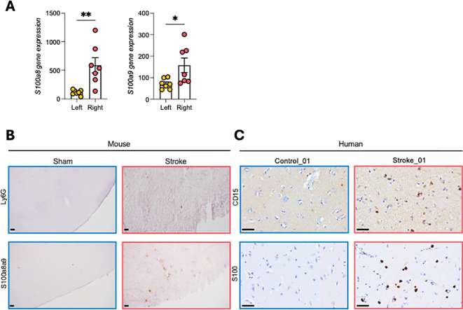 Figure 1
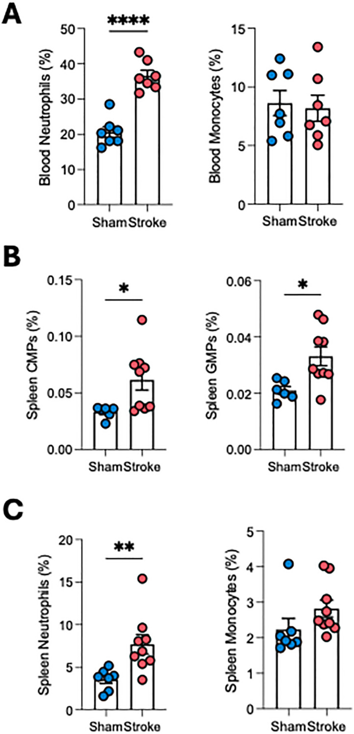 Figure 2
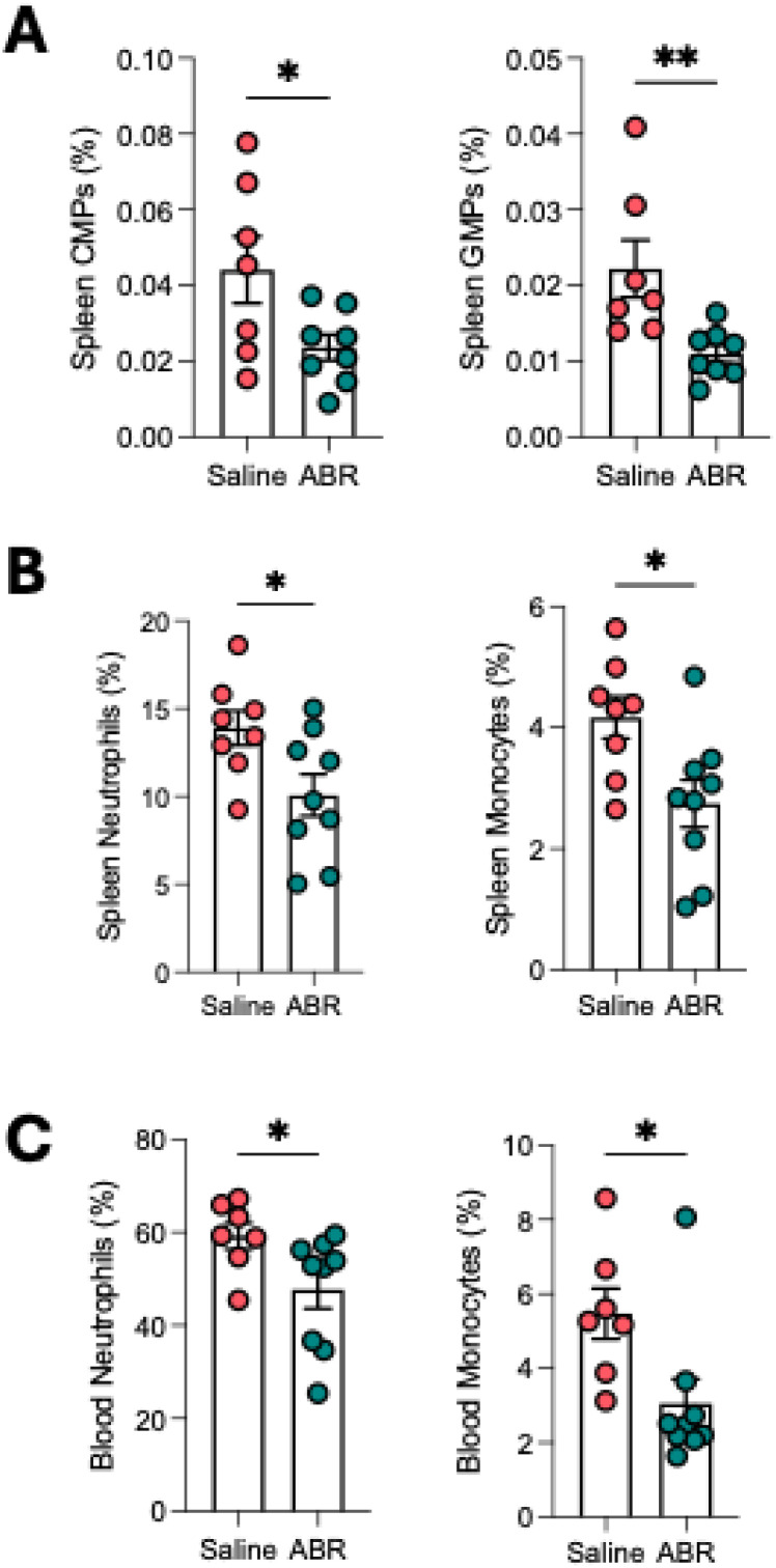 Figure 3
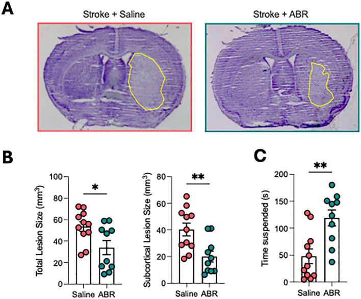 Figure 4
| Case ID | Age (Years) | Sex | Underlying condition | Stroke territory | Surgical intervention |
|---|---|---|---|---|---|
| Control_01 | 0.5 | NA | None reported | Non-stroke | None |
| Control_02 | 40 | M | Severe aplastic anemia; graft failure; multiple organ failure | Non-stroke | None |
| Stroke_01 | 61 | M | Atrial fibrillation | Left MCA territory | IV alteplase; decompressive craniectomy; partial frontal & temporal lobectomy |
| Stroke_02 | 59 | F | Heart failure | Right MCA + ACA territory | Decompressive craniectomy; partial frontal & temporal lobectomy |
| Stroke_03 | 71 | M | Atrial fibrillation; chronic kidney disease | Left ICA + MCA occlusion | Endovascular thrombectomy; decompressive craniectomy; strokectomy |
| Stroke_04 | 42 | M | None reported | Left MCA large territory | Surgical decompression; lobectomy |
- —National Health and Medical Research Council10.13039/501100000925
Peer Reviews
No public reviews on file for this paper yet. If you reviewed it on a platform where reviews are public (OpenReview, ICLR, NeurIPS, ICML), you can paste yours below so the community can read it here.
Videos
No videos yet. Explain this paper in a talk, walkthrough, or lecture? Add one.
Taxonomy
TopicsS100 Proteins and Annexins · Neuroinflammation and Neurodegeneration Mechanisms · Immune Response and Inflammation
Introduction
Ischemic stroke, a leading cause of mortality and long-term disability, is caused by a sudden disruption of cerebral blood flow that leads to neuronal injury and a complex inflammatory response. While restoring blood flow via thrombolysis (tPA) (1), anti-platelet drugs (2), or thrombectomy (3) remain the primary therapeutic approaches, these strategies do not address the secondary inflammation-driven brain injury that worsens stroke outcomes.
Neutrophils and monocytes infiltrate the ischemic brain within the first few hours of stroke onset (4), exacerbating injury through disruption of the blood-brain barrier (BBB), cerebral edema, and neurotoxic cytokine release (5). This pathological mechanism involves factors released by neutrophils, including reactive oxygen species (ROS) (superoxide), proteases (matrix metalloproteinases, elastase), cytokines (interleukin-1β (IL-1β), IL-6, IL-8, tumor necrosis factor-alpha (TNF-α)) and chemokines (CCL2, CCL3, CCL5) (6). Clinically, elevated neutrophil counts correlate with stroke severity (7), infarct size (8), and worse functional outcomes (9). Despite their role in driving inflammation, the source of these myeloid cells and the mechanisms regulating their production remain poorly understood.
Emergency myelopoiesis, the rapid expansion of myeloid progenitors in response to acute inflammation, is a well-characterised phenomenon in several diseases including infection (10) and myocardial infarction (11, 12). Traditionally, the femoral bone marrow was considered the primary site of post-stroke neutrophil and monocyte production. However, recent studies from the Nahrendorf group challenge this view, demonstrating that the skull bone marrow, instead of the femoral bone marrow in mice, serves as the dominant source of neutrophils entering the infarct during the first 24 hours (13). These findings suggest that post-stroke myelopoiesis is compartmentalised, potentially involving multiple distinct hematopoietic reservoirs.
Beyond the bone marrow, the spleen has also emerged as a key immune organ in stroke pathology (14). The marked ability of the spleen to contract in both preclinical models and in stroke patients has been shown to result in the release of stored leukocytes into the circulation, exacerbating inflammation. However, whether the spleen acts as a reservoir or as an active site of myeloid cell production remains unknown.
S100 calcium-binding protein S100A8 and S100A9 are damage-associated molecular pattern (DAMP) molecules, that are present in increased levels in several inflammatory and autoimmune states (15). S100A8 and S100A9 constitute approximately 40% of the cytosolic protein content in human blood neutrophils and 1% in monocytes (16). Once released extracellularly, S100A8/A9 functions as an endogenous agonist to bind to toll-like receptor 4 (TLR4) (17) and the receptor for advanced glycation end products (RAGE) (18, 19), driving emergency myelopoiesis as seen in infection (10) and cardiovascular disease (11). In stroke, clinical studies have demonstrated elevated plasma S100A8/A9 levels that correlate with disease severity (20). While we and others have previously demonstrated that S100A8/A9 promotes emergency myelopoiesis in the bone marrow (12, 21, 22), its role in stroke remains unexplored.
Given the ability of S100A8/A9 to regulate myeloid expansion, we hypothesized that S100A8/A9 may act as a key signal driving neutrophil and monocyte production in response to stroke. Herein, we first assessed the expression of S100A8/A9 and neutrophils in human and mouse brains after ischemic stroke and their role in ischemic brain injury. We then explored stroke-induced myelopoiesis and the impact of S100A8/A9 on the formation of neutrophils and stroke outcomes.
Materials and methods
Human brain
Human brain tissue samples were obtained from two anonymized autopsy non-stroke subjects and four anonymized acute ischemic stroke (AIS) patients receiving decompressive craniectomy and lobectomy at National Taiwan University Hospital with approval from the National Taiwan University Hospital ethics committee. The clinical characteristics of these individuals and samples are summarized in Table 1. The brain tissues were collected within 48 h of death or at the time of surgery. Samples were taken from the temporal lobe within the infarcted area in the AIS patients. Brain tissues were fixed in 4% buffered formalin for at least three weeks before paraffin embedding. Brain sections were processed for immunohistochemical staining using primary antibodies against human CD15 [clone HI98] (Cat# 301902, Biolegend, US) and human S100A8/S100A9 heterodimer (Cat# MAB45701, R&D Systems, US) with a non-biotin-amplified method (Novocastra Laboratories Ltd, UK). Images were acquired using an Olympus microscope.
Animals
This study fully adheres to the Animal Research: Reporting In Vivo Experiments (ARRIVE) guidelines (23). All animal experiments were conducted in accordance with National Health and Medical Research Council of Australia guidelines for the care and use of animals in research and were approved by the Monash University Animal Ethics Committee. Mice had free access to water and food pellets before and after surgery. Three animals (2 Vehicle-treated and 1 ABR-215757-treated) died prior to the scheduled experimental endpoint and were therefore excluded from all subsequent analysis.
ABR-215757 treatments
ABR-215757 was a gift from Active Biotech and was used to inhibit the bioactivity of S100A8/A9. Mice received drinking water containing ABR-215757 (0.5 mg/ml) or normal water from 6 days prior to the induction of ischemia. ABR-215757 (0.5 mg/ml, 100 μl per dose) was injected intraperitoneally 24 h before ischemia and immediately after reperfusion. Control mice were injected with saline. Mice were randomized into different treatment groups and experiments were conducted in a blinded fashion.
Transient focal cerebral ischemia
Focal cerebral ischemia was induced by transient intraluminal filament-induced occlusion of the right MCA, as described previously (24). Mice were anesthetized with ketamine-xylazine (80 and 10 mg/kg, respectively; intraperitoneally). Rectal temperature was monitored and maintained at 37.5°C ± 0.5°C using an electronic temperature controller (Testronics, Kinglake, Victoria, Australia) linked to a heat lamp throughout the procedure and until animals regained consciousness. Briefly, the right proximal common carotid artery was clamped, and a 6–0 nylon monofilament with silicone-coated tip (Doccol Co., Redlands, CA, USA) was inserted and gently advanced into the distal internal carotid artery, 11–12 mm distal to the carotid bifurcation, occluding the MCA at the junction of the Circle of Willis. Severe (typically ~80%) reduction in rCBF was confirmed using transcranial laser-Doppler flowmetry (Perimed, Järfälla, Sweden) in the area of cerebral cortex supplied by the MCA. The filament was then tied in place and the clamp was removed. After 1 h of cerebral ischemia, the monofilament was retracted to allow reperfusion for 23 h. Reperfusion was confirmed by an immediate increase in rCBF, which reached the pre-ischemic level within 5 min. The wound was then closed and the animal was allowed to recover. Regional CBF was recorded for 30 min of reperfusion. All animals were administered 1 mL of sterile saline via a subcutaneous injection for rehydration after surgery. Gel nectar (Able Scientific, WA, Australia) was placed inside the cage and access to chow food and water was provided. All animals’ boxes were placed on heat pads post-surgery until euthanasia. All mice were euthanized at 24 h by isoflurane overdose followed by decapitation.
Neurological assessment
At 23 h after induction of stroke, a hanging wire test was performed in which mice were suspended from a wire 30 cm high for up to 180 s, and the average time of 3 trials with 5-min rest periods in between was recorded. Neurological assessment was evaluated by an observer blinded to experimental groups.
S100A8/9 and neutrophil staining
Mice were euthanized at 24 h by isoflurane overdose, followed by decapitation. The brains were immediately removed and snap frozen with liquid nitrogen. Coronal sections (10 μm) from vehicle-treated mice were fixed with 4% paraformaldehyde, endogenous peroxidase blocked, and stained using primary antibodies; rabbit recombinant multiclonal anti-S100A8+S100A9 [clone RM1038] (cat# ab288715, Abcam, UK) or rat monoclonal anti-mouse Ly-6G (cat# 551459, BD Biosciences, USA). Sections were then stained using horseradish peroxidase-conjugated secondary antibody against rabbit or rat (cat# P0448 or P0450, Dako, Denmark), respectively. Sections were incubated with a 3,3’-diaminobenzidine (DAB) substrate solution (cat# D4418, Sigma-Aldrich, USA) and counterstained with hematoxylin solution. Images were acquired using an Olympus light microscope.
S100 gene expression
A separate cohort of mice was euthanized at 24 h by isoflurane overdose, and perfused with PBS via cardiac puncture. Ipsilateral (right) and contralateral (left) brain hemispheres were processed for RNA extraction. Total RNA was extracted using Trizol and cDNA synthesized using Superscript Vilo (Invitrogen, Thermo Fisher Scientific). Quantitative real time PCR was monitored in real time with an Mx3000 sequence detection system (Stratagene) using SYBR Green PCR Core Reagents (Agilent Technologies) and normalized to 18s.
Flow cytometry
Blood leukocyte counts and analysis
Neutrophils and monocytes were identified using flow cytometry as previously described (25). Blood was collected via tail bleeding into EDTA tubes, which were immediately incubated on ice. BM was harvested from the femurs and tibias. Spleens were flushed through a 40 μm cell strainer with PBS to obtain a single cell suspension (25). For all organs, red blood cells were lysed using 1x RBC lysis buffer (BD pharm Lyse; BD Biosciences) for 15 minutes (blood) or 5 minutes (BM and spleen). Cells were then centrifuged at 400g for 5 minutes and resuspended in 1x HBSS containing 0.1% BSA w/v and 5 mM EDTA. Cells were stained with a cocktail of antibodies. For monocytes and neutrophil analysis, they were stained with anti-CD45 (PB), anti-Ly6-C/G (PerCP-Cy5.5) (BD Biosciences) and anti-CD115 (PE) (eBioscience). Monocytes were identified as CD45^hi^CD115^hi^, neutrophils were identified as CD45^hi^CD115^lo^Ly6-C/G^hi^ (Gr-1). For LSKs, CMPs and GMPs, a cocktail of antibodies to lineage committed cells (CD45R, CD19, CD11b, CD3e, TER-119, CD2, CD8, CD4, and Ly-6G; all FITC; eBioscience) and stem cell markers anti-Sca1 (Pacific Blue) and anti-ckit (APC-Cy7) were used. Hematopoietic stem and progenitor cells were identified as Lin^–^Sca1^+^ckit^+^ (LSKs). Where further identification of myeloid progenitor cells was required, antibodies to CD16/CD32 (FcγRII/III) and CD34 were used to separate common myeloid progenitors (CMPs) (lin^–^Sca1^–^ckit^+^FcγRII/III^int^CD34^+^) and granulocyte-macrophage progenitors (GMPs) (lin^–^Sca1^–^ckit^+^FcγRII/III^hi^CD34^+^). All samples were run on a BD Canto II or BD LSR Fortessa X-20, and analyzed using FlowJo (TreeStar). Gating strategy is shown in Supplementary Figure 1.
Cerebral infarct and edema volumes
Coronal sections (30 μm) separated by ~420 μm were stained with thionin (0.1%) to delineate the infarct. Images of the sections were captured with a CCD camera mounted above a light box. Infarct volume was quantified as described previously (24) using image analysis software (ImageJ, NIH, Bethesda, MD, USA) and corrected for brain edema, estimated using the formula: corrected infarct volume = [left hemisphere area – (right hemisphere area – right hemisphere infarct area) × (thickness of section + distance between sections)] (26). Edema-corrected infarct volumes of individual brain sections were added to give a three-dimensional approximation of the total infarct volume. Total and subcortical infarct volumes were quantified individually.
Statistical analysis. Data are presented as mean ± SEM and were analyzed using the 2-tailed Student’s t test. P less than 0.05 was considered significant. All tests were performed using Prism software (GraphPad Software Inc.).
Results
Stroke increases S100A8/A9 expression in mouse and human ischemic brain
Transient middle cerebral artery occlusion (tMCAO) in mice resulted in robust upregulation of S100a8 and S100a9 mRNA in the ischemic (right) hemisphere at 24 h compared with the contralateral (left) side (Figure 1A). Immunohistochemistry confirmed accumulation of S100A8/A9 together with infiltrating neutrophils in the infarct region (Figure 1B). Similar findings were observed in brain tissue from 4 stroke patients where S100A8/A9 was detected alongside neutrophil infiltration, whereas this was not present in 2 non-stroke controls, (Figure 1C; Supplementary Figure 2), supporting the translational relevance of this pathway.
*S100A8/A9 is upregulated in mouse and human ischemic brain. (A)S100a8/a9 mRNA expression in ipsilateral (right) vs contralateral (left) hemispheres 24 h after tMCAO. (B) Mouse immunohistochemistry shows the expression of S100A8/A9 and neutrophils (Ly6G+) in infarcted cortex. (C) Human post-mortem stroke brain also shows the expression of S100A8/A9 and neutrophil (CD15) in the infarct. Additionally, images from a further 3 stroke brains and 1 control brain are presented in Supplementary Figure 2. Student’s t-test. *p<0.05, *p<0.01. n=6–8 per group. Scale bar = 50µm.
Stroke induces splenic, but not bone marrow, myelopoiesis
Stroke was associated with a marked increase in circulating neutrophils, but no change in monocytes at 24 h (Figure 2A). At 24 h post-stroke, flow cytometry revealed expansion of hematopoietic stem and progenitor cells (LSKs) in femoral bone marrow (Supplementary Figure 3A); however, downstream myeloid progenitors and mature neutrophils were not significantly altered (Supplementary Figures 3A, 2B), consistent with previous reports (27). In contrast, the spleen exhibited a striking expansion of myeloid progenitors (CMPs, GMPs; Figure 2B) as well as increased neutrophils (Figure 2C), indicating that splenic myelopoiesis is a key contributor to systemic neutrophilia after stroke.
*Stroke induces splenic, but not bone marrow, myelopoiesis. (A) Circulating leukocyte counts at 24 h show increased neutrophils, but no change in monocytes. (B) Splenic progenitors (myeloid progenitors, CMPs, and granulocyte–monocyte progenitors, GMPs) are significantly increased after stroke. (C) Splenic mature neutrophils also increased compared with sham, but no change in monocytes. Student’s t-test. *p<0.05, **p<0.01, ***p<0.001. n=6–9 per group.
Inhibition of S100A8/A9 reduces stroke-induced myeloid skewing
Treatment with the S100A8/A9 inhibitor ABR-215757 markedly reduced splenic CMPs and GMPs (Figure 3A), as well as monocytes and neutrophils (Figure 3B), compared with vehicle-treated stroke mice. As expected, bone marrow myelopoiesis and mature myeloid cells remained unaffected (Supplementary Figure 4B). Inhibition of S100A8/A9 also significantly lowered circulating neutrophils and monocytes (Figure 3C), confirming that blocking this pathway mitigates myeloid skewing after stroke.
*ABR-215757 reduces splenic myelopoiesis and mitigates myeloid skewing post-stroke. (A) Splenic progenitors (CMPs, GMPs) are reduced by ABR-215757 treatment. (B) Splenic neutrophils and monocytes are significantly reduced by ABR-215757. (C) Circulating neutrophil and monocyte counts are also reduced by ABR-215757. Student’s t-test. *p<0.05, *p<0.01. n=7–9 per group.
ABR-215757 improves stroke outcomes
Mice treated with ABR-215757 showed a ~35% reduction in infarct volume compared with vehicle-treated controls (Figures 4A, B), along with significantly improved neurological performance in the hanging wire test (Figure 4C). These findings demonstrate that inhibition of S100A8/A9 not only modulates peripheral immune responses but also provides functional neuroprotection in ischemic stroke.
*ABR-215757 improves outcomes after ischemic stroke. (A) Representative thionin-stained brain sections show reduced infarct size in ABR-215757–treated mice. (B) Quantification reveals ~35% reduction in total and subcortical infarct volumes. (C) Hanging wire test shows significantly improved neurological function with ABR-215757. Student’s t-test. *p<0.05, *p<0.01. n=10–11 per group.
Discussion
Ischemic stroke is a leading cause of disability, cognitive impairment, and mortality worldwide. Despite advances in our understanding of cerebral ischemia, current treatments focus almost exclusively on reperfusion strategies such as thrombolysis and thrombectomy, with no approved therapies targeting the immune response. Shortly after cerebral artery occlusion, ischemia triggers a cascade of inflammatory events, including endothelial activation, ROS production, and complement activation, which facilitate leukocyte recruitment and BBB disruption (28, 29). A growing body of evidence suggests that immune cells play complex and multiphasic roles, initially exacerbating tissue injury but later contributing to repair and regeneration (30). Neutrophils are among the earliest immune cells to infiltrate the ischemic brain within minutes, peaking at 24–72 hours (31, 32), worsening injury through BBB disruption, oxidative stress, and release of proteolytic enzymes and their accumulation correlates with stroke severity and worse outcomes (33). Consistent with this, we show an elevated number of neutrophils in the infarcted brain of both tMCAO mice and stroke patients, confirming that neutrophil-driven inflammation is a conserved feature of ischemic stroke. In this study, we demonstrate that S100A8/A9, a neutrophil-derived DAMP, is upregulated in the ischemic brain and promotes myelopoiesis in the spleen, leading to increased circulating neutrophils and monocytes. Importantly, we found that blocking S100A8/A9 with ABR-215757 suppressed splenic myelopoiesis, reduced circulating neutrophils, and significantly improved stroke outcomes. These findings provide novel insight into the regulation of stroke-induced immune responses and identify S100A8/A9 as a potential therapeutic target.
Beyond their role in brain injury, neutrophils contribute to systemic inflammation and myeloid expansion following stroke. We demonstrate that stroke leads to increased expression of S100A8/A9, a neutrophil-derived alarmin, within the infarcted brains of mice and humans. While we acknowledge that our human data on S100A8/A9 expression in the stroke tissue was limited to four samples, previous studies have shown a significant accumulation of S100A8/A9 in human stroke tissues (34). Nevertheless, while these findings are consistent with our data from mice, they are based on a small number of human samples and should be interpreted with caution.
Similarly, we confirmed elevated gene expression of S100a8 and S100a9 in the ischemic hemisphere in mice after tMCAO. S100A8/A9 is constitutively expressed in neutrophils and monocytes, which are its primary cellular sources. During inflammation, these cells actively release S100A8/A9 (15). Given that neutrophils are among the earliest and most numerous immune cells to infiltrate the ischemic brain, they represent the most likely contributors to the elevated S100A8/A9 mRNA observed in our study. S100A9 gene expression is tightly regulated in a differentiation- and lineage-specific manner within the hematopoietic system (35). Previously, we showed that induction of myocardial infarction promoted rapid recruitment of neutrophils and release of S100A8/A9 as alarmins. These complexes bind to TLR4 and prime the nod-like receptor family pyrin domain-containing 3 (NLRP3) inflammasome in neutrophils and promote IL-1β secretion (12). The released IL-1β interacts with IL-1 receptor type 1 expressed on hematopoietic stem and progenitor cells in the bone marrow and stimulates granulopoiesis in a cell-autonomous manner. Whilst the direct mechanistic link has not yet been delineated in stroke, the key upstream components – neutrophil infiltration, S100A8/A9 release, and systemic inflammatory activation – are consistent between the two conditions. Our findings now extend this phenomenon to stroke, showing that brain-derived S100A8/A9 functions as a systemic inflammatory signal driving an activation of extramedullary myelopoiesis.
A central question in post-stroke immunity is the origin of circulating neutrophils. While emergency myelopoiesis has traditionally been attributed to the femoral bone marrow, it has been shown that the skull bone marrow supplies neutrophils to the infarct within the first 24 hours (27). Our study focused on femoral bone marrow and observed no increase in CMPs, GMPs, or mature neutrophils at 24 hours post-stroke. This observation aligns with that of Courties et al. (2015), who similarly reported no early femoral bone-marrow myelopoietic expansion at 24 hours, with increases in progenitor activity emerging only at later time points, particularly around 3 days post-tMCAO (27). These findings suggest that femoral bone-marrow myelopoiesis may follow a delayed temporal pattern after stroke. In contrast, we detected a marked expansion of CMPs, GMPs, and mature myeloid cells in the spleen at 24 h; evidence that stroke triggers this myelopoiesis as a contributing mechanism for increasing circulating neutrophils and monocytes. This aligns with previous reports that splenic contraction post-stroke releases stored leukocytes into the circulation. Further, this observation supports the concept that hematopoietic activity following stroke may involve multiple compartments, including extramedullary sites. While extramedullary myelopoiesis is a known phenomenon in chronic inflammation, such as in atherosclerosis and myocardial infarction, as demonstrated previously by our group (36, 37) and others (11, 38), its involvement in acute settings like stroke has not been previously reported. Our findings provide direct evidence that the spleen can actively generate myeloid cells beginning from CMPs in the acute phase of stroke. Future studies should incorporate cranial bone marrow to fully delineate the relative contributions of different hematopoietic reservoirs.
After stroke, increased activation of the sympathetic nervous system (SNS) leads to reduced spleen size through splenic contraction and release of immune cells (39, 40). Other than this role of the SNS, our data suggest that S100A8/A9 could be a crucial factor that links brain ischemia to systemic myeloid expansion. Given its role in emergency myelopoiesis in myocardial infarction, it is likely that S100A8/A9 released from brain-infiltrating neutrophils acts as a systemic signal to promote myelopoiesis in the spleen. Therefore, we hypothesized that blocking S100A8/A9 signaling could suppress neutrophil production and reduce the severity of stroke outcomes. Using ABR-215757, an S100A8/A9 inhibitor that prevents the interaction with TLR-4 and RAGE (41), we found that treatment significantly reduced CMP and GMP expansion in the spleen, reducing its eventual differentiation into circulating neutrophils and monocytes. Importantly, ABR-215757 treatment significantly reduced infarct volume development and improved neurological function. These findings highlight S100A8/A9 as a promising therapeutic target for reducing post-stroke inflammation and improving functional recovery. Although 24 hours after MCAO represents a relatively early time point, infarct maturation in mice is generally complete by this stage (42). Early improvements, however, do not necessarily indicate sustained benefit, and longer-term follow-up is required to determine whether ABR-215757 confers durable neuroprotection. Our study was designed to focus on early inflammatory mechanisms, including the rapid induction of S100A8/A9, the acute splenic myelopoietic response, and the immediate effects of ABR-215757 on neutrophil dynamics – processes that occur within the first hours after stroke. In addition, while the dosing strategy was based on prior preclinical work, alternative regimens (e.g., repeated or delayed dosing, or extended treatment) may further enhance therapeutic efficacy and warrant investigation in future studies.
A limitation of our study is the lack of genetic validation, e.g. using S100a9-deficient mice. Although ABR-215757 is a known inhibitor of S100A8/A9, it has pleiotropic immunomodulatory effects, including modulation of macrophage and T-cell responses (43), and interactions with other S100 family proteins, including S100a12 (44). These off-target actions could have contributed to the observed reduction in systemic inflammation and spleen myelopoiesis. Future studies using genetic models or alternative inhibitors will be necessary to confirm specificity.
Another limitation of our study is that only male mice were used. Understanding sex-specific differences in stroke immunity is important, as these differences have been extensively characterized (45). Estrogen has been shown to modulate multiple inflammatory pathways, including limiting excessive neutrophil infiltration after injury (46). In particular, estrogen can attenuate neutrophil recruitment (47), which may in turn dampen S100A8/A9-mediated activation of downstream receptors such as TLR4 and RAGE. These hormonal influences have the potential to alter both systemic inflammation and myelopoiesis after stroke. Future studies should therefore include female mice to determine how sex hormones modulate S100A8/A9 signaling and therapeutic efficacy.
Given the growing body of evidence linking neutrophils to infarct expansion and stroke severity, several strategies have been proposed to modulate neutrophil function as a therapeutic approach (48). While direct neutrophil depletion has shown efficacy in preclinical stroke models, this approach carries risks of immunosuppression and increased infection susceptibility (49). Moreover, the failure of clinical trials targeting total neutrophil numbers or adhesion may in part reflect the functional heterogeneity of neutrophils, highlighting the importance of determining whether S100A8/A9 inhibition can modulate neutrophil phenotype in addition to reducing their number. Our findings suggest that targeting upstream regulators of myelopoiesis, such as S100A8/A9, may provide a more selective approach to reducing harmful neutrophilia without compromising host defense. Beyond stroke, S100A8/A9 has been implicated in atherosclerosis, myocardial infarction, and thrombosis, suggesting that its inhibition could have broader implications for cardiovascular disease. Given that plasma S100A8/A9 levels correlate with infarct size and clinical outcomes in stroke patients, future studies should assess whether S100A8/A9 inhibitors could be translated into clinical trials for stroke and other vascular inflammatory disorders.
In conclusion, we identify S100A8/A9 as a key regulator of stroke-induced myelopoiesis, linking brain ischemia to systemic neutrophil production via extramedullary myelopoiesis in the spleen. We also demonstrate that blocking S100A8/A9 suppresses splenic myelopoiesis, reduces circulating neutrophils, and improves stroke outcomes. These findings further identify the spleen as a key organ in stroke and highlight S100A8/A9 as a novel therapeutic target to dampen post-stroke inflammation.
The reference list from the paper itself. Each links out to its DOI / PubMed record.
- 1Xiong Y Campbell BCV Schwamm LH Meng X Jin A Parsons MW . Tenecteplase for Ischemic Stroke at 4.5 to 24 Hours without Thrombectomy. New Engl J Med. (2024) 391:203–12. doi: 10.1056/NEJ Moa 2402980, PMID: 38884324 · doi ↗ · pubmed ↗
- 2Johnston SC Easton JD Farrant M Barsan W Conwit RA Elm JJ . Clopidogrel and aspirin in acute ischemic stroke and high-risk TIA. New Engl J Med. (2018) 379:215–25. doi: 10.1056/NEJ Moa 1800410, PMID: 29766750 PMC 6193486 · doi ↗ · pubmed ↗
- 3Sarraj A Hassan AE Abraham MG Ortega-Gutierrez S Kasner SE Hussain MS . Trial of endovascular thrombectomy for large ischemic strokes. New Engl J Med. (2023) 388:1259–71. doi: 10.1056/NEJ Moa 2214403, PMID: 36762865 · doi ↗ · pubmed ↗
- 4Ross AM Hurn P Perrin N Wood L Carlini W Potempa K . Evidence of the peripheral inflammatory response in patients with transient ischemic attack. J Stroke Cerebrovasc Dis. (2007) 16:203–7. doi: 10.1016/j.jstrokecerebrovasdis.2007.05.002, PMID: 17845917 PMC 2570354 · doi ↗ · pubmed ↗
- 5Segel GB Halterman MW Lichtman MA . The paradox of the neutrophil’s role in tissue injury. J Leukoc Biol. (2011) 89:359–72. doi: 10.1189/jlb.0910538, PMID: 21097697 PMC 6608002 · doi ↗ · pubmed ↗
- 6Kolaczkowska E Kubes P . Neutrophil recruitment and function in health and inflammation. Nat Rev Immunol. (2013) 13:159–75. doi: 10.1038/nri 3399, PMID: 23435331 · doi ↗ · pubmed ↗
- 7Kim J Song TJ Park JH Lee HS Nam CM Nam HS . Different prognostic value of white blood cell subtypes in patients with acute cerebral infarction. Atherosclerosis. (2012) 222:464–7. doi: 10.1016/j.atherosclerosis.2012.02.042, PMID: 22460048 · doi ↗ · pubmed ↗
- 8Buck BH Liebeskind DS Saver JL Bang OY Yun SW Starkman S . Early neutrophilia is associated with volume of ischemic tissue in acute stroke. Stroke. (2008) 39:355–60. doi: 10.1161/STROKEAHA.107.490128, PMID: 18162626 · doi ↗ · pubmed ↗