Endoscopic ultrasound-guided creation of a drainage route combined with third-space endoscopy for esophageal intramural abscess debridement
Tzong-Hsi Lee, Guan-De Li, Chao-Yu Liu, Chung-Tsui Huang, Chi-Chu Lo, Chen-Shuan Chung

Abstract
Genes, proteins, chemicals, diseases, species, mutations and cell lines named across the full text — each resolved to its canonical identifier and authoritative record.
Click any figure to enlarge with its caption.
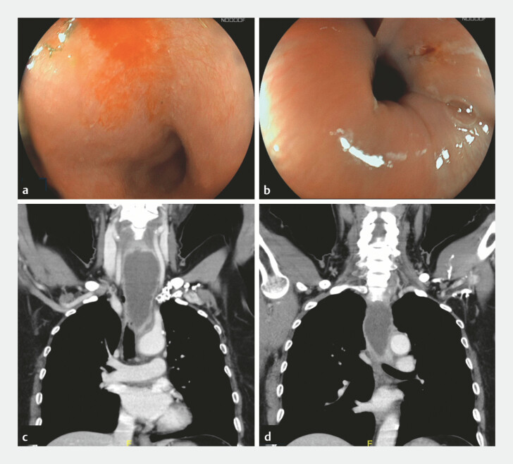 Fig. 1
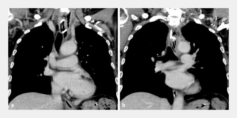 Fig. 2
Peer Reviews
No public reviews on file for this paper yet. If you reviewed it on a platform where reviews are public (OpenReview, ICLR, NeurIPS, ICML), you can paste yours below so the community can read it here.
Videos
No videos yet. Explain this paper in a talk, walkthrough, or lecture? Add one.
Taxonomy
TopicsEsophageal and GI Pathology · Amoebic Infections and Treatments · Foreign Body Medical Cases
A 62-year-old woman presented with odynophagia after accidental fish bone ingestion. Subsequent esophagogastroduodenoscopy (EGD) identified a 2-cm fish bone lodged at the upper esophageal inlet, which was removed with retrieval forceps. Multiple mucosal erosions at the esophageal inlet and mid-esophagus were noted ( Fig. 1 a, b ). Two weeks later, she developed persistent odynophagia, dysphagia and fever, and laboratory tests showed leukocytosis. Computed tomography (CT) revealed an esophageal para-esophageal abscess extending from the upper to mid-esophagus ( Fig. 1 c, d ).
Evolution of esophageal mucosal erosions into an esophageal intramural abscess. a, b Multiple erosions were noted at the esophageal inlet and mid-esophagus during the initial EGD. c, d Contrast-enhanced CT revealed a large (12.8 cm × 4.5 cm × 3.2 cm) fluid-filled collection with marginal enhancement along the para-esophageal region at the C5–T5 spine level, suggestive of a para-esophageal abscess. CT, computed tomography; EGD, esophagogastroduodenoscopy.
Endoscopic ultrasound (EUS) confirmed an abscess located in the submucosal space. EUS-guided transmural drainage was performed using a 19-gauge needle (Expect Slimline Needle, Boston Scientific, Marlborough, MA) and guidewire-assisted tract creation, followed by the placement of a 10-Fr double-pigtail stent (Advanix Biliary Stents, Boston Scientific, Marlborough, MA) for continuous drainage. A mucosal incision was then made to access the submucosal tunnel, allowing endoscopic entry into the abscess cavity for debridement ( Video 1 ). A nasogastric tube was inserted for postoperative feeding.
EUS-guided creation of a drainage route combined with third-space endoscopy for esophageal intramural abscess debridement. EUS, Endoscopic ultrasound.Video 1
Symptoms improved markedly after the procedure, and cultures obtained from the abscess cavity grew Streptococcus anginosus , Streptococcus constellatus and Eikenella corrodens , consistent with oral flora. Follow-up CT and EGD 3 days later demonstrated the complete resolution of the abscess ( Fig. 2 a, b ). The stent was removed 1 week later, and the mucosal entry site was closed with endoclips (MANTIS Clip, Boston Scientific, Marlborough, MA and SureClip, Micro-Tech Endoscopy, USA; Video 1 ). The patient resumed normal oral intake without further complications.
Resolution of the esophageal intramural abscess after EUS-guided double-pigtail stent placement combined with submucosal endoscopic debridement. a, b A CT scan performed 3 days later showed the complete resolution of the esophageal abscess. CT, computed tomography; EUS, endoscopic ultrasound.
Submucosal esophageal abscess is a rare condition that may arise from mucosal injury without full-thickness perforation. EUS can delineate both the depth and the layer of involvement with greater accuracy as demonstrated in this case. While antibiotics are typically first-line therapy, extensive abscesses may require intervention 1 . This case highlights a novel, minimally invasive approach combining endoscopic ultrasound-guided drainage and third-space endoscopic debridement, achieving rapid recovery without surgical intervention.
Endoscopy_UCTN_Code_CCL_1AB_2AC_3AZ
The reference list from the paper itself. Each links out to its DOI / PubMed record.