Colorectal lymph node harvest in cancer surgery, adequacy and treatment implication: a 5-year retrospective analysis from a tertiary hospital in Ethiopia
Sirna Emana Jaleta, Abdo Kedir Abafogi, Tamirat Godebo Woyimo, Gashahun Mekonnen Disassa, Sultan Jebel Usman, Abduletif Haji-Ababor Abagojam, Kedir Negesso Tukeni

TL;DR
This study from Ethiopia shows that most colorectal cancer patients are young and only 23% had enough lymph nodes collected during surgery, which is crucial for proper cancer staging and treatment.
Contribution
The study identifies surgeon expertise, tumor depth, and specimen length as key factors affecting lymph node harvest adequacy in colorectal cancer surgery in Ethiopia.
Findings
Only 23% of colorectal cancer patients had adequate lymph node retrieval.
GI oncologic surgeons were significantly associated with improved lymph node harvest adequacy.
Tumor depth and specimen length also influenced lymph node retrieval outcomes.
Abstract
Colorectal cancer is one of the common malignancies, and obtaining sufficient lymph nodes after surgeries is critical for staging and subsequent treatment planning. While guidelines advocate collecting at least 12 lymph nodes, insufficient lymph node sampling can have catastrophic consequences. This was a retrospective study that looked at the parameters influencing lymph node retrieval during colorectal cancer surgery in one of tertiary hospital in Ethiopia. In this study, data from 85 patients’ records for stages I-III were analyzed and divided into two groups: adequately harvested and inadequately harvested. The association between potential factors impacting optimal harvests was analyzed. The study found that the majority of cancer patients were between the ages of 34 and 53 years, in which the adequate lymph node retrieval was achieved only in 23% of cases. Procedures being…
Genes, proteins, chemicals, diseases, species, mutations and cell lines named across the full text — each resolved to its canonical identifier and authoritative record.
Click any figure to enlarge with its caption.
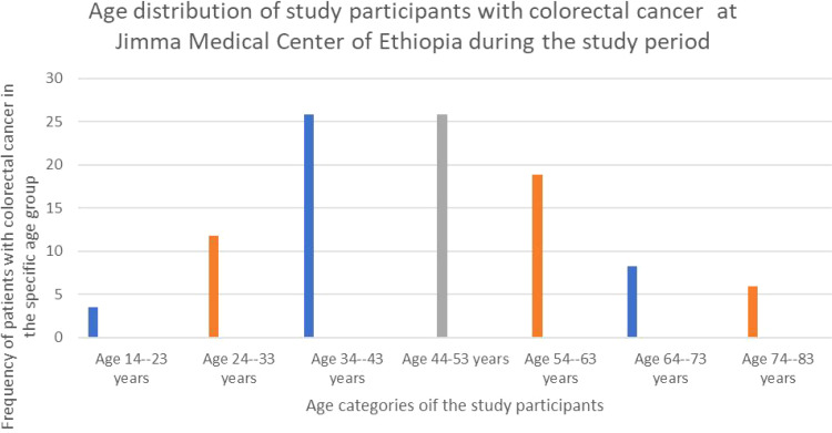 Figure 1
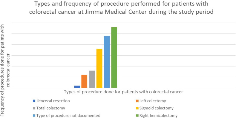 Figure 2
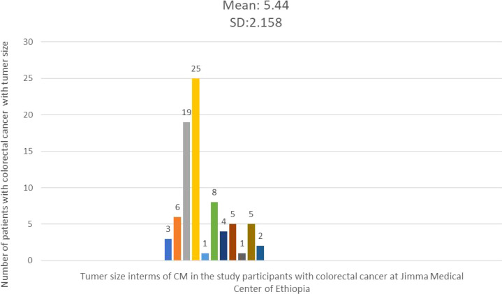 Figure 3
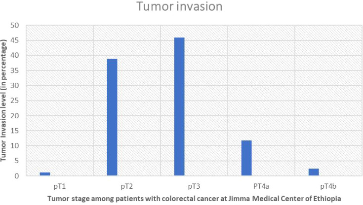 Figure 4
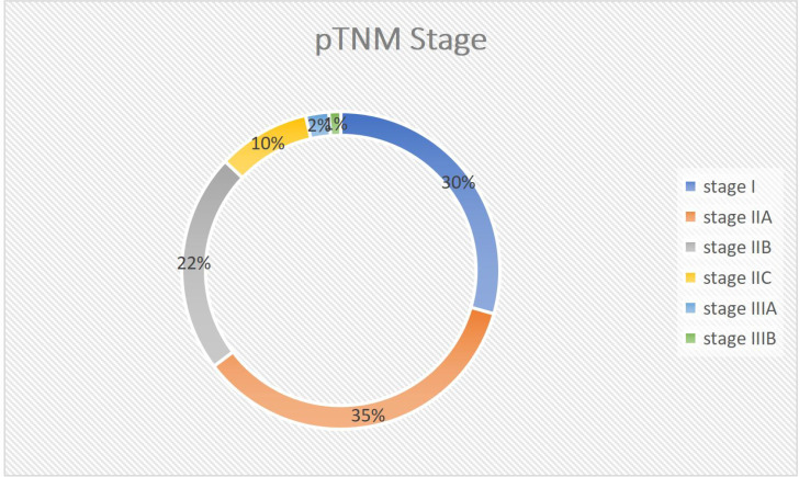 Figure 5
| Tumor Location | N (%) |
|---|---|
| Cecum | 16 (18.8%) |
| Ascending colon | 9 (10.60%) |
| Transverse colon | 1 (1.20%) |
| Descending colon | 1 (1.20%) |
| Sigmoid colon | 26 (30.60%) |
| Rectum | 10 (11.80%) |
| Not documented | 20 (23.50%) |
| Rectosigmoid junction | 2 (2.40%) |
| Type of Resection | |
| Ileocecal resection | 1 (1.20%) |
| Not documented | 24 (28.20%) |
| Right hemicolectomy | 28 (32.90%) |
| Left colectomy | 6 (7.10%) |
| Total colectomy | 8 (9.40%) |
| Sigmoid colectomy | 18 (21.20%) |
|
| 85 (100.00%) |
| Feature | Value | Frequency | Percentage |
|---|---|---|---|
| Proximal margin | Free | 83 | 97.60% |
| Involved | 2 | 2.40% | |
| distal margin | Free | 81 | 95.30% |
| Involved | 4 | 4.70% | |
| radial or another margin | Free | 54 | 63.50% |
| Involved | 2 | 2.40% | |
| Not documented | 29 | 34.10% | |
| histologic type | Adenocarcinoma | 75 | 88.20% |
| Mucinous carcinoma | 8 | 9.40% | |
| Signet ring carcinoma | 1 | 1.20% | |
| Adenoma like carcinoma | 1 | 1.20% | |
| histologic grade | Well-differentiated | 58 | 68.20% |
| Moderately differentiated | 18 | 21.20% | |
| Poorly differentiated | 9 | 10.60% | |
| pT stage | Depth | Frequency | Percentage |
| pT1 | 1 | 1.20% | |
| pT2 | 33 | 38.80% | |
| pT3 | 39 | 45.90% | |
| pT4a | 10 | 11.80% | |
| pT4b | 2 | 2.40% |
| Variable | OR | At 95% CI | P-value |
|---|---|---|---|
| Tumor-negative lymph node | 0.622 | 0.375 - 1.034 | 0.622 |
| Tumor positive LN | 1.986 | 1.137 - 3.47 | 0.024 |
| Procedure: General Surgeon | 0.709 | 0.489 - 1.029 | 0.709 |
| Procedure: GI surgeon | 2.6 | 1.188 - 5.689 | 0.015 |
| Specimen ≤30cm | 0.487 | 0.243 - 0.978 | 0.487 |
| Specimen >30cm | 1.82 | 1.195 - 2.772 | 0.013 |
| Depth invasion ≤pT2 | 0.433 | 0.174 - 1.082 | 0.433 |
| Depth invasion: >p2 | 1.486 | 1.085 - 2.034 | 0.037 |
| Age: ≤50 | 1.486 | 1.085 - 2.034 | 0.03 |
| Age: >50 | 0.433 | 0.174 - 1.082 | 0.433 |
| Variables | Adequate LN harvested (%) | Inadequate LN harvested (%) | Exp (B) | 95% C.I. | P value |
|---|---|---|---|---|---|
| Depth invasion > pT2 | 16(31) | 35(69) | 2.704 | 14.942,101.399) | 0.006 |
| Length of specimen > 30 cm | 14(35.8) | 25(64,2) | 5.365 | (1.041, 27.644) | 0.045 |
| Age > 50 | 4(11.7% | 30(88.3) | 0.126 | (0.025, 0.644) | 0.013 |
| High-grade tumor | – | – | 3.42 | (0.378,30.945) | 0.274 |
| LN positivity | – | – | 0.895 | (0.158, 5.076) | 0.901 |
| Right side tumor | – | – | 0.988 | (0.151, 6.474) | 0.99 |
| GI surgeon | 10(50) | 10(50) | 26.402 | (2.613,266.737) | 0.006 |
| Constant | -1.722 | 0.179 | (0.027,1.329) | 0.296 |
Peer Reviews
No public reviews on file for this paper yet. If you reviewed it on a platform where reviews are public (OpenReview, ICLR, NeurIPS, ICML), you can paste yours below so the community can read it here.
Videos
No videos yet. Explain this paper in a talk, walkthrough, or lecture? Add one.
Taxonomy
TopicsColorectal Cancer Surgical Treatments · Colorectal and Anal Carcinomas · Gastric Cancer Management and Outcomes
Introduction
Colorectal cancer (CRC) is a cancer of the colon or rectum. Cancer begins as a tiny development in the mucosal layer of the colon that is referred as polyps, and are benign (1). Over time, the polyps may develop into malignant (cancerous) tumors known as colorectal cancer (2). It is among the most common types of cancer worldwide, the third most frequent type of cancer in men and the second in women (3). Furthermore, it accounts for 8% of all cancer fatalities, is the fourth leading cause of mortality in Central and Eastern Europe, outpacing tuberculosis, malaria, and HIV/AIDS combined (3, 4). It occurs in hereditary, sporadic, or familial forms (1, 5).
CRC is the third most common malignancy in Ethiopia’s adult population, and patients frequently present in advanced stages of the disease, the symptoms vary depending on the location and stages of the disease (6, 7). Patients may present with symptoms such as bowel habit changes, intestinal obstructions, pain with abdominal mass, unexplained weight loss, blood in the stool, or anemia (7). Furthermore, patients with colorectal cancer on the right side of the colon predominantly exhibit signs of anemia, weight loss, or abdominal pain, while those with cancer on the left side of the colon frequently report an alteration in bowel habit change or rectal bleeding (8).
Surgical resection is the only option for a cure while also providing significant palliation in those with advanced disease. Surgical management should favor adequate resection for cure or palliation rather than bypass or diversion (9). The en-bloc excision of the presenting tumor, including appropriate margins and the lymphatic nodal basin, remains the hallmark of curative surgery. Adequacy of resection is now universally acknowledged to comprise a 10-cm proximal bowel margin and at least a true 2-cm distal margin, together with complete resection of the primary and secondary nodal basin, based on the blood supply of the affected intestinal segment (9, 10).
The involvement of lymph nodes by cancer cells in CRC is a significant step toward systemic tumor dissemination and hence a strong predictor of poor prognosis. Lymph node involvement is a determining characteristic in the AJCC/UICC TNM system, which is now the most important prognostic classification and serves as the foundation for further therapeutic decisions (11–13). Adequate lymphadenectomy and lymph node retrieval from of resected specimen is critical to ensuring staging accuracy, particularly to avoid under diagnoses of lymph node involvement by tumor cells (14, 15). Furthermore, a higher number of sampled lymph nodes has emerged as an independent predictive factor for better survival in several prior investigations, specifically in stage II CRC (14–18).
The current guidelines indicate that at least 12 lymph nodes should be checked to ensure appropriate sampling. Because detecting any positive lymph node is crucial for predicting patient outcomes, a sufficient number of lymph nodes must be investigated as inadequate lymph node sampling has severe consequences. It can result in positive lymph nodes being overlooked and patients being incorrectly categorized as having lymph node-negative disease and hence these patients might be denied the benefits of adjuvant therapy (10). Various studies have shown that the number of retrieved lymph nodes is influenced by a variety of parameters, including surgical radicality and devoted pathological work-up, as well as patient- and tumor-specific characteristics (14, 15). Little information is available in parameters that influence lymph node harvest in CRC resection specimens in Africa are not well documented. Furthermore, the status and characteristics influencing lymph node harvest following colon cancer surgery in Ethiopia are lacking. The purpose of this study is therefore to investigate the factors that influence lymph node harvest during colon cancer surgery at Ethiopia’s Jimma medical Center between September 2018 and August 2023.
Methods
Study area, design and period
A hospital-based retrospective study was done at Jimma Medical Centre of Ethiopia in the department of pathology from September 30 to December 20, 2023. The medical center serves about 20 million people in the Jimma zone and southwestern region of Ethiopia. Aside from offering therapeutic services to patients, the Centre offers a variety of undergraduate and postgraduate degrees in basic sciences and clinical medicine. The pathology together with other clinical departments offers cytopathology, surgical pathology, and hematopathology services.
Inclusion and exclusion criteria
While all patients who underwent resection for colorectal carcinoma during the study period and had a specimen submitted to the pathology department with a histologic diagnosis of adenocarcinoma were included, those with incomplete specimens or pathology reports were excluded.
Sample size and sampling technique
From September 2018 to August 2023, all sequential colectomy specimens having a histologic diagnosis of colorectal cancer that satisfied the inclusion criteria were identified retrospectively using a non-probability convenient sampling method. This procedure entailed analyzing all biopsy hard copy reports and choosing appropriate cases. Of the identified patients, 95 specimens (81 elective and 14 emergency) with confirmed adenocarcinoma were obtained. Six instances with no lymph node information and four stage IV cases were excluded, leaving 85 cases for analysis.
Data collection tools and procedures
A comprehensive checklist with sociodemographic, clinical, and microscopic data was developed. The data was extracted by three technical assistants, two first-year pathology residents and one lab technician, following the checklist and under careful supervision and support from the primary investigator.
Operational definitions
Colorectal cancer: carcinoma arising from large intestines from the cecum to the rectumRight side tumor: tumor located from the cecum to the splenic flexure of the colonLeft side tumor: tumor located in splenic flexure to distal RectumTumor histologic type: based on WHO GI tumor classification 5^th^ EditionTumor stage: based on AJCC 8 TNM stagingTumor differentiation: based on WHO for 2019 Two-tier grading systemIn adequate LN: LN number <12Surgeon level of training: specialty or sub-specialtyPathologist level of training: pathology resident or pathologist
Data quality control
The data collectors received two days of instruction on how to retrieve, categorize, and record data. The principal investigator constantly monitored and guided the data collectors as they extracted and recorded biopsy results from the pathology department archive using prepared checklists. Every day, the principal investigator rechecked each data point for completeness, consistency, and accuracy to ensure data quality. Furthermore, any data that did not satisfy the defined inclusion criteria or was found as incorrect was carefully noted and removed.
Data processing and analysis
The acquired data was coded and input into Epidata 3.1, then cleaned and exported to SPSS version 26 for analysis. Descriptive statistical analysis was performed, and categorical variables’ results were presented using frequency, percentages, tables, and graphs, while continuous variables were summarized using mean, standard deviation, and histograms. To examine the relationship between independent factors and dependent variables, inferential statistical analysis was performed using bivariate and logistic regression with multivariable analysis. The Chi-square/Fisher Exact test was used to determine the significance of study parameters on a categorical scale comparing two or more groups, with significance set at P value < 0.05.
Ethical consideration
The Jimma University Institutional Review Board provided ethical approval, and the Department of Pathology granted authorization to conduct the study.
Results
Socio-demographic characteristics of study participants
Out of the eighty-five participants in the study, forty-six (54.1%) was female with a male-to-female ratio of roughly 1:1.12. The participants’ ages ranged from 14 to 80 (average was 47.52 with SD of 15.233) years. Interestingly, the majority 44 (51.76%) were between the ages of 34 and 53 years. The next most common age group was 54-65 years, accounting for 16 (18.82%) participants (Figure 1).
The bar graph showing the age distribution of study participants of patients with colorectal cancer at Jimma Medical Center of Ethiopia, from September 2018 to August 2023.
Clinical characteristics of the study participants
Of the eighty-five study participants, sixty-five (76.5%) had reported tumor location information. The sigmoid colon was the most common tumor site among recorded cases, accounting for 26 cases (30% of the total). The cecum was the second most common site, accounting for 16 instances (18.8%). None of the cases had undergone neoadjuvant chemoradiotherapy (Table 1).
Out of the eighty-five subjects, sixty-one (71.8%) had documented surgical procedures. The most common operation among those with recorded procedures was right hemicolectomy, which was performed on twenty-eight (32.2%) of the patients. The second most common procedure was sigmoid colectomy, which was performed on eighteen patients (21.2 percent) (Figure 2).
Graph showing the type and percentage of resection done for CRC in the study participants with colorectal cancer at Jimma Medical Center of Ethiopia, from September 2018 to August 2023.
Among the eighty-five individuals analyzed, the majority (76.5%) had surgery performed by general and colorectal surgeons while all of them had their tissue inspected by pathologists. Furthermore, 83.5 percent of surgeries were elective.
Tumor size, length of colon resected
The average tumor size in 79 cases with documented tumor sizes was 5.44 cm, with a standard deviation of 2.18 cm. The smallest tumor measured was 2 cm, while the largest was 15 cm. The majority of cases (67 patients) had tumor sizes less than 5.44 cm in diameter (Figure 3). The average length of the resected colon was 29.67 cm, with a standard deviation (SD) of 14.725. While the minimum and greatest lengths removed were 10 cm and 80 cm, respectively, the majority of cases (57.1%) comprised colon resections of less than 29.67 cm in length. The right and left colons had average lengths of 39.19 cm with an SD of (15.5) and 24.21 cm with an SD of 13.27, respectively.
Histogram showing the frequency distribution of tumor size in the study participants with colorectal cancer at Jimma Medical Center of Ethiopia, from September 2018 to August 2023.
Tumor gross morphology, microscopic report of margin status
Regarding the tumors gross appearance, the majority (63.5%) were classified as polypoid masses, followed by circumferential constrictive thickening (31.8%) pattern. In this study, the majority of proximal (83, 97.6%) and distal (81, 95.3%) resection margins were tumor-free, indicating satisfactory surgical margins in most cases. However, a small number of cases had positive margins, with two cases involving the proximal margin and four involving the distal edge. The analysis of radial margins revealed that more than half (54, 63.7%) were clear of tumor involvement, with only two cases implicated. Furthermore, twenty-nine cases (34.1%) lacked verified radial margin status. In terms of histologic features, conventional adenocarcinoma was the most common type observed in this investigation, accounting for 75 (88.2%) cases. There were eight cases of mucinous carcinoma. The majority of tumors (58, 68.2%) were classed as well-differentiated, whereas just a minor percentage (18, 21.2%) were classified as moderately differentiated (Table 2).
Depth of invasion
The most prevalent pathologic tumor stage, or depth of invasion to the wall, was pT3 stage, which was found in 39 cases (45.9%), followed by PT2 which also was found in 33 cases (38.8). Furthermore, of the 45 cases with established lymph vascular infiltration (LVI) status, only 20 (44.45%) had LVI (Figures 4, 5).
Showing percentage of pT staging and level of invasion of the tumor in study participants with colorectal cancer at Jimma Medical Center of Ethiopia, from September 2018 to August 2023.
Pie chart showing TNM Stage Distribution of the tumor stage in the study participants with colorectal cancer at Jimma Medical Centre of Ethiopia, from September 2018 to August 2023.
Pathology report of number lymph nodes
This study found an average of 7.01 lymph nodes removed following surgery, with a standard deviation (SD) of 4.79. The smallest number of lymph nodes retrieved was zero, while the greatest was 18. The majority of cases (61.2%) had less than 7.01 lymph nodes detected. The average number of extracted lymph nodes grew annually, from 4.5 ± 3.5 SD in 2011EC to 8.94 ± 5.9 SD in 2014. The mean of lymph nodes extracted varied significantly with different predictors, which were highest when the surgery was done by a GI surgery fellowship trained surgeon and grossing done by a pathologist (9.33 ± 4.3 SD) and lowest when the specimen was shorter (below 30CM) (5.41 ± 4 SD).
The average size of the largest lymph node found was 1.5cm ±0.6 SD. However, only 20 cases (23.5%) were classified as sufficiently harvested lymph nodes (identified LN>11). The most common pathologic stage is IIA (35.3%), followed by stage I (29.4%). Adequate lymph node harvest was only achieved in 8 of 30 stage IIA cases and 1 of 25 stage I instances.
Factors influencing lymph node harvest
The age, depth of tumor invasion, tumor laterality, surgeon’s level of training, histologic grade, tumor size, lymph node positivity, and specimen length were factors influencing the lymph node harvest all with (P-value < 0.05). There were no significant associations found for tumor size, sex, or size of the biggest lymph node (Table 3).
Binary logistic regression was used to assess lymph node adequacy and independent factors that had significant relationships in the univariate study. The data revealed that GI oncologic surgeons (26 times more likely), deeper tumor infiltration (pT2 and above, 14 times more likely), and longer specimens (>30 cm, 5 times more likely) significantly increased the likelihood of attaining appropriate lymph node harvest (≥12 nodes) in colon cancer patients. However, age over 50 was found to be a negative predictor of adequate harvest (87% reduction in probabilities). Notably, there was no significant link between tumor laterality, lymph node positivity, and sufficient harvest (Table 4).
Discussion
Inadequate lymph node sampling has severe consequences. It can result in positive lymph nodes being overlooked and patients being incorrectly categorized as having lymph node-negative disease. Such patients may not have the opportunity to benefit from adjuvant therapy. Furthermore, inadequate lymph node sampling may fail to remove relevant lymph nodes, increasing the likelihood of local recurrence; it may also be a sign of poor surgical or pathologic care, both of which are linked to poorer long-term results for colon cancer patients.
This study aimed to identify parameters influencing lymph node retrieval of at least 12 LNs from patients with CRC as specified by AJCC recommendations. In this study, 85 patients who met the criteria were enrolled. The study population had a mean age of 47.52± 13.08 years, with the range from fourteen to eighty. The majority (51.76%, or 44 individuals) were between the ages of 34 and 53 years. According to study in Addis Abeba, the average age was 47.52 ± 16 (SD), with 36% of the population under 40 (19).
Our study found a high percentage of colorectal cancer in people under the age of 40 years (38.8%), which is much higher than the European (3%) (20) and US (20%) rates. However, this is consistent with the study (19),which identified 36% of patients who were under-40 years. This study was also similar with that of Nancy J’s research in India (21), which found 35.5% of young adults with CRC. Furthermore, the mean (SD) number of LNs extracted was 7.01 ± 4.8, with a range of 0 to 18 LNs, that was comparable to the Wright et al. research in Canada 2003, which revealed a mean of 7.0 LNs, and slightly lower than another study (22) who exhibited a mean of 8.3 LNs. However, it was much lower than other studies (10, 19, 23),which reported a mean of 9, 10.1, and 11.7 harvested LNs, respectively. This was also lower than two studies in Nepal (24), which found a mean of 9.8 LNs, and 14.5 LNs, respectively. This could be from the fact that both of them did not include rectal cancer, which has been associated with a lower rate of LN harvest compared to colonic cancer in different studies.
This study found that only 23% of patients achieved acceptable LN harvest (≥12), which is slightly lower than the percentages reported by the other studies. Furthermore, GI surgery fellowship training was found to be strongly related with adequate lymph node harvest, with a 26-fold increase in the likelihood of being adequately harvested. This is consistent with one study (25), which found an adequacy rate of (77 vs. 63) for fellowship-trained surgeons and general surgeons. However, the bigger discrepancy in this study could be attributed to the smaller sample size. Furthermore, specimen length was substantially related to proper lymph node harvest, which was consistent with another studies (26, 27). This makes sense, as a longer colon segment has more lymph nodes holding mesocolon, leading in a higher yield.
Our study, like others (10, 28–32), indicated that patients under 50 achieved considerably higher sufficient lymph node harvest (≥12 LNs). This could be owing to a stronger immune response in younger people, making the nodes more visible to surgeons and pathologists. Additionally, age-related involution of lymphoid tissue may contribute to reduced harvest rates. While these are plausible theories, more research is required to fully comprehend the intricate interaction of factors impacting LN recovery.
This study found a substantial correlation between the depth of tumor invasion and the number of lymph nodes retrieved. This is consistent with previous study, which suggested that deeper penetration of the gut wall in T3 and T4 tumors causes a larger antigenic immunological and inflammatory response inside the nearby lymph nodes, making them more visible during pathological examination (10, 31–33). In a population-based study, one study found that the T stage was independently linked with the number of investigated LNs (33). Similarly, research conducted in India and other one (24, 34–36), in the Netherlands indicate that advanced TNM stage can result in larger LN size, making them simpler to recognize and thus contributing to higher LN harvest.
Our results did not show a statistically significant relationship between lymph node yield and tumor size. This is also consistent with a single-institution study in Nepal which enrolled 87 study participants and found no relationship, in contrast to data from multiple other studies indicating a higher yield of lymph nodes in patients with bigger tumors (10, 22, 31, 37). This could be due to the fact that the majority of research that found a favorable relationship between tumor size and lymph node yield focused on rectal cancer. Furthermore, the cancer location was more diversified in our study, which could lead to a weaker correlation. The small sample size may have hampered our ability to detect a statistically significant link as well. As a result, more study with bigger and more diverse patient populations is required to completely understand the complex link between tumor size and lymph node yield in colorectal cancer.
Unlike prior studies (10, 30, 31, 38, 39), this study was unable to reveal a link between tumor site and lymph node harvest. This gap could be explained in part by the presence of a high number of unrecorded cases (15%) at the sites. More study with larger data sets and various patient populations is required to determine the true association between right colon tumor site and lymph node yield. Current study on the association between lymph node (LN) positive and lymph node harvest (LNH) yield produces inconsistent results. This study revealed no significant link between the two, which is consistent with other studies done elsewhere (39).
However, some studies (22, 23), reported opposing findings, indicating a positive relationship between nodal positivity and increased LNH yield. This could be because specimens with numerous LN metastases were not as thoroughly tested for further LNs. Another possible reason is that our small sample size is insufficient to reveal a genuine association. As a result, further research with a large sample size is needed to investigate the association between lymph node positive and appropriate lymph node yield. Similarly, this study revealed no statistically significant correlation between histologic subtype/grade and lymph node yield, unlike some earlier investigations (22–24). This could be owing to the small sample size in our study as well, and more research with a larger population size is required to demonstrate the association between histologic/grade and lymph node adequacy. Previous investigations (10, 15, 23, 24, 26), discovered that pre-operative chemotherapy or radiotherapy has a significant effect on lymph node yield, which could not be measured because all of the patients had no history of prior treatment in this study.
Limitation of study
Our findings provide light on factors influencing colon cancer lymph node yield, but limitations necessitate additional research. Its small size and single-institution setup limit generalizability, while inadequate data hampered the assessment of particular characteristics such as tumor location. Nonetheless, this study establishes the foundation for future research with larger, more comprehensive datasets to definitively unravel the complex interaction of variables influencing lymph node yield in colorectal cancer.
Conclusion
This study provides an overview of the incidence, prevalence, and age/gender distribution of colorectal cancer at a tertiary hospital center in Ethiopia. Through a thorough retrospective data analysis, we evaluated the appropriateness of lymph node sampling, which has a significant clinical and prognostic impact on patient outcomes. Based on the results, we emphasize the importance of obtaining adequate sample sizes that accurately represent lymph node status. An additional recommendation is to involve qualified, when demanded additional trained operators. This is expected to enhance the quality of lymph node sampling. This, in turn, could substantially improve future treatment prospects and clinical decision-making.
The reference list from the paper itself. Each links out to its DOI / PubMed record.
- 1Watanabe T Itabashi M Shimada Y Tanaka S Ito Y Ajioka Y. Japanese Society for Cancer of the Colon and Rectum (JSCCR) guidelines 2010 for the treatment of colorectal cancer. Int J Clin Oncol. (2012) 17:1–29. doi: 10.1007/s 10147-011-0315-2 22002491 · doi ↗ · pubmed ↗
- 2Society AC. Cancer facts & figures 2017. J Consum. Health Internet. (2012) 16:366–7.
- 3Favoriti P Carbone G Greco M Pirozzi F Pirozzi RE Corcione F. Worldwide burden of colorectal cancer: a review. Updates surgery. (2016) 68:7–11. doi: 10.1007/s 13304-016-0359-y 27067591 · doi ↗ · pubmed ↗
- 4Bray F Piñeros M. Cancer patterns, trends and projections in Latin America and the Caribbean: a global context. Salud publica Mexico. (2016) 58:104–17. doi: 10.21149/spm.v 58i 2.7779 27557369 · doi ↗ · pubmed ↗
- 5Boutayeb A. The double burden of communicable and non-communicable diseases in developing countries. Trans R Soc Trop Med Hygiene. (2006) 100:191–9. doi: 10.1016/j.trstmh.2005.07.021 16274715 · doi ↗ · pubmed ↗
- 6Memirie ST Habtemariam MK Asefa M Deressa BT Abayneh G Tsegaye B. Estimates of cancer incidence in Ethiopia in 2015 using population-based registry data. J Global Oncol. (2018) 4:1–1. doi: 10.1200/JGO.17.00175 PMC 622344130241262 · doi ↗ · pubmed ↗
- 7Cuffy M Abir F Audisio RA Longo WE. Colorectal cancer presenting as surgical emergencies. Surg Oncol. (2004) 13:149–57. doi: 10.1016/j.suronc.2004.08.002 15572097 · doi ↗ · pubmed ↗
- 8Simpson J Scholefield JH. Treatment of colorectal cancer: surgery, chemotherapy and radiotherapy. Surg (Oxford). (2008) 26:329–33. doi: 10.1016/j.mpsur.2008.06.003 · doi ↗