Establishment and research progress of animal models for intervertebral disc degeneration
Cong Zhang, Rui Sun, Qing Jiang

TL;DR
This review summarizes current animal models for studying intervertebral disc degeneration and highlights challenges and future directions to improve research and treatment development.
Contribution
The paper provides a systematic synthesis of existing IDD animal models and outlines future research directions to enhance translational outcomes.
Findings
Current IDD animal models include injury, spontaneous, mechanical, and chemical models with distinct advantages and limitations.
Existing models struggle to replicate the progressive and heterogeneous nature of human IDD.
Advances in bioengineering and molecular imaging offer new opportunities for improved model development and evaluation.
Abstract
Low back pain associated with intervertebral disc degeneration (IDD) is a prevalent condition in clinical practice, significantly impacting patients’ work and quality of life. Animal models are indispensable for IDD research, offering crucial tools to investigate the molecular mechanisms of disease onset and progression, as well as to evaluate potential therapeutic interventions. Current animal models for IDD include intervertebral disc injury, spontaneous degeneration, mechanically induced, and chemically induced models, each exhibiting unique strengths and limitations in mimicking the pathological features of human IDD. Despite these advancements, existing models continue to struggle with replicating the long-term, progressive nature of degeneration and the heterogeneity observed in human patients. With the emergence of bioengineering techniques and molecular imaging, novel approaches…
Genes, proteins, chemicals, diseases, species, mutations and cell lines named across the full text — each resolved to its canonical identifier and authoritative record.
Click any figure to enlarge with its caption.
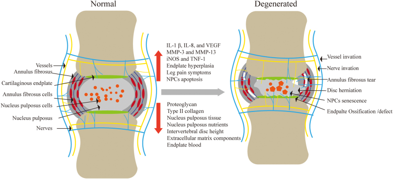 Figure 1
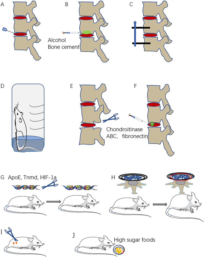 Figure 2
| Induction method | Concrete method | Study | Speciman | Observed onset of degeneration | Advantages | Limitations | Suitable research fields |
| Intervertebral disc injury model | Needle puncture | Glaeser [ | SD rat | 4 and 8 weeks | 1. Relatively simple and reproducible | 1. Long observation period | Regenerative therapy |
| Needle puncture | Gianluca [ | sheep | 1, 3, and 6 months | Regenerative therapy | |||
| Needle puncture | Zhang [ | SD rat | 4 weeks | Regenerative therapy/pharmacological testing | |||
| Needle puncture | Zhang [ | SD rat | 4 weeks | Regenerative therapy | |||
| Needle puncture and ethanol injection | Yuan [ | SD rat | 1, 2, 3, and 6 months | Regenerative therapy | |||
| Cement in CEP | Kang [ | Pig | 3 months | Regenerative therapy/pharmacological testing | |||
| Pingyangmycin in CEP | Wei [ | Monkey | 1, 3, 6, 9, 12, and 15 months | Regenerative therapy/pharmacological testing | |||
| Pingyangmycin in CEP | Wei [ | Rabbit | 1, 3, and 6 months | Regenerative therapy/pharmacological testing | |||
| Abnormal mechanical stimulation model | Immobilization | Wang [ | Sheep | 6 and 26 weeks | 1. Without acute injury | 1. Surgical procedure exacerbates spinal instability | Regenerative therapy/pharmacological testing |
| Compression load | Ji [ | SD rat | 14 days | Regenerative therapy/pharmacological testing | |||
| Compression load | Li [ | Mice | 2 months | Regenerative therapy | |||
| Tail-looping | Sakai [ | Mice | 4, 8, and 12 weeks | Regenerative therapy | |||
| Bipedal animal model | Forelimbs amputation | Kong [ | SD rat | 6 months | Simulate the upright state of the human | 1. Muscle tissue damage worsens | Regenerative therapy |
| Limited water-containing space | Ao [ | Mice | 6 and 10 weeks | Regenerative therapy/pharmacological testing | |||
| Limited water-containing space | Jin [ | Mice | 10 weeks | Regenerative therapy | |||
| Spinal instability models | Paraspinal musculature was exposed and excised | Yao [ | Mice | 4 and 8 weeks | 1. Creating static and dynamic posterior instability | 1. Modelling time relatively long | Regenerative therapy/pharmacological testing |
| Lumbar facetectomy | Xiao [ | Mice | 12 weeks | Regenerative therapy/pharmacological testing | |||
| Cutting off interspinous ligaments | Liu [ | Mice | 1, 2 and 16 weeks | Regenerative therapy/pharmacological testing | |||
| Resection of facet joints, supra- and interspinous ligaments | Oichi et al. [ | Mice | 2 and 12 weeks | Regenerative therapy | |||
| Chemically induced model | Monosodium iodoacetate | Najah [ | SD rat | 6 weeks | 1. Less invasive, | 1. Chemical reagents cannot replicate the natural progression of IDD | Regenerative therapy/pharmacological testing |
| Chondroitinase ABC | Rustenburg [ | Caprine | 1week | Regenerative therapy/pharmacological testing | |||
| Trypsin | Rosenzweig [ | Bovine | 1week | ||||
| Fibronectin fragment | Liu [ | Rabbit | 4, 8, 12, and 16 weeks | Regenerative therapy | |||
| Spontaneous degeneration model | Spontaneous degeneration | Gruber [ | Sand rat | 2 months | Closer to human disc physiopathology | Long time consumption | Regenerative therapy |
| Spontaneous degeneration | Vincent [ | Mice | 24 months | Regenerative therapy | |||
| Spontaneous degeneration | Choi [ | Mice | 8 and 17 weeks | Regenerative therapy | |||
| Zhang [ | SD rat | 50 weeks | Regenerative therapy | ||||
| Gene knockout models | APOE knockout | Beierfuβ [ | Rabbit | 24 months | 1. Revealing the role of specific genes in IDD | 1. Knockout of certain genes may lead to embryonic death or defects | Regenerative therapy/pharmacological testing |
| Tnmd knockout | Lin [ | Mice | 6 months | Regenerative therapy/pharmacological testing | |||
| HIF-1 alpha knockout | Wu [ | Mice | 6 and 12 weeks | Regenerative therapy/pharmacological testing | |||
| SPARC knockout | Magali [ | Mice | 78 weeks | Regenerative therapy/pharmacological testing | |||
| Others | Smoking | Wang [ | Mice | 6 months | Further support tobacco smoke as a contributor to spinal degeneration | 1. The molding time is long. | Regenerative therapy |
| Ovariectomy | Najah [ | SD rat | 3 and 6 weeks | 1. Ease of establishment | 1. Non-physiological | Pharmacological testing | |
| Type 2 diabetes | Rosenberg [ | SD rat | 68 days | Further validates the causality between type 2 diabetes and IDD | 1. Not a full mimic of human IDD | Regenerative therapy/pharmacological testing |
Peer Reviews
No public reviews on file for this paper yet. If you reviewed it on a platform where reviews are public (OpenReview, ICLR, NeurIPS, ICML), you can paste yours below so the community can read it here.
Videos
No videos yet. Explain this paper in a talk, walkthrough, or lecture? Add one.
Taxonomy
TopicsSpine and Intervertebral Disc Pathology · Medical Imaging and Analysis · Cervical and Thoracic Myelopathy
Introduction
1
IDD is a common degenerative disorder of the musculoskeletal system and a major cause of chronic low back pain, severely affecting the quality of life of patients and imposing a significant economic burden on families and society [1], 2]. About 20 % of adolescents have mild IDD, and 80 % have experienced back pain during their lifetime [3]. Currently, the pathogenesis of IDD remains incompletely understood, resulting in a lack of standardized, effective treatments for IDD-related diseases. Due to the limited availability of human experimental materials, establishing reliable animal models of IDD is crucial. These models offer a vital approach for elucidating the underlying mechanisms of disc degeneration and serve as valuable platforms for evaluating potential therapeutic interventions [4], [5], [6].
This review provides a comprehensive overview of currently employed animal models for IDD, comparing their respective advantages, disadvantages, applicability, and limitations. We recommend that researchers carefully select the most appropriate animal model for their IDD studies, based on factors such as the experimental species, the desired experimental duration, and the specific pathological features being investigated. Future research should focus on refining model construction standards, incorporating multi-omics technologies to elucidate the intricate mechanisms of IDD, and developing more clinically relevant evaluation systems for emerging intervertebral disc regeneration therapies.
In vitro model
2
Intervertebral disc cell model
2.1
The intervertebral disc cells were cultured in vitro by cell biology technology, and the characteristics of their growth, proliferation and differentiation were observed. The model was treated with external conditions to explore the effect of single factor on IDD. Wang et al. used lipopolysaccharide and adenosine triphosphate to induce nucleus pulposus cells (NPCs) degeneration model in vitro [7]. Herrera et al. used monolayer and three-dimensional culture to study human intervertebral disc cells, which showed a significant increase in type I and type II collagen content in three-dimensional culture. Compared to monolayer culture, three-dimensional culture provides a more complex culture environment and can reduce the loss of cell viability and unstable protein expression [8]. The co-culture of NPCs with BMSCs in a three-dimensional environment can increase inflammatory resistance and autoimmune regulation [9].
However, neither monolayer nor three-dimensional in vitro culture fully replicates the complex human environment, potentially overlooking the influence of mechanical and other crucial factors on IDD [10], 11]. The absence of a functional extracellular matrix in in vitro settings hinders cell proliferation, compromises the maintenance of a stable cellular phenotype, and can even lead to dedifferentiation or a complete loss of original cellular function [12], 13].
Intervertebral disc tissue model
2.2
In vitro models of intervertebral disc (IVD) tissue can be broadly categorized into those that incorporate the cartilage endplate (CEP) and those that do not. The IVD comprises the nucleus pulposus (NP), annulus fibrosus (AF), and CEP. The CEP serves as a crucial pathway for IVD nutrition and metabolism, maintaining IVD integrity and limiting tissue expansion. Preserving the CEP in in vitro models can limit tissue swelling and ensure adequate nutrient supply, thereby better mimicking the in vivo environment. For instance, researchers have successfully infected rat IVD tissue in vitro with an adenovirus vector carrying green fluorescent protein (GFP), observing GFP expression in both the CEP and outer AF for at least 14 days. This underscores the potential of using adenoviruses as vectors for gene therapy in IVD repair and slowing disc degeneration, offering a more precise approach for managing and treating degenerative disc disease [14]. A novel ex vivo goat IDD model involves pre-loading under simulated physiological load conditions during the day, followed by enzymatic degradation of lumbar goat intervertebral disc through injection of collagenase and chondroitinase ABC. After digestion, simulated physiological loading was performed on intervertebral disc for 7 days, and it was found that the extracellular matrix components decreased, while interleukin-1 β, −8, and VEGF continued to increase. The observed changes are consistent with the changes in human IDD [15].
The comprehensive tissue culture model offers the advantage of utilizing the inherent structure of the IVD, allowing cells to reside within their natural matrix and preserving the morphological integrity of the tissue, including cell-cell interactions [16]. Functioning as a bridge between in vivo and cellular in vitro models, the IVD tissue model provides a valuable experimental platform. This allows researchers to study the tissue’s response to external stimuli and observe the histological changes occurring in both healthy and degenerated discs [4], 17], 18].
In vivo models
3
Intervertebral disc injury model
3.1
This model predominantly induces IDD by surgically damaging the NP, AF, and CEP. The fine needle puncture method is a common choice, favored for its ease of use, consistent results, and the ability to finely control the extent of the induced damage.
Annulus fibrosus and nucleus pulposus injury
3.1.1
Glaeser et al. showed that anterior disc injury using an 18G needle induced severe IDD and mechanical hypersensitivity, whereas the 21G needle resulted in moderate degeneration without increased pain sensitivity [19]. In a separate approach, a 2 mm channel was drilled through the CEP into the NP to perform a partial nucleus pulpectomy, creating an animal model of progressive IDD while maintaining an intact AF. This model is beneficial for investigating NP regeneration and repair, as well as early-stage, mild IDD [20]. In our previous experiments, we targeted the rat coccygeal 5–6, 6–7, 7–8, and 8–9 discs. These were percutaneously punctured using a 21 G needle with a stopper set to a depth of 5 mm; the needle was then rotated 360° and held for 30 s. Four weeks post-operatively, we observed significant loss of disc height, accompanied by an increase in Pfirrmann grades [21], 22].
In addition, as the structural integrity of the disc is disrupted, nerves and blood vessels can enter the NP through the torn AF [23], 24]. This nerves and blood vessels invasion is associated with low back pain (Figure 1). It was found that after AF puncture in mice, tears in the AF occurred, with loss of NP tissue and an increase in radiating leg pain symptoms. Increased nerve distribution and painful nerve fibres density in degenerating discs were also found, suggesting that the process of disc degeneration is accompanied by nerves and blood vessels invasion [25].
Degenerated intervertebral discs show the ingrowth of blood vessels and nerves, aging of nucleus pulposus cells, tearing of the annulus fibrosus, and increased levels of inflammatory factors.
The method of creating animal models through NP and AF puncture is relatively simple, highly reproducible, and successfully mimics the human processes of NP injury, AF injury, and subsequently IDD. This makes it suitable for investigating the pathophysiological mechanisms following AF injury and acute intervertebral disc herniation (Figure 2A). However, this method has limitations, including the need for long observation periods, the potential for infection after needle insertion, and the inherent possibility that the puncture itself may trigger an immune inflammatory response.
Examples of animal models for intervertebral disc degeneration. (A) Annulus fibrosus and nucleus pulposus injury model. (B) Cartilage endplate injury model. (C) Compression model. (D) Upright model. (E) Spinal instability model. (F) Chemically induced model. (G) Gene knockout model. (H) Spontaneous degeneration model. (I) ovariectomy model. (J) High sugar diet induced type 2 diabetes triggered intervertebral disc degeneration model.
Cartilage endplate injury
3.1.2
The intervertebral disc is one of the largest non-vascular and nerve organs in the vertebrate body, with nutrients permeating from the CEP and outer AF to the NP tissue. As the IDD, the supply of nutrients decreases, limiting intervertebral disc cell activity and viability [26]. Therefore, a model of disc degeneration constructed by blocking the CEP blood supply to reduce the nutrient supply to the disc can be used to explore the relationship between nutrient supply and IDD (Figure 1).
Injection of anhydrous alcohol beneath the caudal CEP of rat discs induced a transformation of the NPCs, shifting their morphology from cord-like to cartilage-like, and finally into fibrocartilage cells. This process was associated with rupture of the annulus fibrosus, followed by degradation and eventual disappearance of the CEP growth plate [27]. Another study utilizing an immature animal model investigated the effects of blood supply blockade using bone cement on IDD (Figure 2B). They found that injecting bone cement into the CEP effectively induced IDD [28].
Researchers have established IDD models by administering the chemical drugs pingyangmycin into the subchondral bone of the lumbar disc in rhesus monkeys and rabbits under CT guidance, thereby blocking nutrient supply. Postoperative observations revealed the formation of bony endplates, a reduction in intervertebral disc height, and alterations in MRI signals [29], 30]. These findings suggest that CEP is essential for maintaining the viability of intervertebral disc cells.
Abnormal mechanical stimulation model
3.2
Biomechanics is fundamental to intervertebral disc homeostasis, and abnormal mechanical loading is a primary driver of disc degeneration. For instance, individuals engaged in heavy labor face a significantly elevated risk of disc degeneration. Disrupting the spine’s biomechanical equilibrium can alter the morphological and physiological characteristics of the disc, mirroring the early stages of disc degeneration observed in humans.
Compression model
3.2.1
Fixation of lumbar vertebrae through pedicle screws and rods leads to changes in the intervertebral disc load environment, thereby inducing IDD [31] (Figure 2C). Examination of the 1.8 N and 4.5 N load groups identified structural disorganization within both the NP and AF, occurring on both convex and concave surfaces. MRI and histological findings demonstrated that the degree of IDD was amplified by greater compression forces. Moreover, the compression model exhibited increased mRNA expression of MMP-3 and MMP-13, with a concurrent significant decrease in collagen type II-α1 expression [32]. Piezo1 expression was upregulated by excessive compression loading, resulting in ECM degradation and increased NP cell apoptosis. Inhibition of Piezo1 attenuated these pathological processes [33].
By circumferentially coiling and fixing rat tails with steel wire, Sakai et al. were able to subject each intervertebral disc to varying degrees of compressive stress. This resulted in the development of IDD models with different levels of degeneration [34]. The study by Zak et al. demonstrated that long-term cyclic compression-flexion loading (100,000 cycles) leads to degenerative changes in both the structural and mechanical properties of intervertebral discs, while highlighting the posterior column’s crucial role in preserving the integrity and biomechanical characteristics of the annulus fibrosus [35].
Bipedal animal model
3.2.2
The upright walking posture of humans is thought to be an important mechanical factor contributing to disc degeneration (Figure 2D). In mice, the movement pattern during standing and jumping is similar to that of humans. In addition, some researchers induce IDD by forcing animals to stand on both feet to mimic human bipedal gait after amputation of their forelimbs [36]. As the time and biomechanical load increases, the fissure at the AF gradually increases, the height of both the disc and CEP decreases, and the extracellular matrix composition changes, forming an IDD model [37]. Researchers leveraged mice’s innate fear of water to induce them to adopt a bipedal stance within a water-filled enclosure. The water temperature was maintained at 24 °C with a depth of 5 mm to ensure the animals’ fur remained dry and their body temperature stayed normal. After 10 weeks, the mice exhibited severe IDD, primarily affecting the AF and facet joints. This method established a non-invasive and effective IDD model that closely mimics the human condition. However, it necessitates continuous monitoring of water temperature and volume, and the model development period is relatively long [38].
Spinal instability models
3.2.3
This model induced spinal instability by disrupting the dynamic and static equilibrium of the spine and damage to peri-vertebral disc structures, causing excessive spinal motion (Figure 2E). The resulting biomechanical imbalance, caused by removing the paravertebral musculature and posterior ligaments, led to spinal instability. This instability subsequently drove CEP hyperplasia, increased NP apoptosis, and a reduction in disc height, successfully establishing an IDD model [39]. After 12 weeks following facetectomy (mechanical instability), intervertebral disc degeneration occurred, with a decrease in type II collagen expression, while the model promoted the collapse of subchondral bone trabeculae [40]. Liu et al. reported a mouse model of lumbar instability, in which L3–L5 spinous processes were surgically exposed, paraspinal muscles were separated, articular processes and ligaments were removed, and degenerative changes began to appear in the L4/5 intervertebral discs one week after surgery [41]. Oichi et al. created a lumbar instability model by surgically excising the lumbar facet joints, supraspinous ligament, and interspinous ligaments of mice. Imaging studies showed a decrease in disc height beginning 2 weeks after surgery, with continued decline over the next 12 weeks. Furthermore, histological analysis confirmed the presence of IDD in the lumbar vertebrae [42].
However, the modelling time for this model is relatively long, the cost of the experiment is high, and it remains controversial whether its degenerative factors include surgical stimulation. Unlike models that directly injure the disc, these animal models preserve the integrity of the annulus fibrosus and nucleus pulposus. The slow progression of IDD makes these models more representative of adjacent segment degeneration seen after human lumbar fusion surgery [43].
Chemically induced model
3.3
Biochemical reagents have been used to induce animal models for disc degeneration. Intralesional injection of monosodium iodoacetate into the NP of the rat intervertebral disc revealed a narrowing of the intervertebral space and a decrease in the signal intensity of magnetic resonance T2-weighted images after 6 weeks, suggesting degeneration of the intervertebral disc [44]. Rustenburg et al. injected collagenase and chondroitinase ABC into the intervertebral disc of caprine, causing degeneration and necrosis of the NP, AF, CEP. It was found that extracellular matrix components decreased, while degradative enzymes and catabolic proteins (IL-1β, IL-8, and VEGF) continued to increase [15]. Another method was to inject 100 μg of trypsin into the intervertebral disc of bovine to lyse the proteoglycan and collagen (Figure 2F). Once in the disc centre, the trypsin solution was slowly injected and the needle gradually pulled out to avoid flow [45]. Liu et al. used a minimally invasive injection technique to inject fibronectin fragment into the intervertebral disc of rabbits. IDD began to occur 4 weeks after surgery, with increased expression of MMPs, iNOS and TNF-1 (Figure 1). With prolonged postoperative observation, the NP was completely replaced by fibrous tissue, successfully establishing a reproducible, simple and cost-effective model of disc degeneration [46].
Although chemical induced models are less invasive, reproducible, and allow for easy drug administration, their limitations stem from the primarily physiological nature of IDD. The natural progression of degeneration is not effectively mimicked by the injection of chemical reagents. Moreover, the distribution of the injected drugs throughout the intervertebral disc and their impact on subsequent experimental assessments must be carefully evaluated.
Gene knockout model
3.4
In recent years, with the development of genetic engineering technology and the progress of gene knockout technology, it provides a new method for animal models of many diseases. In particular, crispr-cas9 gene editing technology is becoming more and more mature, and it is possible to make a large number of degenerative disease models [47], [48], [49], [50]. After ApoE gene knockout in rabbits, it was found that the inflammatory factors in the intervertebral disc were increasing and the metabolism in the intervertebral disc was disordered, resulting in the symptoms of IDD [51]. Knockout of the mice tenomodulin (tnmd) gene promoted the upregulation of p65 and matrix metalloproteinases, induced angiogenesis and macrophage infiltration in the outer AF, and increased chondroid NPCs in the NP [52]. Some scholars have studied the role of HIF-1α gene in IDD in mice (Figure 2G). The results show that glycosaminoglycan, type II collagen and the expression of vascular endothelial growth factor are reduced in the gene knockout HIF-1α group [53]. The secreted protein, acidic, rich in cysteine (SPARC)-null mice exhibited decreased tolerance to axial stretching, hindpaw cold hypersensitivity, motor impairment, and the degree of IDD was more severe compared with age-matched control mice [54].
Spontaneous degeneration model
3.5
Some animals show disc changes with age, which have many common characteristics with human disc degeneration (Figure 2H). Sand rat are a classic model of spontaneous disc degeneration. It is found that the disc degeneration of 2-month-old sand rat is similar to that of humans [55]. Vincent et al. also found that 24-month-old mice exhibited typical disc degeneration compared to 3- and 12-month-old mice [56]. Choi et al. found in SM/J mice a progressive decrease in NPCs, changes in stromal composition and a decrease in disc height with age, similar to many of the distinguishing features of human disc degeneration, a new small animal model of disc degeneration [57].
Our early experiments found that spontaneous degeneration of intervertebral disc also existed in rats. Compared with the 4W groups, the level of vacuolated nochordal cells (50 weeks) increased gradually over time, and nucleus pulposus cell clusters and serpentine AF were found, intervertebral disc height decreased significantly, Pfirrmann grade increased, indicating disc generation [58].
Other IDD models
3.6
Exposure to tobacco smoke reduces proteoglycan content in the intervertebral discs of mice and significantly enhances matrix metalloproteinase activity. This smoke exposure also resulted in a two-fold increase in senescent cells within the intervertebral discs of the mice [59]. Removal of ovaries in rats reduce disc height, water content, and histologic score [60] (Figure 2I). In addition, in rats with type 2 diabetes, metabolic disorders in intervertebral disc can lead to disc degeneration [61] (Figure 2J).
Conclusions and the future
4
IDD is one of the main causes of chronic low back pain and neurological dysfunction. Its pathological mechanisms are complex, involving inflammation, oxidative stress, extracellular matrix degradation, and cellular aging. Animal models serve as important tools for studying IDD, playing a crucial role in revealing disease mechanisms and evaluating potential therapies. Regarding the selection of experimental animals, from the perspective of human lifestyle and spinal morphology, the spinal physiological structure and biomechanical environment of large animals (such as goats, pigs, primates) share a high degree of similarity with humans [62], [63], [64]. Factors like spontaneous degeneration and spinal mechanical stress also closely resemble human conditions. However, due to considerations such as cost, maintenance requirements, and ethical constraints, large primates are difficult to become routine models for intervertebral disc degeneration. Currently, rodents and rabbits, benefiting from their low husbandry costs and readily available supply, remain the preferred experimental animals for intervertebral disc degeneration models. Existing animal models of IDD each have their unique advantages and limitations, making it difficult to fully replicate the natural course and heterogeneity of human disc degeneration (Table 1). Various intervertebral disc degeneration models still lack standardized protocols (e.g., puncture depth, enzyme dosage, gene knockout strategies, etc.), which leads to discrepancies in results and makes inter-study comparisons difficult. Researchers need to develop detailed standardized operating procedures and unified reporting guidelines specifically for different animal models (e.g., rabbits, mice, etc.).
To evaluate the effectiveness of novel regenerative strategies for the NP, preclinical models are needed that preserve the AF of the intervertebral disc. With this in mind, the study was to characterize a preclinical ovine model of IDD. This model was induced by damaging and repairing the EP via a transpedicular approach [20]. In cases of mild disc degeneration, defined by biochemical imbalances and microenvironmental disruption without structural collapse, biomaterial-based strategies aim to restore disc homeostasis. Hydrogels, such as methacrylated cellulose derivatives or alginate-pNIPAAm composites, are employed for injection due to their ability to mimic the nucleus pulposus (NP) and annulus fibrosus (AF). Their mechanism of action involves regulating hypoxia, acidity, and inflammation by localized delivery of anti-inflammatory agents or growth factors. This delivery suppresses catabolic enzymes like MMP-3 and ADAMTS-4, while simultaneously encouraging the synthesis of extracellular matrix components, including aggrecan and collagen II [65], [66], [67]. Many treatment strategies, such as stem cell therapy and biomaterial repair, have shown significant effects in animal experiments but have failed to meet expectations in clinical trials [67]. With the advancement of biomedical technology and the deepening understanding of the IDD mechanism, some new models have been proven to have good advantages in regenerative research. At the same time, new models will be created in the future, such as organoid models of nucleus pulposus [68], microfluidic chemotaxis intervertebral disc organ-on-a-chip simulating intervertebral disc microenvironments [69], humanized animal models or patient-derived xenografts for personalized medicine.
Therefore, ongoing refinement of IDD animal models will promote a deeper understanding of the disease mechanisms and lay the foundation for the development of novel therapeutic approaches.
The reference list from the paper itself. Each links out to its DOI / PubMed record.
- 1Melrose J Guilak F Diverse and multifunctional roles for perlecan (HSPG 2) in repair of the intervertebral disc JOR Spine 20247 e 136210.1002/jsp 2.136239081381 PMC 11286675 · doi ↗ · pubmed ↗
- 2Ambrosio L Schol J Ruiz-Fernández C Tamagawa S Joyce K Nomura A Getting to the core: exploring the embryonic development from notochord to nucleus pulposus J Dev Biol 2024121810.3390/jdb 1203001839051200 PMC 11270426 · doi ↗ · pubmed ↗
- 3Yang S Zhang F Ma J Ding W Intervertebral disc ageing and degeneration: the antiapoptotic effect of oestrogen Ageing Res Rev 20205710097810.1016/j.arr.2019.10097831669486 · doi ↗ · pubmed ↗
- 4Wang Y Kang J Guo X Zhu D Liu M Yang L Intervertebral disc degeneration models for pathophysiology and regenerative therapy-benefits and limitations J Invest Surg 2022359355210.1080/08941939.2021.195364034309468 · doi ↗ · pubmed ↗
- 5Tian T Wang H Li Z Yang S Ding W Intervertebral disc degeneration induced by needle puncture and ovariectomy: a rat coccygeal model Bio Med Res Int 20212021551012410.1155/2021/551012434055972 PMC 8147532 · doi ↗ · pubmed ↗
- 6Wang D Lai A Gansau J Seifert AC Munitz J Zaheer K Lumbar endplate microfracture injury induces Modic-like changes, intervertebral disc degeneration and spinal cord sensitization - an in vivo rat model Spine J 20232313758810.1016/j.spinee.2023.04.01237086976 PMC 10524828 · doi ↗ · pubmed ↗
- 7Wang J Fan C Zhang Y Hua D Ji Z He W (Z)-ligustilide alleviates intervertebral disc degeneration by suppressing nucleus pulposus cell pyroptosis via Atg 5/NLRP 3 axis Sci China Life Sci 20256829658110.1007/s 11427-024-2907-740614011 · doi ↗ · pubmed ↗
- 8Herrera Quijano MA Sharma N Morissette Martin P Séguin CA Flynn LE Development of 2-D and 3-D culture platforms derived from decellularized nucleus pulposus Front Bioeng Biotechnol 20221093723910.3389/fbioe.2022.93723936237211 PMC 9551564 · doi ↗ · pubmed ↗