Fabrication and Characterization of ZnO Nanoparticle‐Coated NiTi Orthodontic Wires With Enhanced Friction‐Reducing Surfaces
Mona Gholami, Zahra Kachoei, Mahdiyeh Esmaeilzadeh, Mojgan Kachoei

TL;DR
This paper shows that coating orthodontic wires with ZnO nanoparticles can reduce friction and kill bacteria, potentially improving orthodontic treatments.
Contribution
A novel polymer composite coating method for ZnO nanoparticles on NiTi wires is introduced, offering both high antibacterial activity and reduced friction.
Findings
Polymer composite coating provided the highest reduction in frictional forces at 0° wire/bracket angle.
Polymer-coated samples showed 89% reduction in cell viability against Streptococcus mutans.
CVD-coated samples had 98% antibacterial activity but insufficient friction reduction.
Abstract
The study is aimed at modifying the surface of nickel–titanium (NiTi) wire with novel coating techniques of zinc oxide (ZnO) nanoparticles (NPs) for reduction in friction and improving antibacterial activity. NiTi orthodontic wires were coated with ZnO NPs by the chemical vapor deposition (CVD) and polymer composite coating. Physicochemical properties of ZnO NPs were assessed by field emission scanning electron microscopy (FE‐SEM), energy‐dispersive x‐ray (EDX), and x‐ray diffraction (XRD) analysis. Besides, the surface characterization of NiTi substrates was analyzed with atomic force microscopy (AFM). The antibacterial activity of the coated samples against Streptococcus mutans was assessed using the colony count method. Polymer coating method demonstrated the most durable and well‐adhered ZnO coating among the groups with a relatively high antibacterial activity (reduction in cell…
Genes, proteins, chemicals, diseases, species, mutations and cell lines named across the full text — each resolved to its canonical identifier and authoritative record.
Click any figure to enlarge with its caption.
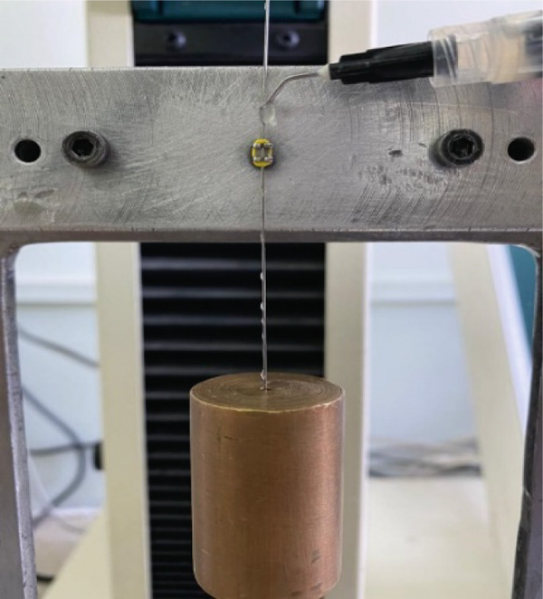 Figure 2
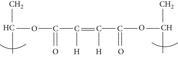 Figure 3
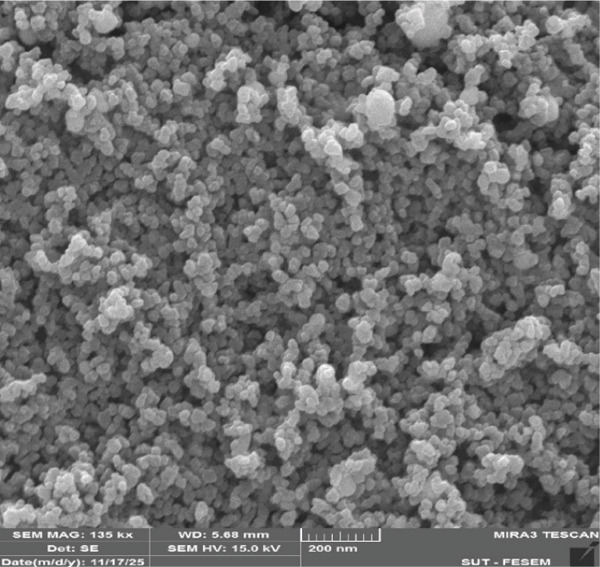 Figure 5
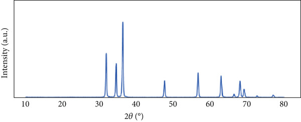 Figure 6
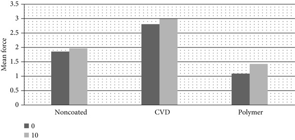 Figure 16
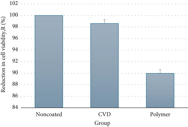 Figure 17
|
|
|
|
|
|
|
|
|
|
|---|---|---|---|---|---|---|---|---|
| Noncoated | — | — | −18.7 ± 0.5 | 42.2 ± 1.5 | 1.8 ± 0.2 | 76.4 ± 2.1 | 68.0 ± 1.4 | −0.6 ± 0.2 |
| Polymer coated | — | — | −7.9 | 24.3 | 2.54 | −11.8 | −24.2 | −3.3 |
| CVD coated | 9.3 | −7.2 | −6.2 | 28.7 | 13.8 | −20.5 | −38.1 | −10.6 |
|
|
|
|
|---|---|---|
| Control | 324.0 ± 0.00 | |
| CVD | 4.5 ± 2.12 | 98.61 ± 0.65 |
| Polymer | 32.5 ± 2.12 | 89.97 ± 0.65 |
- —Tabriz University of Medical Sciences10.13039/501100004366
Peer Reviews
No public reviews on file for this paper yet. If you reviewed it on a platform where reviews are public (OpenReview, ICLR, NeurIPS, ICML), you can paste yours below so the community can read it here.
Videos
No videos yet. Explain this paper in a talk, walkthrough, or lecture? Add one.
Taxonomy
TopicsShape Memory Alloy Transformations · Advanced Sensor and Energy Harvesting Materials · Electrodeposition and Electroless Coatings
1. Introduction
For effective tooth movement, the archwire must be capable of sliding within the bracket slot. However, this motion is opposed by sliding resistance [1]. Sliding resistance typically arises from a combination of frictional forces and binding effects [2]. Several factors influence sliding resistance, among which the most critical are the surface characteristics and the material composition of both the archwire [3] and the bracket slot [4, 5].
Among the types of wires, nickel–titanium wire has been introduced as the ideal orthodontic archwire in the first stage of comprehensive orthodontic treatment due to the production of light force in the range of large performance. However, the major deficiency of nickel–titanium wire is its high coefficient of friction and surface roughness, which increases the frictional resistance [6, 7].
During treatment, resistance to sliding can significantly reduce the efficiency and effectiveness of fixed orthodontic treatment [8]. Accordingly, greater orthodontic forces must be applied to achieve the desired tooth movement and to overcome both sliding resistance and unintended dental movements [9, 10]. These inordinate forces lead to the loss of anchorage, which is crucial in orthodontic treatment. Moreover, it multiples the incidence of root resorption [11] and lengthens the treatment span, which leads to an additional amount of pain throughout the course of treatment [12, 13].
So far, different methods have been evaluated to control resistance to sliding by changing surface characterization, size, shape, and metallic material used in brackets and wires [14]. Changing the surface characterization like coating NiTi wires with various substances like Teflon [15], silver–rhodium [14], inorganic fullerene‐like nanoparticles of tungsten disulfide (IF‐WS2) [16, 17], and ZnO [18, 19] nanoparticles has been useful to some degree.
Recent investigations have expanded the range of effective coatings, demonstrating that nanoparticle‐based modifications including TiO₂ and SiO₂ can significantly alter frictional behavior and surface morphology. For example, SiO₂ nanoparticle coatings were shown to reduce frictional resistance in both dry and wet conditions while also decreasing surface roughness compared to TiO₂ and uncoated wires [20]. Likewise, graphene oxide/silver (GO/nAg) nanocomposite coatings deposited on NiTi alloys have improved tribological performance, corrosion resistance, and antibacterial activity, especially when smaller GO sheets promote uniform silver dispersion [21]. In addition, comprehensive reviews highlight that many functional coating systems—including metal, carbon‐based, polymeric, and bioactive layers—can enhance orthodontic appliances, though an ideal balance of friction reduction, antibacterial action, and corrosion resistance has not yet been achieved [22].
Reducing friction [19, 23] and antimicrobial effect of zinc oxide versus Streptococcus mutans (S. mutans) [18, 24], a predominant bacterium in the oral environment, makes ZnO nanoparticles a complete coating substance. This antimicrobial activity inhibits white spot lesions (WSLs) by preventing the aggregation of S. mutans around wires and brackets.
The success of decreasing friction is in relation to the penetration of ZnO nanoparticles as solid lubricants into the interface between the wire and bracket. Thus, having a slow‐release mechanism to stop coated nanoparticles from being washed out instantly after the application of force in the presence of saliva is a primary factor [18].
Therefore, the primary aim of the current study was to change different nanocoating processes of ZnO to correct the adhesion properties of ZnO nanoparticles without disrupting the slow‐release mechanism and to evaluate the effectiveness of the NiTi‐coated wires in reducing frictional forces through a simulated tooth sliding model. Besides, the antibacterial activity of different synthesized ZnO NPs coated on the wires was also tested.
2. Materials and Methods
2.1. Materials
Potassium hydroxide and ethanolamine (EA) were sourced from Merck (Darmstadt, Germany). Polyvinyl alcohol (PVA) (average Mw = 13, 000–23,000), maleic anhydride 99%, zinc acetate dihydrate ≥ 98*%, isopropanol ≥ 99.5%*, and polyvinyl pyrrolidone (PVP) (average MW = 400,000) were prepared from Sigma–Aldrich (Baden‐Württemberg, Germany). Commercially available 0.016‐in. round nickel–titanium orthodontic wires (Ortho Technology, Florida, United States), each measuring 18 cm in length, were utilized as substrates for the deposition of ZnO nanoparticles.
2.2. Synthesis Methods
2.2.1. Chemical Vapor Deposition (CVD) Method
For the CVD procedure, thin films were deposited onto NiTi wire substrates. For this, high‐purity metallic Zn powders (99.99%) and oxygen gas were applied as Zn and oxygen sources, respectively. Following, 0.3 g of Zn powder was placed in a quartz vessel and located at the center of a tube heater. The NiTi wire was then placed 1 cm above the zinc source. The chamber was evacuated to a base pressure of roughly 10 Pa using a mechanical vacuum pump. Argon gas, with high purity, was introduced as a carrier at a flow rate of 100 sccm, and oxygen was supplied at 30 sccm into the reaction zone. The deposition was conducted at a maintained temperature of 650°C for a duration of 30 min.
2.2.2. Polymer Composite Coating
For the polymer coating method, PVA was dissolved in distilled water at 70°C under continuous stirring to prepare a solution of 5% w/v. Following, maleic anhydride (1.5 g) was added into PVA solution while stirring. Then, 0.1 g of ZnO nanoparticles (prepared according to our previous study [25]) was attached to the solution. Stirring was continued for 5 min. The wires were placed into the obtained solution. The coating wires were dried at room temperature and consequently cured in the oven at 90°C for 90 min.
2.3. Antibacterial Procedure
Each piece of NiTi wire was cut into 8 cm segments and cleaned multiple times with a 70% ethanol solution. To evaluate the antibacterial effect of the samples against Streptococcus mutans (ATCC 35668), the colony count method was performed as follows.
The microbial strains used in this study were cultivated aseptically on brain–heart infusion (BHI) agar at 37°C for 24 h. After incubation, a bacterial suspension was prepared in BHI broth, adjusted to the 0.5 McFarland standard, which corresponds to approximately 1.5 × 10^8^ CFU/mL. Under sterile conditions, 1 mL of this standardized suspension was introduced into microtubes containing the CVD‐coated, polymer composite, and uncoated control samples, followed by incubation at 37°C for 24 h. Postincubation, 20 μL aliquots from each microtube were transferred onto agar plates to monitor bacterial growth. For samples showing visible growth, serial dilutions (10^−1^, 10^−2^, 10^−3^, etc.) were prepared to enable colony counting. The spread plate technique was employed, whereby 10 μL of each diluted sample was evenly distributed across the surface of an agar plate using a sterile glass spreader and then incubated at 37°C for 24 h. Plates displaying between 30 and 300 colonies were selected for counting, and the bacterial load was calculated in CFU/milliliter [25].
The reduction in microbial cell count (R %) was determined using the following formula:
CFU_control_ refers to the sample without ZnO coating, while CFU_sample_ represents the number of colony‐forming units per milliliter for the ZnO‐coated samples [26].
2.4. Characterization of ZnO NPs
The dimension and morphology of ZnO NPs coated on the NiTi wires were analyzed using a field emission scanning electron microscope, FE‐SEM (MIRA3 FEG‐SEM Company: Tescan, Czech) equipped with energy‐dispersive x‐ray (EDX) spectroscopy. EDX analysis revealed the composition of the NPs. Characterizing the crystalline structure of ZnO NPs was done by using the x‐ray diffraction (XRD) technique (Siemens D5000, Germany).
2.5. Surface Characteristics of ZnO‐Coated and Noncoated Wires
Surface morphology of both ZnO‐coated and uncoated wires was analyzed using atomic force microscopy (AFM; Nanowizard 2, JPK Instruments, Berlin, Germany). This technique was employed to evaluate the topographical features and to quantify the surface roughness parameters, including root mean square (RMS) and average roughness (Ra) values.
2.6. Differential Scanning Calorimetry
Differential scanning calorimetry of the samples was done with a DSC instrument (DSC; Netzsch 404 C, Pegasus, Germany) equipped with refrigerated cooling in order to measure austenite start (As), austenite finish (Af), martensite start (Ms), and martensite finish (Mf) temperatures and enthalpies of heating (ΔHH) and cooling (ΔHC). The samples were heated up to 100°C and were kept for 3 min to reach thermal equilibrium. The DSC analysis commenced with cooling down the samples to −100°C, followed by heating back to 100°C with a rate of 10°C/min [27].
2.7. Friction Test
Coated NiTi wires (0.016‐in., Ortho Technology, Lutz, Florida, United States) from the CVD and polymer composite groups (each comprising 10 samples), along with 10 uncoated samples (each 18 cm in length), were subjected to a frictional test. Thirty stainless steel brackets (0.018 standard edgewise system, Ultratrimm, Dentaurum, Germany) for upper central incisors were bonded to a bracket‐mounting device at angles of 0° and 10° relative to the long axis of the wire to simulate bracket‐wire binding during sliding. Five samples were tested at each angle (Figure 1).
Figure 1. Universal testing machine set in (a) 0° and (b) 10° angulations.(a)(b)
The testing device was mounted on a universal testing machine (UTM) (Hounsfield Test Equipment, Model H5KS, Redhill, United Kingdom; sampling rate 15 Hz) to conduct the friction test. A 150 g weight was attached to the free end of the wire to initiate sliding, while the upper end was connected to the tension load cell (5000 N, HTE, Interface, Arizona, United States) of the UTM [18]. To simulate intraoral conditions, human saliva was continuously applied using a saliva injector during the friction measurement (Figure 2) [28]. A heater was also employed to maintain a constant temperature of 37°C throughout the test.
Injection of human saliva.
2.8. Statistical Analysis
Descriptive statistics, including mean and standard deviation, were used to summarize the data. The Kolmogorov–Smirnov test assessed the normality of the friction force distribution, and Levene’s test was applied to examine the homogeneity of variances. A two‐way ANOVA was conducted to evaluate the effects of bracket angulation and coating condition, as well as their interaction, on frictional forces. When significant differences were observed, post hoc comparisons were performed using the Games–Howell test due to unequal variances. The significance level was set at p < 0.05.
3. Results
3.1. Structure and Morphology
The scheme of structure of PVA cross‐linked with maleic anhydride is shown in Figure 3.
Structure of cross‐linked PVA.
The FTIR spectrum of the PVA composite coating and ZnO nanoparticles is revealed in Figure 4. FTIR spectra of cross‐linked PVA composite (Figure 4a) containing ZnO nanoparticle expression characteristic absorption peaks at 3299 cm^−1^ (O–H stretching), 2914 cm^−1^ (symmetric stretching of CH2), 1712 cm^−1^ (strong carbonyl band due to esterification reaction with maleic anhydride), 1424 cm^−1^ (CH2 bending), 1370 cm^−1^ (δ (OH), rocking with CH wagging), 1244 cm^−1^ for (C–O) stretch mode of ester group, 1083 cm^−1^ (stretching of C–O and bending of OH) (amorphous sequence of PVA), and 1062 cm^−1^ related to Zn–O stretching (corresponds to absorption peak of ZnO nanoparticles, Figure 4b).
Figure 4FTIR spectrum of (a) PVA composite and (b) ZnO nanoparticles.(a)(b)
The FE‐SEM image of ZnO nanoparticles synthesized by chemical precipitation is shown in Figure 5, with an average particle size of 30 nm.
FE‐SEM image of ZnO sample synthesized from chemical precipitation.
The XRD pattern of the synthesized ZnO nanoparticles, presented in Figure 6, confirms the formation of the hexagonal wurtzite crystal structure. The characteristic diffraction peaks appeared at 2θ values corresponding to the specific crystallographic planes of wurtzite ZnO. All observed reflections show complete agreement with JCPDS card No. 36‐1451 [29], with the most intense peak, indexed to the (101) plane [30].
XRD pattern of ZnO synthesized from chemical precipitation.
The FE‐SEM images of CVD samples show homogeneous NPs of almost 90 nm, which are tightly gathered (Figure 7a). Figure 7b represents no more NPs after the frictional test, which is confirmed by the EDX analysis with 0 at% of zinc and 0 at% of oxygen elements (Figure 8a,b). It refers to poor adhesion of NPs in the CVD technique and desorption of NPs during the 20 cycles of the friction test.
Figure 7FE‐SEM of ZnO NPs deposited by CVD (a) before frictional test and (b) after frictional test.(a)(b)
Figure 8EDX analysis of CVD coating samples (a) before frictional test and (b) after frictional test.(a)(b)
Figure 9 illustrates outspread and nonuniform ZnO NPs in the PVA matrices of the nanocomposite. Since entrapping chemical precipitation synthesized ZnO NPs (Figures 5 and 6), it is possible to estimate the size of ZnO NPs. Subsequent EDX analysis also confirms that the atomic percent of zinc was 0.09% and oxygen was 28.43%. This was the lowest amount of zinc among the samples and got even lower after undergoing the friction test. EDX analysis also did not detect any Ni and Ti before performing the friction test, which means a perfect coverage of NiTi wire with the polymer composite technique. FE‐SEM images and EDX analysis of polymer composite coated wires also demonstrate the durability of ZnO NPs after 20 cycles of friction test (Figure 10a,b).
Figure 9FE‐SEM of ZnO NPs synthesized by polymer composite coating (a) before frictional test and (b) after frictional test.(a)(b)
Figure 10EDX analysis of polymer composite coating samples (a) before frictional test and (b) after frictional test.(a)(b)
The XRD pattern of the CVD‐coated samples (Figure 11a) further reveals the crystalline nature of ZnO nanoparticles with an average crystallite size of 89 nm, calculated from the (111) reflection. Notably, the peak intensities of the CVD coating are significantly reduced compared to those obtained from the chemical precipitation method (Figure 6), indicating a substantial reduction in particle agglomeration.
Figure 11XRD pattern of (a) CVD coating sample and (b) polymer composite coating sample.(a)(b)
In contrast, the XRD pattern of the polymer composite coating (Figure 11b) displays the amorphous character of PVA, with only slight contributions from ZnO nanoparticles. Two distinct diffraction peaks at 2θ = 42.4^°^ and 77.6° correspond to the (111, 202) planes of ZnO, respectively. The amorphous nature of PVA arises from its cross‐linking with maleic anhydride, which eliminates the characteristic PVA diffraction peaks. This effect can be attributed to the formation of ester bonds that replace intermolecular hydrogen bonding, with the cross‐linker effectively enhancing the amorphousness of the polymer matrix.
3.2. Differential Scanning Calorimetry
The DSC curves of the samples are shown in Figure 12. CVD demonstrated two peaks during heating, which are related to the R (rhombohedral) phase. The absence of the R phase in the polymer composite coating group may be related to low enthalpy changes, resulting in direct martensite to austenite transformation. TG (glass transition temperature) was also obtained from the DSC curve of the polymer, referring to PVA of polymeric matrices. Phase transformation temperatures and enthalpy changes are listed in Table 1.
Figure 12. The (a) DSC‐cooling curve of CVD coating sample, (b) DSC‐heating curve of CVD coating sample, (c) DSC‐cooling curve of polymer composite coating sample, (d) DSC‐heating curve of polymer composite coating sample, (e) DSC‐cooling curve of noncoated wire sample, and (f) DSC‐heating curve of noncoated wire sample.(a)(b)(c)(d)(e)(f)
3.3. Surface Characteristics of Coated Wires
AFM topography images (Figures 13, 14, and 15) show that the surface quality of the coated wires differs from each other and is obviously varied from the control group (noncoated wires). The Ra of CVD and polymer is 55.51 and 77.75, respectively. Moreover, compared to noncoated wires with a surface roughness of 109.21, CVD and polymer display a smoother surface. Rms value for CVD is 68.19 nm and for polymer is 96.43 nm in comparison to 136.51 nm for noncoated wires.
Figure 13AFM images of CVD coating sample: (a) 2D AFM images; (b) 3D AFM images; (c) histogram.(a)(b)(c)
Figure 14AFM images of polymer composite coating sample: (a) 2D AFM images; (b) 3D AFM images; (c) histogram.(a)(b)(c)
Figure 15AFM images of noncoated NiTi wire: (a) 2D AFM images; (b) 3D AFM images; (c) histogram.(a)(b)(c)
3.4. Friction Test
Overall, there was a remarkable dissimilarity in friction between groups for the surface modification method they received (p < 0.001). Mean frictional forces were significantly lower in the 0° angle than in the 10° angle (p < 0.05). The greatest friction force was recorded in the CVD group with the 10° angle through the bracket and wire (2.99 ± 0.9 N), and the minimum friction force was measured in the 0° polymer composite group (1.08 ± 0.15 N). Tribological measurements of coated and noncoated wires are shown in Figure 16.
Tribological measurements of coated and noncoated wires.
3.5. Antibacterial Activity
All coated wires demonstrated a significant antibacterial activity due to the colony count method compared to the control group (noncoated wire). Reduction in cell viability (R %) against S. mutans is demonstrated in Figure 17 and Table 2. Different synthesis methods offer various tailoring of the shapes and sizes of ZnO NPs, and this was successfully achieved. Reduction in cell viability differs among the groups based on the size and shape of the NP. The CVD and polymer composite coating had a 98.6% and 89.97% percentage of microbial cell reduction, respectively [25].
Reduction in cell viability, R (%) against S. mutans.
4. Discussion
This study introduces novel coating methods by which ZnO nanoparticles were successfully deposited onto NiTi wires to reduce friction. NiTi wires are widely used in orthodontics due to their ability to deliver light, continuous forces over a broad range of activation. However, a major limitation of NiTi wires is their inherent surface roughness and high coefficient of friction, which contribute to excessive sliding resistance and hinder efficient tooth movement. Therefore, enhancing the surface quality of NiTi wires can significantly improve the efficiency of the initial phase of orthodontic treatment [31, 32].
In addition, the surface deterioration of NiTi wires provides favorable sites for bacterial colonization, thereby increasing the risk of WSLs. WSLs are a prevalent side effect of fixed edgewise orthodontic appliances and may appear as early as the initial months of treatment, particularly during the leveling and alignment phase when NiTi wires are primarily inserted [33, 34]. Numerous efforts have been made to enhance the antibacterial properties of NiTi wires; however, few studies have evaluated the deposition of ZnO nanoparticles for the dual purpose of improving antibacterial activity and reducing friction [18, 35].
In this study, CVD and polymer composite coating techniques were applied and compared with the chemical deposition method previously reported by Kachoei et al. [18]. Kachoei et al. synthesized ZnO NPs with an average crystallite size of 25 ± 5 nm using a chemical deposition method. FE‐SEM images revealed uniformly spherical ZnO NPs produced by this technique. EDX analysis indicated a reduced Zn:Ni ratio, and mechanical testing demonstrated a 21% reduction in friction for ZnO‐coated wires. These findings are consistent with the authors’ findings, with the exception of the CVD samples, which exhibited an increase in frictional forces.
In the authors’ study, crystalline ZnO NPs with an average diameter of 89 nm were synthesized via the CVD method, as determined by XRD analysis and FE‐SEM imaging. Postfriction EDX analysis revealed a decline in Zn and O content across all subgroups following 20 sliding cycles. Notably, the CVD group showed 0% Zn and O content in the EDX spectra after testing, corroborating the FE‐SEM results. This suggests poor coating adhesion in the CVD samples, as the ZnO layer was completely removed after 20 sliding cycles using a UTM. This loss of coating is likely the primary factor contributing to the increased friction observed in the CVD‐treated samples.
Krishan et al. [14] enhanced the surface characteristics of NiTi archwires using coatings such as resin, Teflon, titanium, silver, rhodium, and various nitrides, all of which were effective in reducing surface roughness. In the present study, AFM and FE‐SEM analyses of the polymer composite coating also demonstrated a uniform, defect‐free surface, confirming the effectiveness of the coating in achieving a smooth topography.
Redlich et al. [16] coated stainless steel wires with fullerene‐like NPs of tungsten disulfide (WS₂) embedded in a nickel–phosphorus (Ni‐P) matrix. Frictional force measurements using an Instron testing machine demonstrated a reduction of up to 54% in the coated wires. In the present study, frictional forces at 0° decreased from 1.85 ± 0.59 in the uncoated group to 1.08 ± 0.15 in the polymer composite‐coated group.
Behroozian [23], similar to the present study, reported a reduction in friction following ZnO deposition on both stainless steel wires and ceramic brackets. Data obtained from a UTM demonstrated a significant decrease in frictional forces in the coated groups.
ZnO NPs were also used by Hammad and El‐Wassefy [35] to evaluate the antibacterial and mechanical properties of NiTi wires. Their findings indicated enhanced bacteriostatic performance and reduced friction in the coated NiTi wires.
Wei et al. [36] reported a considerable reduction in frictional forces in CNx‐coated SS orthodontic wires. This effect was seen in both air and artificial saliva.
This study represents the first attempt to evaluate frictional force reduction under conditions simulating the oral environment, including the presence of human saliva and physiological temperature. Given the thermoresponsive nature of the NiTi alloy, maintaining a constant temperature is essential to prevent undesired phase transformations. Therefore, the friction tests were conducted at 37°C to ensure thermal stability and reflect intraoral conditions. According to DSC thermograms, both the CVD and polymer‐coated samples exhibited a single, upright peak during the heating cycle. In contrast, the noncoated wires showed two inverted peaks, corresponding to the martensite‐to‐R and R‐to‐austenite phase transformations. During cooling, the CVD samples displayed two peaks, associated with the austenite‐to‐R and R‐to‐martensite transformations; however, these peaks often overlapped to the extent that they became indistinguishable. A similar pattern was observed in the polymer‐coated group, which presented a single prominent peak during both endothermic (heating) and exothermic (cooling) transitions. To estimate the start and finish temperatures of transformation, a straight line was fitted to the steepest slope of the bell‐shaped peak. The intersection of this line with the baseline was used to determine the transformation temperature [37].
From both clinical and manufacturing perspectives, A _ f _ is a critical parameter in selecting NiTi alloys for orthodontic applications [37–39]. All coated samples exhibited A _ f _ values below oral temperature, indicating that martensitic transformation can occur under physiological conditions. In contrast, the noncoated wires displayed A _ f _ values above body temperature. Between the two coated groups, the polymer sample exhibited the A _ f _ temperature closest to the intraoral range (24.3°C), followed by the CVD sample (28.7°C). As a result, the likelihood of martensitic transformation in the polymer group is relatively high. Since tooth movement is initiated after A _ f _ during the unloading phase, this type of NiTi wire may be particularly suitable for cases with severe malalignment, where lower force and a broader range of action are desirable [38, 39]. Additionally, the enthalpy change in the polymer composite‐coated wire was lower than that observed in the CVD‐coated group.
The antibacterial activity of ZnO NPs has been confirmed in previous studies [31, 40]. Pasquet et al. [41] studied the antimicrobial effect of ZnO NPs against C. albicans, A. brasiliensis, E. coli, P. aeruginosa, and S. aureus. Antimicrobial efficacy was assessed using the disc diffusion method and minimum inhibitory concentration (MIC). They reported that ZnO powders with smaller platelet‐shaped nanoparticles (ZnO‐1 and ZnO‐2; specific surface areas 39 and 47 m^2^/g) demonstrated higher dissolution rates and stronger antibacterial activity than larger rod‐shaped particles (ZnO‐3; specific area 8.25 m^2^/g). Their findings confirmed that antibacterial efficacy is strongly size and morphology‐dependent due to enhanced Zn^2+^ release from smaller nanoparticles with higher surface area. In the present study, ZnO nanoparticles synthesized by the CVD method had an average crystallite size of approximately 89 nm, with a predominantly spherical morphology, as confirmed by XRD and FE‐SEM. The CVD method generated a highly dense and continuous ZnO layer, compensating for the relatively larger particle dimensions. In contrast, the polymer composite coating contained a much lower density of ZnO nanoparticles, as the majority of the matrix consisted of polymer rather than inorganic ZnO. These results align with the observations of Pasquet et al., emphasizing that both particle size and surface availability, determined here by coating density and morphology, play decisive roles in antibacterial performance.
Additional studies [33, 34] concluded that TiO_2_ NPs coated on SS wire and bracket had a bactericidal impact on S. mutans and L. acidophilus.
Ramazanzadeh and Jahanbin [42] reported a reduction in S. mutans growth in the presence of CuO and ZnO NPs coated on orthodontic brackets. In addition, Azam et al. [43] evaluated the antibacterial effect of ZnO, CuO, and Fe_2_ O_3_ nanoparticles on Gram‐positive and Gram‐negative bacteria and concluded that ZnO NPs have the highest activity.
Kachoei et al. [18] coated NiTi wires with ZnO nanoparticles using the chemical deposition method and reported significant antibacterial activity, attributing it primarily to the size of the nanoparticles. However, as demonstrated in the present study, antibacterial activity is not solely size‐dependent but also strongly influenced by the coating method. Different coating techniques result in variations in particle size, surface area, and morphology, all of which can significantly affect the antibacterial performance. In the polymer group, the ZnO nanoparticles did not develop distinct crystallites, which may explain the relatively low bactericidal effect observed in this group compared to others. This limitation may be addressed by modifying the stoichiometric composition, as the atomic percentages of zinc and oxygen were significantly lower in the polymer composite coating, according to the EDX analysis. Increasing the content of Zn and O within the PVA matrix could enhance not only the antibacterial activity but also the mechanical properties of the NiTi wire.
5. Conclusion
Comparison of the mechanical, biological, and antibacterial characteristics among the samples highlighted the polymer composite group’s favorable bacteriostatic activity and excellent tribological performance. Embedding ZnO NPs in a PVA matrix appears to be a promising approach for modifying NiTi archwires in orthodontic applications, owing to its simplicity, biocompatibility, and cost‐effectiveness. Furthermore, the polymer‐coated wires retained their superelasticity and shape memory properties—features that were compromised in the CVD group, likely due to the high temperatures involved in the synthesis process.
Ethics Statement
Ethical approval for this study was obtained from the Ethics Committee of Tabriz University of Medical Sciences (Approval Code IR.TBZMED.VCR.REC.1400.164).
Conflicts of Interest
The authors declare no conflicts of interest.
Funding
The Vice‐Chancellor for Research at Tabriz University of Medical Sciences granted financial support for this study.
The reference list from the paper itself. Each links out to its DOI / PubMed record.
- 1Nik T. H. , Hooshmand T. , and Farhadifard H. , Effect of Different Types of Toothpaste on the Frictional Resistance Between Orthodontic Stainless Steel Brackets and Wires, Journal of Dentistry. (2017) 14, no. 5.PMC 574845529296113 · pubmed ↗
- 2Jejurikar H. , Contractor T. , Nene S. , Kalia A. , Patil W. , and Khan N. , A Comparison of Surface Characteristics, Coating Stability and Friction Coefficients of Esthetic Archwires: A Comparative Study, Journal of Indian Orthodontic Society. (2021) 55, no. 1, 56–63, 10.1177/0301574220959883. · doi ↗
- 3Mittal R. , Attri S. , Batra P. , Sonar S. , Sharma K. , and Raghavan S. , Comparison of Orthodontic Space Closure Using Micro-Osteoperforation and Passive Self-Ligating Appliances or Conventional Fixed Appliances, Angle Orthod. (2020) 90, no. 5, 634–639, 10.2319/111119-712.1, 33378478.33378478 PMC 8032271 · doi ↗ · pubmed ↗
- 4Kumari S. , Singh A. , and Das P. , Orthodontic Inventory for Management of Nickel-sensitive Patients: An In Vitro Study, Journal of Contemporary Dental Practice. (2020) 21, no. 6, 645–650, 10.5005/jp-journals-10024-2826. · doi ↗
- 5Yousif A. A. and Abdel Karim U. M. , Assessment of Friction Resistance of Four Orthodontic Arch Wires Using Three Ligation Methods in Dry and Wet Conditions, Egyptian Orthod Journal. (2020) 50, no. 12, 85–102, 10.21608/eos.2016.78675. · doi ↗
- 6Chandel N. , Dogra A. , Thakur T. , and Mandhotra P. , Evaluation of the Hardness of Different Orthodontic Wires: A Comparative Study, International Journal of Applied Dental Sciences. (2020) 6, no. 3, 517–519, https://www.oraljournal.com/archives/2020/vol 6issue 3/Part H/6-3-73-520.pdf.
- 7Nanda R. , Biomechanics and Esthetic Strategies in Clinical Orthodontics, 2005, John Wiley & Sons.
- 8Savoldi F. , Papoutsi A. , Dianiskova S. , Dalessandri D. , Bonetti S. , Tsoi J. K. H. , Matinlinna J. P. , and Paganelli C. , Resistance to Sliding in Orthodontics: Misconception or Method Error? A Systematic Review and a Proposal of a Test Protocol, Korean Journal of Orthodontics. (2018) 48, no. 4, 268–280.30003061 10.4041/kjod.2018.48.4.268PMC 6041452 · doi ↗ · pubmed ↗