Dermoscopy of cutaneous metastasis of colorectal adenocarcinoma in a patient with phototype V
Esther Verónica Echeverry Rodriguez, Janeth del Pilar Villanueva-Reyes, Carolina Concha Arango, Laura Manuela Pulgarin

Abstract
Genes, proteins, chemicals, diseases, species, mutations and cell lines named across the full text — each resolved to its canonical identifier and authoritative record.
Click any figure to enlarge with its caption.
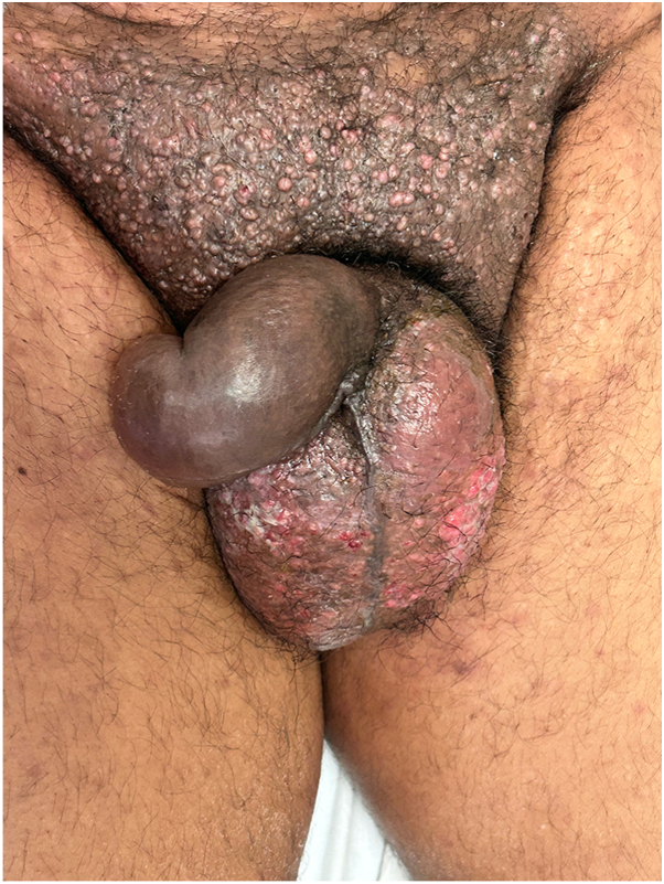 Figure 1
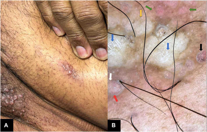 Figure 2
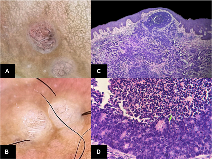 Figure 3
Peer Reviews
No public reviews on file for this paper yet. If you reviewed it on a platform where reviews are public (OpenReview, ICLR, NeurIPS, ICML), you can paste yours below so the community can read it here.
Videos
No videos yet. Explain this paper in a talk, walkthrough, or lecture? Add one.
Taxonomy
TopicsCancer Diagnosis and Treatment · Cutaneous Melanoma Detection and Management · Cancer and Skin Lesions
A 52-year-old man presented with a 4-month history of pink pruritic papules in the pubic area. He was undergoing treatment for stage IIIB colorectal adenocarcinoma, diagnosed 2 years earlier. On physical examination, he had skin phototype V and showed pink papules coalescing into an indurated, hyperpigmented plaque on his abdomen, scrotum, and right leg (Figs 1 and 2, A). He also had lymphedema of the legs, penis, and scrotum.Fig 1. Clinical photo. Grouped papules on the pubis forming a hardened, hyperpigmented plaque with a shell-like appearance. Some lesions are excoriated.Fig 2. Abdomen papules. (A) Clinical Photo. (B) Dermoscopy on polarized light. Globular structures with bright white septa forming a saccular pattern (blue arrows), surrounded by hyperpigmentation, dotted pigment with a pepeering-like (black arrows), with a fine network. White areas without structure (yellow arrow). Pale pink globular structures (green arrows). Punctate vessels (white arrow). Short, irregular linear vessels (red arrow).
Dermoscopy revealed globular structures with a saccular pattern, associated with bright white septa and brown borders, on a structureless white background with peppering-like pigmentation. The lesions were surrounded by pale pink globular areas, giving them an erythematous appearance. Scattered punctate vascular structures, short linear vessels, and irregular linear vessels were also identified (Figs 2 and 3, A, B).Fig 3. Cutaneous metastases from colorectal carcinoma**. (A)** Dermoscopy on polarized light. Close-up of globular structures with peppering-like punctate pigment. (B) Dermoscopy on polarized light. Areas with punctate vascular structures. (C) Malignant tumor infiltrating the dermis, proliferation of atypical glands, cells with irregular cytoplasm, pleomorphism, prominent nuclei, and atypical mitotic figures. Areas of central necrosis and desmoplastic stroma. (D) Accumulation of mucinous cells in the papillary dermis, (green arrow), generating a circular neoformation with central necrosis. (C and D, H&E stain; original magnifications: C, ×10; D, ×40).
Histopathological examination showed lymphatic invasion and infiltration of malignant glandular epithelial cells in the desmoplastic dermis, with morphology similar to the primary colorectal tumor (Fig 3), confirming the diagnosis of cutaneous metastasis.
Cutaneous metastases from colorectal carcinomas (CMCCs) are rare.1 Dermatoscopic reports of CMCC in light-skinned patients describe the main dermatoscopic findings as a polymorphic vascular pattern and a white, pinkish, or reddish background without defined structures.2, 3, 4, 5, 6 Since vascular patterns are more difficult to observe in dermoscopy of high-phototype patients, the description of new structures in these individuals could lead to earlier diagnoses and improved prognosis.2 Our patient presented saccular structures with bright white septa and peppering-like pigmentation, adding new CMCC findings. This case highlighted the need for further dermatoscopic documentation in patients with skin of color. The correlation of dermatoscopy structures is described in Table I.2^,^7^,^8Table ICorrelation of dermoscopy structures and histopathological findings of colorectal metastasisDermoscopy findingsHistopathological correlationReferenceBright white partitions that form saccular structuresThickened collagen bundles/desmoplastic stroma, associated with tumor infiltration in the dermisMarghoob et al7 (2009) Massone et al8 (2021)Structureless white area on a pink to red backgroundNew collagen remodeling in the dermisChernoff, et al2 (2014)Marghoob et al7 (2009)Vascular structuresDisorganized neo-angiogenesisChernoff et al2 (2014)∗Saccular structures with peppering-like pigmentMelanophages or melanin deposition adjacent to tumor nestsAuthors’ theory, based on assone et al8 (2021)∗Patients with dark-skin.
Conflicts of interest
None disclosed.
The reference list from the paper itself. Each links out to its DOI / PubMed record.
- 1Udkoff J.Cohen P.R.Adenocarcinoma of the colon presenting with scrotal metastasis: case report and review of the literature Dermatol Online J 22201613030/qt 1jg 0t 4kw 26990476 · pubmed ↗
- 2Chernoff K.A.Marghoob A.A.Lacouture M.E.Deng L.Busam K.J.Myskowski P.L.Dermoscopic findings in cutaneous metastases JAMA Dermatol 150201442943310.1001/jamadermatol.2013.850224430974 · doi ↗ · pubmed ↗
- 3Al Subait N.A.Bin Jadeed H.F.Al Saleh M.R.Al Faifi F.S.Al Saif F.M.Arafah M.A.Dermoscopy of scalp cutaneous metastasis of sigmoid adenocarcinoma JAAD Case Rep 14202111611910.1016/j.jdcr.2021.06.00434337122 PMC 8318904 · doi ↗ · pubmed ↗
- 4Manganoni A.M.Fusano M.Pavoni L.A solitary cutaneous metastasis from colon adenocarcinoma: dermoscopic and confocal microscopy features Indian J Dermatol Venereol Leprol 84201858910.4103/ijdvl.IJDVL_304_1730073979 · doi ↗ · pubmed ↗
- 5Ito T.Yoshida Y.Yamada N.Furue M.Yamamoto O.Dermoscopy of peristomal polyps and metastasis of colon cancer Acta Derm Venereol 942014969710.2340/00015555-164623756615 · doi ↗ · pubmed ↗
- 6Kamińska-Winciorek G.Wydmański J.Januszewski K.Silny W.Dermoscopy of nodular skin metastases from gastrointestinal primary cancer Postepy Dermatol Alergol 32201531231610.5114/pdia.2015.4804326366159 PMC 4565833 · doi ↗ · pubmed ↗
- 7Marghoob A.A.Cowell L.Kopf A.W.Scope A.Observation of chrysalis structures with polarized dermoscopy Arch Dermatol 145200961810.1001/archdermatol.2009.2819451524 · doi ↗ · pubmed ↗
- 8Massone C.Hofman-Wellenhof R.Chiodi S.Sola S.Dermoscopic criteria, histopathological correlates and genetic findings of thin melanoma on non-volar skin Genes (Basel)122021128810.3390/genes 1208128834440462 PMC 8391530 · doi ↗ · pubmed ↗