A rare case of subclavian steal phenomenon: when a dialysis arm arteriovenous fistula robs the brain
Leonardo Furtado Freitas, Tate Hodges, Charif Sidani, Kevin J. Abrams

Abstract
Genes, proteins, chemicals, diseases, species, mutations and cell lines named across the full text — each resolved to its canonical identifier and authoritative record.
Click any figure to enlarge with its caption.
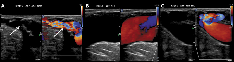 Figure 1
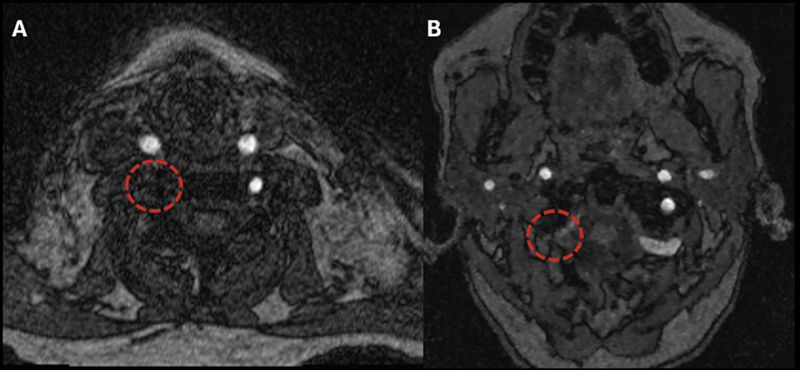 Figure 2
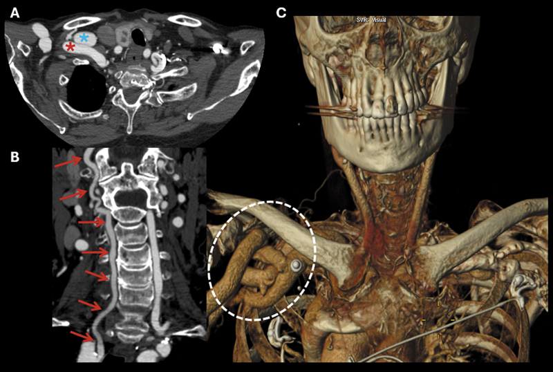 Figure 3
Peer Reviews
No public reviews on file for this paper yet. If you reviewed it on a platform where reviews are public (OpenReview, ICLR, NeurIPS, ICML), you can paste yours below so the community can read it here.
Videos
No videos yet. Explain this paper in a talk, walkthrough, or lecture? Add one.
Taxonomy
TopicsCentral Venous Catheters and Hemodialysis · Vascular Anomalies and Treatments · Vascular Procedures and Complications
A 79-year-old woman on hemodialysis via a right arm arteriovenous fistula (AVF), as shown in Figure 1 , presented with recurrent falls and transient dizziness. Neurovascular imaging ( Figures 2 3 ) revealed reversed flow in the right vertebral artery without subclavian artery stenosis, along with aneurysmal dilation of the right subclavian and axillary vessels. This case illustrates a rare subclavian steal phenomenon in the absence of arterial stenosis, highlighting the importance of recognizing dialysis access-related hemodynamic steal in patients presenting with vertebrobasilar symptoms. 1 2 3
Color Doppler ultrasound images demonstrating the dialysis-related arteriovenous fistula in the right upper extremity, including the arterial (A), anastomotic (B), and venous (C) segments. The fistula was patent, with less than 50% stenosis in the arterial segment, as evidenced by color aliasing (white arrows).
Magnetic resonance angiography of the cervical (A) and intracranial (B) arteries using three-dimensional (3D) time-of-flight (TOF) sequence. The absence of flow-void signal in the cervical and intracranial segments of the right vertebral artery (red dashed circles) suggested slow flow, absence of flow, or retrograde flow from a superior-to-inferior direction.
Cervical arterial computed tomography (CT) angiography. Axial (A), coronal (B), and volume-rendering (VR) reconstruction (C). Aneurysmal dilation of the right subclavian artery (red asterisk) and vein (blue asterisk). Patency of the entire right cervical vertebral artery (red arrows), with relatively reduced opacification compared to the contralateral side. On the VR image, the aneurysmal dilations of the right subclavian and axillary vessels are more conspicuous (white dashed circle), related to the presence of the dialysis-related arteriovenous fistula (AVF) in the right upper extremity.
The reference list from the paper itself. Each links out to its DOI / PubMed record.
- 1Maiodna E Ambekar S Johnson J N Elhammady M S Dialysis arteriovenous fistula causing subclavian steal syndrome in the absence of subclavian artery stenosis Case Rep Vasc Med 2015201572068410.1155/2015/72068425960914 PMC 4415454 · doi ↗ · pubmed ↗
- 2Agarwal S Schwartz L Kwon P Subclavian steal syndrome due to dialysis fistula corrected with subclavian artery stenting Neurol Clin Pract 2018805 e 23e 2510.1212/CPJ.000000000000051030564504 PMC 6276325 · doi ↗ · pubmed ↗
- 3Bucktowarsing B An Overview of Dialysis Access-associated Steal Syndrome Renal Fellow Network. June 30,2019. Accessed on: October 13, 2025 from:https://www.renalfellow.org/2019/06/30/an-overview-of-dialysis-access-associated-steal-syndrome/