Chylothorax as an Unusual Initial Manifestation of Diffuse Large B-Cell Lymphoma in an 86-Year-Old Woman: A Reflection of Diagnostic Reasoning and Age in Therapeutic Decisions
Diogo Dias Ramos, Amanda Hirschfeld, Inês Fiúza M. Rua, André Valente, Ana M Serrano

TL;DR
An 86-year-old woman with chylothorax was diagnosed with diffuse large B-cell lymphoma and showed significant improvement with targeted treatment.
Contribution
This case highlights the importance of considering lymphoma in elderly patients with chylothorax and the potential for effective treatment despite advanced age.
Findings
The patient's chylothorax resolved with a low-fat diet, octreotide, and chest drainage.
Corticosteroid therapy improved clinical status and performance in an elderly patient.
The case demonstrates that age should not limit treatment in elderly patients with lymphoma.
Abstract
Chylothorax is a rare presentation of lymphoproliferative disorders, responsible for only a small proportion of non-traumatic pleural effusions in large series. In older adults, its recognition can be challenging, often delaying diagnosis and treatment. We report an 86-year-old woman admitted with progressive dyspnea and a large right pleural effusion. Thoracentesis yielded a milky fluid with high triglycerides, confirming chylothorax. Cytology was negative for malignant cells, and a CT scan revealed an extensive retroperitoneal mass encasing the aorta and inferior vena cava, highly suggestive of lymphoproliferative disease. An inguinal lymph node biopsy established the diagnosis of diffuse large B-cell lymphoma. The patient was managed with a low-fat diet enriched with medium-chain triglycerides, subcutaneous octreotide, and chest drainage, leading to complete resolution of the…
Genes, proteins, chemicals, diseases, species, mutations and cell lines named across the full text — each resolved to its canonical identifier and authoritative record.
Click any figure to enlarge with its caption.
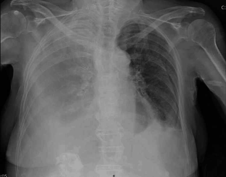 Figure 1
| Blood Work | Patient Results | Normal Range |
| Haemoglobin | 13.5 g/dL | 12.0 – 15.0 |
| Hematocrit | 41.60% | 35 – 46 |
| Mean Corpuscular Volume (MCV) | 87.9 fL | 78.0 – 96.0 |
| Mean Corpuscular Haemoglobin (MCH) | 28.5 pg | 26.0 – 33.0 |
| White Blood Cells (WBC) | 12.09 ×10⁹/L | 4.5 – 11.0 |
| Neutrophils (Absolute Count) | 10.65 ×10⁹/L | 2.0 – 8.5 |
| Lymphocytes (Absolute Count) | 0.78 ×10⁹/L | 0.9 – 3.5 |
| Monocytes (Absolute Count) | 0.45 ×10⁹/L | 0.2 – 1.0 |
| Eosinophils (Absolute Count) | 0.17 ×10⁹/L | 0.0 – 0.6 |
| Basophils (Absolute Count) | 0.04 ×10⁹/L | 0.0 – 0.1 |
| Platelets | 194 ×10⁹/L | 150 – 450 |
| Prothrombin Time (PT) | 11.4 s | 9.4 – 12.5 |
| INR (International Normalized Ratio) | 0.95 | 0.80 – 1.20 |
| aPTT (Activated Partial Thromboplastin Time) | 24.5 s | 25.1 – 36.5 |
| Urea | 54 mg/dL | 21 – 43 |
| Creatinine | 0.78 mg/dL | 0.57 – 1.11 |
| Estimated GFR (CKD-EPI) | 69 mL/min/1.73 m² | ≥ 90 |
| Sodium | 131 mEq/L | 136 – 145 |
| Potassium | 4.0 mEq/L | 3.5 – 5.1 |
| Calcium | 8.5 mg/dL | 8.40 – 10.20 |
| Uric Acid | 4.1 mg/dL | 2.60 – 6.00 |
| Total Bilirubin | 0.73 mg/dL | 0.20 – 1.20 |
| AST (Aspartate Aminotransferase) | 15 U/L | 5 – 34 |
| ALT (Alanine Aminotransferase) | 11 U/L | 0 – 55 |
| GGT (Gamma-Glutamyl Transferase) | 212 U/L | 9 – 36 |
| Alkaline Phosphatase | 239 U/L | 40 – 150 |
| LDH (Lactate Dehydrogenase) | 244 U/L | 125 – 220 |
| C-reactive Protein (CRP) | 36.4 mg/L | < 5.0 mg/L |
| Pleural Fluid Analysis | Patient Results | Normal Range |
| Macroscopic Appearance | Turbid, Light Yellow | Clear, Straw-Colored |
| pH (Pleural Fluid) | 7.5 | ~7.60 |
| Leukocytes (Pleural Fluid) | 801 /µL | < 1000 (Transudate) |
| Polymorphonuclear Cells | 63 /µL | — |
| Mononuclear Cells | 738 /µL | — |
| Erythrocytes | 5.0 ×10³ /µL | — |
| Total Proteins (Pleural Fluid) | 44.6 g/L | < 30 |
| Albumin (Pleural Fluid) | 24.8 g/L | — |
| Glucose (Pleural Fluid) | 291 mg/dL | > 60 |
| LDH (Pleural Fluid) | 142 U/L | < 200 |
| Triglycerides (Pleural Fluid) | 364 mg/dL | < 50 |
| Total Cholesterol (Pleural Fluid) | 87 mg/dL | < 60 |
| Amylase (Pleural Fluid) | 9 U/L | < 1.5 × Serum Value |
| Total Bilirubin (Pleural Fluid) | 1.0 mg/dL | Pleural/Serum Ratio < 0.6 |
| Adenosine Deaminase (ADA) | 15.9 U/L | < 31 |
| Microbiological Cultures (Aerobic) | No Growth after Five Days | Sterile |
| Microbiological Cultures (Anaerobic) | No Growth after Five Days | Sterile |
Peer Reviews
No public reviews on file for this paper yet. If you reviewed it on a platform where reviews are public (OpenReview, ICLR, NeurIPS, ICML), you can paste yours below so the community can read it here.
Videos
No videos yet. Explain this paper in a talk, walkthrough, or lecture? Add one.
Taxonomy
TopicsLymphatic Disorders and Treatments · Vascular Malformations and Hemangiomas · Pleural and Pulmonary Diseases
Introduction
Chylothorax is defined as the accumulation of lymphatic fluid within the pleural space due to disruption or obstruction of the thoracic duct. While trauma and postoperative injury are classical causes, malignancy accounts for approximately one-third of cases, with lymphoma, particularly non-Hodgkin subtypes, as the leading aetiology [1,2].
However, presentation with chylothorax as the first and sole manifestation of lymphoma remains rare, especially in very elderly patients. Such cases pose diagnostic and ethical challenges: the rarity of the presentation often delays tissue diagnosis, and therapeutic decisions may be influenced by chronological age rather than biological fitness. Early recognition is critical, as delayed diagnosis may lead to significant respiratory morbidity, nutritional compromise, and infectious complications, while in aggressive lymphomas such as diffuse large B-cell lymphoma (DLBCL), timely treatment is a major determinant of prognosis, particularly in older adults.
We present a case of DLBCL initially manifesting as chylothorax in an 86-year-old woman, which illustrates the need for comprehensive evaluation and individualised management in older adults.
Case presentation
An 86-year-old woman presented to the emergency department with progressive dyspnea and lower-limb oedema. Her relevant past medical history included a history of hypertension, dyslipidaemia, untreated type 2 diabetes mellitus, depressive disorder, suspected major neurocognitive disorder with behavioural disturbances, and severe diabetic retinopathy with significant visual impairment. She also had a history of pulmonary embolism (2009) and right femoral neck fracture treated with hip prosthesis (2014). Her chronic medication included duloxetine, perindopril, acenocoumarol, simvastatin, alprazolam, and zolpidem.
In the emergency department, she was conscious and cooperative, eupneic on oxygen therapy (6 L/min via face mask), with oxygen saturation of 90-92%, blood pressure of 119/54 mmHg, heart rate of 89 bpm, and afebrile. Pulmonary auscultation revealed absent breath sounds over the lower half of the right hemithorax and reduced sounds at the left base. Peripheral oedema grade 2+/3+ with bilateral pitting was present, and abdominal examination was unremarkable.
Initial laboratory evaluation showed mild leukocytosis with neutrophilia, an elevated C-reactive protein, mild renal impairment with a slight hyponatremia, and a cholestatic pattern of liver enzyme elevation (Table 1).
Urinalysis revealed glycosuria (200mg/dL), mild proteinuria (50mg/dL), and leukocyturia (1687/µL) with no other findings.
Chest X-ray showed near-complete right-sided opacification suggestive of massive right pleural effusion and a small left-sided pleural effusion (Figure 1).
Chest X-ray showing right-sided opacification and small left-sided pleural effusion
A presumed diagnosis of acute decompensated heart failure secondary to respiratory infection was made, and the patient started empirical antibiotic therapy with amoxicillin-clavulanate (1,200 mg three times daily) and intravenous furosemide.
Thoracentesis was performed for symptomatic relief and diagnostic purposes, evacuating 1,300 mL of turbid, milky fluid. Biochemical analysis confirmed chylothorax (pH 7.5, protein 44.6 g/L, triglycerides 364 mg/dL, cholesterol 87 mg/dL), which was inconsistent with the initial working diagnosis of acute decompensated heart failure. These findings, together with the right-sided predominance of the effusion, were anatomically consistent with disruption or obstruction of the thoracic duct along its intrathoracic course. Cytology showed reactive mesothelial cells without malignancy and microbiological cultures were negative (Table 2).
Due to an iatrogenic pneumothorax, a chest drain (28 Fr) was inserted, and the patient was started on a low-fat diet enriched with medium-chain triglycerides and subcutaneous octreotide 100µg t.i.d. Daily drainage progressively decreased, allowing removal of the drain after eight days with radiographic resolution of the effusion.
As part of the etiological investigation of the chylothorax, a CT thoraco-abdomino-pelvic imaging was ordered, revealing a 13 × 10 × 7 cm retroperitoneal mass surrounding the aorta and inferior vena cava and extending to the mesenteric root, compatible with a large conglomerate of lymph nodes. Beta-2-microglobulin was also elevated (3.2 mg/L; reference range: 0.97 - 2.64mg/L).
An ultrasound-guided inguinal lymph node biopsy was performed confirming the diagnosis of DLBCL. The hospital course was further complicated by a transient nosocomial pneumonia and candiduria, both successfully treated.
Given her preserved functional status and improvement, she was started on oral prednisolone 20 mg daily, with excellent tolerance, increased appetite, and weight gain of 4.5 pounds. At follow-up, she was clinically stable, functionally independent (ECOG performance status of 0), and was referred to Haematology, where she was proposed for chemotherapy.
Discussion
Chylothorax secondary to malignancy is most often associated with lymphomas, particularly non-Hodgkin subtypes such as DLBCL [1-3]. Obstruction or infiltration of the thoracic duct leads to lymphatic leakage into the pleural cavity, producing a milky exudate rich in triglycerides. The right-sided predominance observed in this patient suggests ductal involvement below the fifth thoracic vertebra [4].
Chylothorax remains an uncommon initial manifestation of lymphoma, accounting for less than 5% of non-traumatic cases in large series [1,2].
The conservative strategy combining dietary fat restriction with medium-chain triglycerides, octreotide, and pleural drainage remains first-line therapy [3,4]. This approach aims to reduce chyle flow, promote healing of the duct and allow spontaneous resolution, as observed here. Surgical or interventional management is reserved for persistent high-output chylothorax.
In older adults, diagnosis and treatment are often constrained by perceptions of futility. However, evidence from large registries demonstrates that even patients aged ≥85 years with DLBCL can benefit from therapy with curative intent when functionally fit [5]. Few case reports describe chylothorax as the presenting feature of lymphoma in the very elderly, but similar outcomes have been documented when early recognition and multidisciplinary coordination occur [6].
Conclusions
This case highlights chylothorax as a rare initial manifestation of DLBCL in a very elderly patient, emphasising the diagnostic challenges associated with non-traumatic chylothorax and negative pleural fluid cytology. In this context, a high index of suspicion and a structured diagnostic approach are critical for timely identification of the underlying lymphoproliferative disorder. Conservative management, including dietary modification, octreotide, pleural drainage, and corticosteroid therapy, resulted in complete resolution of the effusion and marked functional improvement.
Finally, advanced age should not, in isolation, limit therapeutic decisions. Careful assessment of functional status and biological fitness, supported by a multidisciplinary and patient-centred approach, can allow meaningful recovery and access to disease-directed treatment even in very old patients.
The reference list from the paper itself. Each links out to its DOI / PubMed record.
- 1Chylothorax: aetiology, diagnosis and therapeutic options Respir Med Mc Grath EE Blades Z Anderson PB 181042010 https://doi.org/10.1016/j.rmed.2009.08.0101976647310.1016/j.rmed.2009.08.010 · doi ↗ · pubmed ↗
- 2Etiology of chylothorax in 203 patients Mayo Clin Proc Doerr CH Allen MS Nichols FC 3rd Ryu JH 867870802005 https://doi.org/10.4065/80.7.8671600789110.4065/80.7.867 · doi ↗ · pubmed ↗
- 3Non-traumatic chylothorax: diagnostic and therapeutic strategies Breathe (Sheff) Ur Rehman K Sivakumar P 2101631820223633713410.1183/20734735.0163-2021 PMC 9584559 · doi ↗ · pubmed ↗
- 4Chylothorax: pathophysiology, diagnosis, and management-a comprehensive review J Thorac Dis Bhatnagar M Fisher A Ramsaroop S Carter A Pippard B 164516611620243850502710.21037/jtd-23-1636 PMC 10944732 · doi ↗ · pubmed ↗
- 5Diffuse large B-cell lymphoma in octogenarians aged 85 and older can benefit from treatment with curative intent: a report on 129 patients prospectively registered in the Elderly Project of the Fondazione Italiana Linfomi (FIL)Haematologica Tucci A Merli F Fabbri A 108310911082023 https://doi.org/10.3324/haematol.2022.2814073638424710.3324/haematol.2022.281407 PMC 10071130 · doi ↗ · pubmed ↗
- 6Nontraumatic chylothorax secondary to lymphoma and filariasis J Surg Case Rep Barillas S Rodas A Ardebol J MartíJL 020202020 https://doi.org/10.1093/jscr/rjaa 30910.1093/jscr/rjaa 309PMC 750230932983405 · doi ↗ · pubmed ↗