Pneumorrhachis of Infectious Origin in a Patient with Advanced HIV and Uncontrolled Diabetes: A Case Report and Review of the Literature
Thenell Van der Westhuizen, Abdullah E Laher

TL;DR
A rare case of pneumorrhachis caused by an infectious origin in a patient with HIV and diabetes is reported, highlighting the need for urgent diagnosis and treatment.
Contribution
This case expands the limited literature on pneumorrhachis caused by infection, not trauma or pneumomediastinum.
Findings
Pneumorrhachis was caused by disseminated MRSA infection in a patient with HIV and diabetes.
Most reported cases of infectious pneumorrhachis are linked to emphysematous infections from gas-forming organisms.
Pneumorrhachis is a rare but significant indicator of severe underlying disease requiring urgent antimicrobial therapy.
Abstract
Pneumorrhachis, the presence of air within the spinal canal, is an uncommon radiological finding, typically associated with trauma or pneumomediastinum. Non-traumatic causes, particularly infective ones, are exceedingly rare. We describe a woman with advanced HIV infection and poorly controlled diabetes mellitus who presented with confusion and meningism in the context of diabetic ketoacidosis (DKA). Computed tomography (CT) revealed cervical extradural pneumorrhachis. Blood, urine, sputum, and cerebrospinal fluid (CSF) cultures confirmed disseminated methicillin-resistant Staphylococcus aureus (MRSA) infection. This case adds to the limited literature describing pneumorrhachis of infectious origin unrelated to trauma or pneumomediastinum. A review of published cases shows that most are associated with emphysematous infections (epidural abscesses, pyelonephritis, cystitis, or…
Genes, proteins, chemicals, diseases, species, mutations and cell lines named across the full text — each resolved to its canonical identifier and authoritative record.
Click any figure to enlarge with its caption.
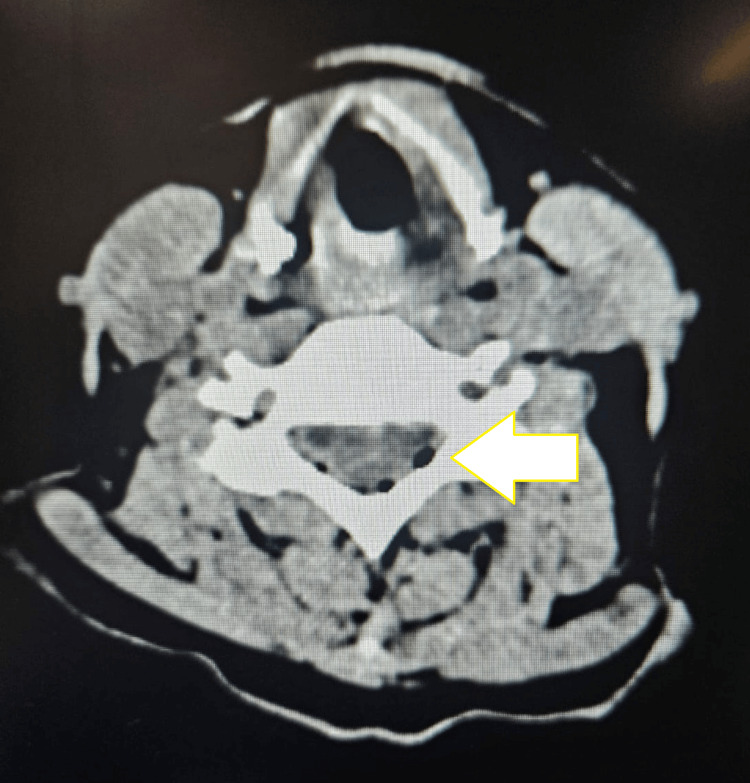 Figure 1
| Author | Date of Publication | Gender | Age (Years) | Risk Factors | Symptoms | Outcome | Pathogen |
| Kirzner et al. [ | 1988 | F | 68 | Diabetes mellitus | Epidural abscess, back pain, bilateral leg paralysis, fever | Completely recovered | Staphylococcus aureus |
| Shintani et al. [ | 1992 | F | 71 | Contaminated epidural catheter | Epidural abscess, headache, fever | Completely recovered | Staphylococcus aureus |
| Kökes et al. [ | 1993 | M | 52 | Diabetes mellitus | Epidural abscess, back pain, paraparesis | Demised secondary to septic shock | Streptococcus, Peptostreptococcus, Bacteroides |
| Fujisawa et al. [ | 1998 | Epidural abscess, neck pain, fever, paraparesis | Completely recovered | Staphylococcus aureus | |||
| Nakatani et al. [ | 1998 | F | 54 | Nil | Epidural abscess, fever, decubitus ulcer | Paralysis of both legs | Bacteroides fragilis, Peptostreptococcus |
| Jomir et al. [ | 2009 | M | 27 | Paraplegia | Sacral pressure ulcer, orthostatic headache, and nausea | Discharged |
|
| Hur et al. [ | 2012 | M | 68 | Anterior cervical surgery | Epidural abscess, neck pain, paraparesis, urinary retention | Died secondary to septic shock | Streptococcus anginosus |
| Hur et al. [ | 2012 | M | 52 | Anterior cervical surgery | Neck pain, epidural abscess | Discharged | Methicillin-resistant |
| Lee et al. [ | 2013 | M | 72 | Vertebroplasty | Paralysis of both legs, back pain | Completely recovered | Aeromonas hydrophila |
| Amara et al. [ | 2013 | M | 21 | Headache, fever, vomiting, dyspnea | Died | Bacterial meningitis (unspecified) | |
| Akagawa et al. [ | 2015 | F | 78 | Diabetes mellitus | Epidural abscess, back pain, paralysis of the legs | Completely recovered | Clostridium perfringens, Escherichia coli, Enterococcus faecalis |
| Kim and Kim [ | 2017 | M | 56 | Nil | Epidural abscess, neck pain, fever | Paralysis and urinary incontinence | Streptococcus anginosus |
| Matsuo et al. [ | 2019 | F | 70 | Diabetes mellitus | Epidural abscess, back pain, left leg paralysis | Completely recovered | Escherichia coli |
| Miranda et al. [ | 2021 | F | 67 | Diabetes mellitus, Hypertension, Alzheimer’s, bedbound | Sacral decubitus ulcer, confusion | Died | Streptococcus anginosus |
| Navriya et al. [ | 2021 | M | 65 | Diabetes mellitus | Emphysematous pyelonephritis, flank pain, fever, hypotension | Completely recovered | Escherichia coli |
| Maegawa et al. [ | 2022 | M | 68 | Parkinson’s disease, bedbound | Fever | Discharged | Clostridium perfringens |
| Ehret et al. [ | 2022 | M | 88 | Diabetes mellitus, Crohn’s disease with chronic steroid use | Back pain and confusion, emphysematous cystitis | Discharged to hospice | Escherichia coli |
| Sumitro et al. [ | 2023 | M | 57 | Hepatitis C | Fever, back pain, weakness, dysuria, prostate abscess | Discharged | Klebsiella pneumoniae |
| Allena et al. [ | 2023 | F | 77 | Diabetes mellitus, Hypertension | Emphysematous cystitis, paraspinal abscess, leg weakness | Discharged to hospice | Klebsiella pneumoniae |
Peer Reviews
No public reviews on file for this paper yet. If you reviewed it on a platform where reviews are public (OpenReview, ICLR, NeurIPS, ICML), you can paste yours below so the community can read it here.
Videos
No videos yet. Explain this paper in a talk, walkthrough, or lecture? Add one.
Taxonomy
TopicsPneumothorax, Barotrauma, Emphysema · Infectious Disease Case Reports and Treatments · Infectious Diseases and Tuberculosis
Introduction
Pneumorrhachis, defined as the presence of air within the spinal canal, is an exceptional radiological finding first described in 1977 [1]. The condition is broadly classified as traumatic or non-traumatic. Non-traumatic pneumorrhachis may arise spontaneously or from iatrogenic, infectious, or neoplastic processes. In most cases, air reaches the spinal canal secondarily, typically via pneumomediastinum, infected sacral ulcers, or invasive procedures such as lumbar puncture. In the case of infectious pneumorrhachis, air created by gas-forming organisms enters the spinal canal via direct extension, dural breach, or inflammatory dissection of tissue between the source and the spinal canal. All of these cases involved patients with immunocompromised or epidural abscesses [1,2]. Diagnosis is radiological. It is often an incidental finding when imaging is requested for another concern, such as delirium, pyelonephritis, or epidural abscess. CT of the spine is the most sensitive modality, whereas MRI is useful when concomitant pathology (e.g., abscess or tumor) is suspected. Plain radiographs are rarely diagnostic but may help identify pneumomediastinum as a potential source. Once identified, investigations should aim to determine the underlying cause, for instance, chest radiography for pneumomediastinum, urinalysis for urinary tract infections, or cerebrospinal fluid (CSF) analysis for meningitis. Management should be individualized. Most patients can be treated conservatively through management of the underlying condition, supplemental oxygen, and supportive care. Surgical decompression is reserved for rare cases complicated by neurological deficit or cord compression. While pneumorrhachis itself is self-limiting, it is an indicator of severe underlying disease. The following case highlights an infective cause of pneumorrhachis secondary to disseminated methicillin-resistant Staphylococcus aureus (MRSA) infection. It also illustrates the severity and rapid disease progression often associated with pneumorrhachis.
Case presentation
A woman in her mid-fifties presented to the emergency department with a two-week history of confusion and generalized weakness. She had known HIV infection and poorly controlled diabetes mellitus. Her CD4 count was 89, and HbA1c was 15.4%. On examination, she appeared acutely ill, with a Glasgow Coma Scale score of 11 and marked neck stiffness. There was no history or clinical evidence of trauma, sacral ulcers, or prior lumbar puncture. Initial investigations demonstrated sepsis complicated by diabetic ketoacidosis (DKA) and acute kidney injury. A non-contrast CT of the brain was performed because of her altered level of consciousness. The scan revealed pneumorrhachis with gas locules in the extradural spinal canal extending from C1 to C4 (Figure 1). Additional small air locules were seen in the cervical paravertebral space, with minimal fat stranding but no muscular edema or fluid collections to suggest an abscess. Scant air was also seen in the retropharyngeal space without associated swelling or effusion. A chest radiograph excluded pneumothorax or pneumomediastinum as a possible source.
Non-contrast CT of the brain (axial plane) showing cervical extradural pneumorrhachis (C1–C4) with associated retropharyngeal and paravertebral air locules. Pneumorrhacis is indicated by the white arrow.
Lumbar puncture revealed elevated protein and pleocytosis with both polymorphonuclear and lymphocytic predominance. Blood, urine, sputum, and CSF cultures all grew MRSA, confirming disseminated MRSA infection. The patient was resuscitated with intravenous fluids, started on ceftriaxone, and treated for DKA in the emergency department. Blood, CSF, and urine cultures performed on day 1 of presentation indicated MRSA and vancomycin sensitivity. Antibiotic therapy was changed from ceftriaxone to intravenous vancomycin on day 2. The patient was given a fluid preload with each vancomycin dose, and urea and creatinine were monitored daily. Despite appropriate fluids and antibiotic therapy, she developed progressive renal failure and nosocomial pneumonia. Blood results showed an estimated glomerular filtration rate that diminished from normal to 10 ml/min/1.73 m², procalcitonin that climbed from 0.4 to 1.79 ng/ml, and C-reactive protein that continued to increase despite culture-directed antibiotics. On day 10 in the medical ward, she was intubated and ventilated for worsening encephalopathy. A repeat CT demonstrated resolution of the pneumorrhachis with no new findings. Unfortunately, she died from disseminated MRSA sepsis on day 11 of hospitalization.
Discussion
The true incidence of pneumorrhachis remains unknown due to its rarity and diverse etiologies. However, reported cases have increased in recent years, likely reflecting advances in imaging and greater awareness among clinicians. Infectious pneumorrhachis is exceptionally uncommon. Signs and symptoms are mainly related to the underlying infection; however, the patient may complain of back pain or neck stiffness. Due to the vagueness of signs and symptoms, the diagnosis is exclusively made on imaging while investigating the underlying infection. Air may be introduced into the spinal canal through several mechanisms: direct extension from adjacent emphysematous infections such as osteomyelitis, pyelonephritis, or cystitis; meningitis due to gas-forming organisms; or fistulous tracts from infected pressure ulcers or malignancy, resulting in CSF leakage and negative-pressure air entry. The most frequently reported organisms include Escherichia coli, *Klebsiella *spp., Clostridium spp., Staphylococcus aureus, *Streptococcus *spp., *Citrobacter *spp., *Morganella *spp., and other enteric pathogens. In this case, disseminated MRSA infection was implicated, a rare finding. A summary of previously published cases is presented in Table 1. A systematic narrative review was performed to identify published cases and studies describing pneumorrhachis of infectious origin. PubMed/Medical Literature Analysis and Retrieval System Online (MEDLINE) and Scopus were searched from inception to the date of manuscript preparation.
Of reported cases, the average age was 61.7 years, 57.9% of patients were male (11 males and eight females). The most common comorbidity was diabetes mellitus, while four patients presented after an invasive procedure. Epidural abscess was present in 52.6% of reported cases. The most common symptoms reported were back or neck pain (57.9%), fever (47.4%), and leg paraparesis or paralysis (42%). Outcomes varied: 36.8% completely recovered, while 21% of patients died.
A review of reported cases demonstrates that air enters the spinal canal via contiguous spread from adjacent emphysematous infections, gas-forming meningitis, or secondary to invasive procedures. The microbiological spectrum is broad and predominantly involves gas-forming or enteric organisms, while Staphylococcus aureus, and especially MRSA, remains an infrequent cause. Outcomes vary widely, ranging from complete neurological recovery to death from overwhelming sepsis, highlighting the importance of early recognition, prompt imaging, and aggressive management of the underlying cause.
Conclusions
Non-traumatic pneumorrhachis is a rare but clinically significant entity. When identified, it should trigger an urgent search for a possible infective source, particularly in immunocompromised patients. As demonstrated by this case, though the intraspinal air may resolve spontaneously, its presence often signifies severe underlying disease. Early recognition, diagnosis, and aggressive management of sepsis are essential for improving outcomes. Further research is needed to clarify optimal management strategies and long-term prognosis for non-traumatic, infection-related pneumorrhachis.
The reference list from the paper itself. Each links out to its DOI / PubMed record.
- 1Pathogenesis, diagnosis and management of pneumorrhachis Eur Spine J Oertel MF Korinth MC Reinges MH Krings T Terbeck S Gilsbach JM 63664315 Suppl 520061683573510.1007/s 00586-006-0160-6PMC 1602196 · doi ↗ · pubmed ↗
- 2Pneumorrhachis: an uncommon radiological entity SA J Radiol Vanmali A Daji KD 22552520213491741110.4102/sajr.v 25i 1.2255 PMC 8661299 · doi ↗ · pubmed ↗
- 3Intraspinal air: a CT finding of epidural abscess AJR Am J Roentgenol Kirzner H Oh YK Lee SH 121712181511988326377510.2214/ajr.151.6.1217 · doi ↗ · pubmed ↗
- 4Iatrogenic acute spinal epidural abscess with septic meningitis: MR findings Clin Neurol Neurosurg Shintani S Tanaka H Irifune A Mitoh Y Udono H Kaneda A Shiigai T 253255941992138291210.1016/0303-8467(92)90099-o · doi ↗ · pubmed ↗
- 5Epidural spinal abscess containing gas: MRI demonstration Neuroradiology KökeşF Iplikçioğlu AC Camurdanoğlu M Bayar MA Gökçek C 497498351993823287210.1007/BF 00588704 · doi ↗ · pubmed ↗
- 6Intraspinal air: an unusual finding of cervical epidural abscess Acta Neurochir (Wien) Fujisawa H Hasegawa M Tsukada T Kita D Tachibana O Yamashita J 2872881401998963826710.1007/s 007010050097 · doi ↗ · pubmed ↗
- 7A case report of epidural abscess due to anaerobic bacteria, producing a mass of gas (Article in Japanese)Rinsho Shinkeigaku Nakatani S Hoshi K Yuasa T Sato T Tauchi T 224227381998 https://pubmed.ncbi.nlm.nih.gov/9711118/9711118 · pubmed ↗
- 8Pneumorrhachis and pneumocephalus due to a sacral pressure sore after paraplegia Neurorehabil Neural Repair Jomir L Fuentes S Gélis A Labauge P 7457462320091938046110.1177/1545968309332926 · doi ↗ · pubmed ↗