From Glaphene to Glaphynes: A Hybridization of Two-Dimensional Silica Glass and Graphynes
Guilherme S. L. Fabris, Raphael B. de Oliveira, Marcelo L. Pereira, Robert Vajtai, Pulickel M. Ajayan, Douglas S. Galvão

TL;DR
This paper introduces glaphynes, a new class of 2D materials combining silica glass and graphynes, and studies their stability and electronic properties.
Contribution
The novel contribution is the proposal and computational analysis of glaphynes, a hybrid of silica and graphynes.
Findings
Glaphynes show energetic and structural stability.
The electronic proximity effect opens a band gap in some glaphyne structures.
Not all glaphyne configurations exhibit band gap opening despite Si–O–C bond formation.
Abstract
Hybrid two-dimensional (2D) materials have attracted increasing interest as platforms for tailoring electronic properties through interfacial design. Very recently, a hybrid 2D material termed glaphene, which combines monolayers of 2D silica glass and graphene, was experimentally realized. Inspired by glaphenes, we proposed a class of similar structures named glaphynes, which are formed by stacking SiO2 monolayers onto α-, β-, and γ-graphynes. Graphynes are 2D carbon allotropes with the presence of acetylenic groups (triple bonds). The glaphynes’ structural and electronic properties were investigated using the self-consistent-charge density functional tight-binding (SCC-DFTB) method, as implemented in the DFTB+ package. Our analysis confirms their energetic and structural stability. We have observed that in the case of glaphynes, the electronic proximity effect can indeed open the…
Genes, proteins, chemicals, diseases, species, mutations and cell lines named across the full text — each resolved to its canonical identifier and authoritative record.
Click any figure to enlarge with its caption.
 1
1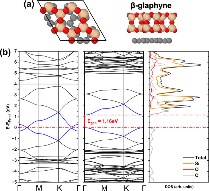 2
2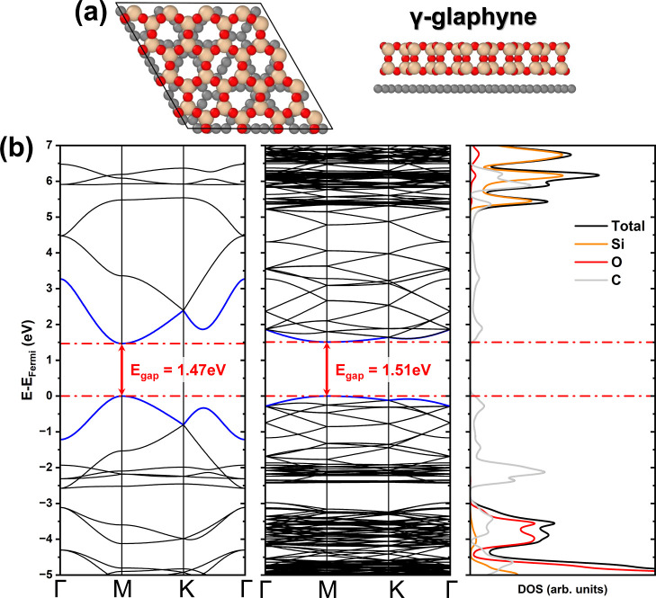 3
3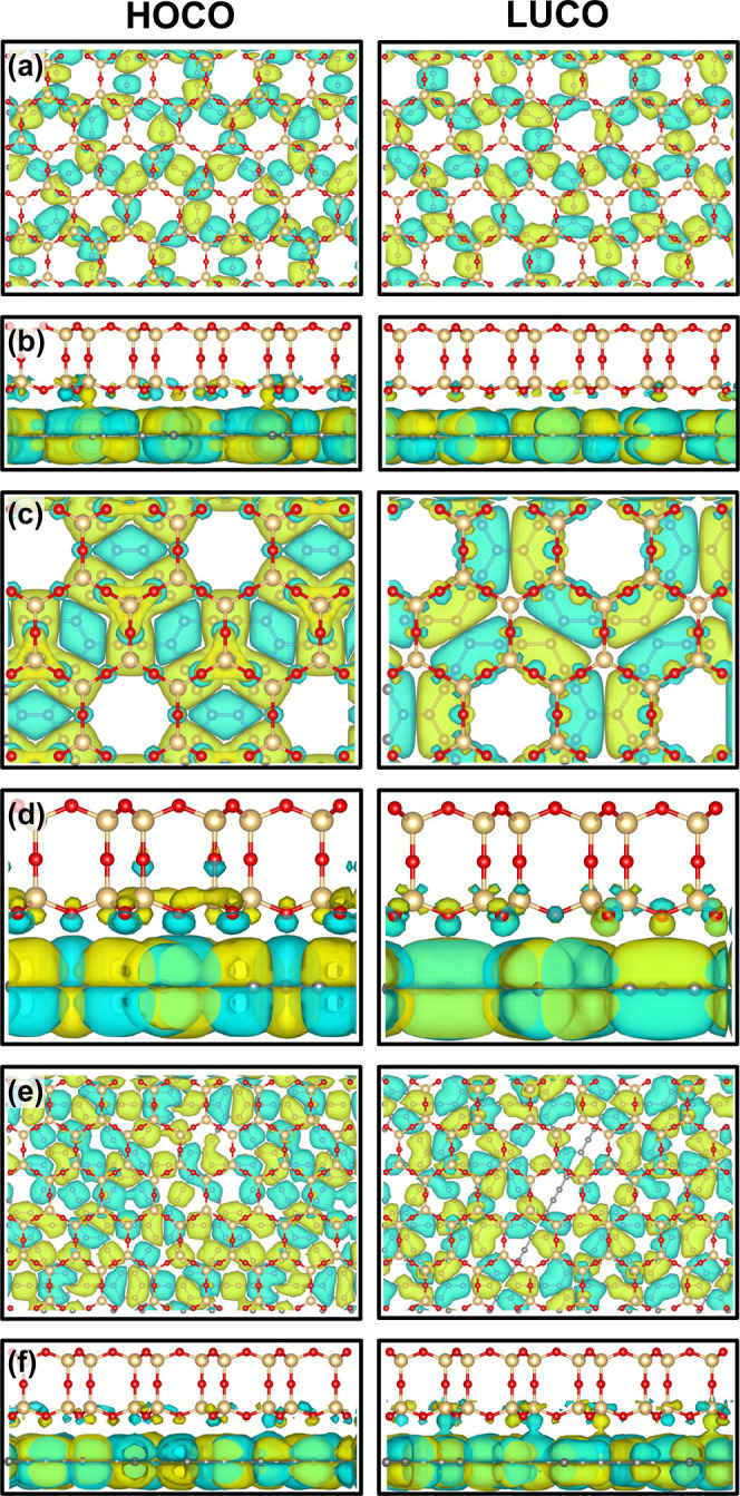 4
4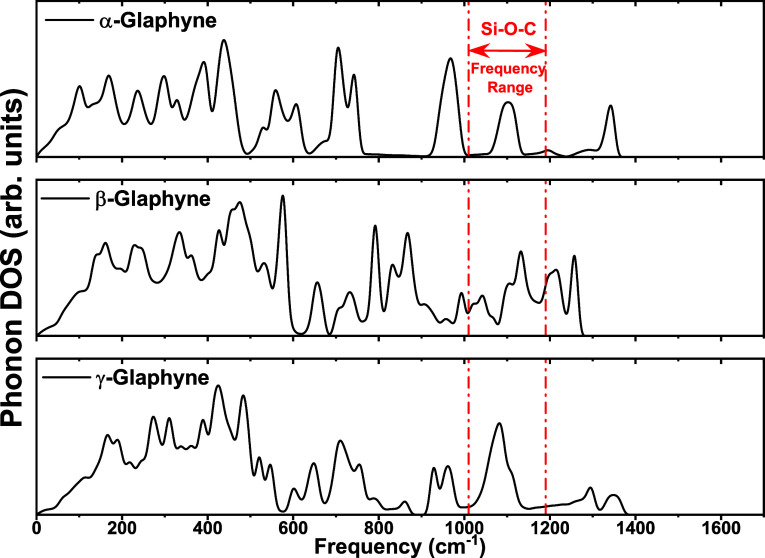 5
5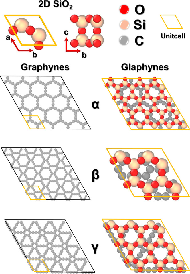 6
6| material |
|
| γ (°) |
|
|
|---|---|---|---|---|---|
| α-graphyne | 8 | 7.02 (6.98 | 119.9 (120 | 0.0 (0.0 | –8.13 |
| β-graphyne | 18 | 9.57 (9.50 | 119.9 (120 | 0.0 (0.0 | –8.20 |
| γ-graphyne | 12 | 6.93 (6.88 | 119.9 (120 | 1.47 (1.32 | –8.41 |
| α-glaphyne | 264 | 21.19 | 120.01 | 0.01 | –8.74 |
| β-glaphyne | 66 | 10.27 | 119.98 | 1.16 | –8.65 |
| γ-glaphyne | 300 | 21.02 | 119.99 | 1.51 | –8.77 |
- —Funda??o de Amparo ? Pesquisa do Estado de S?o Paulo10.13039/501100001807
- —Funda??o de Amparo ? Pesquisa do Estado de S?o Paulo10.13039/501100001807
- —Funda??o de Amparo ? Pesquisa do Estado de S?o Paulo10.13039/501100001807
- —Coordena??o de Aperfei?oamento de Pessoal de N?vel Superior10.13039/501100002322
- —Conselho Nacional de Desenvolvimento Cient?fico e Tecnol?gico10.13039/501100003593
- —Conselho Nacional de Desenvolvimento Cient?fico e Tecnol?gico10.13039/501100003593
- —Conselho Nacional de Desenvolvimento Cient?fico e Tecnol?gico10.13039/501100003593
- —Conselho Nacional de Desenvolvimento Cient?fico e Tecnol?gico10.13039/501100003593
- —Funda??o de Apoio ? Pesquisa do Distrito Federal10.13039/501100005668
Peer Reviews
No public reviews on file for this paper yet. If you reviewed it on a platform where reviews are public (OpenReview, ICLR, NeurIPS, ICML), you can paste yours below so the community can read it here.
Videos
No videos yet. Explain this paper in a talk, walkthrough, or lecture? Add one.
Taxonomy
TopicsGraphene research and applications · 2D Materials and Applications · Surface Chemistry and Catalysis
Introduction
Few fields have experienced such rapid growth in recent decades as nanoscience. This growth can be partly attributed to the prediction and subsequent synthesis of two-dimensional (2D) nanostructures, such as graphene.? Owing to its remarkable properties, including high mechanical strength? and thermal conductivity,? graphene has inspired the search for new 2D materials with similarly outstanding characteristics. In this perspective, graphynes ?−? ? represent a class of 2D carbon allotropes composed of sp and sp^2^ hybridized carbon atoms, resulting from the insertion of acetylenic linkers between the carbon atoms of graphene.? The proportion and arrangement of these acetylenic chains give rise to distinct types of graphyne, such as α, β, and γ-graphyne, ?,? among others. Since the experimental synthesis of γ-graphyne, ?−? ? ? several studies have proposed its use in supercapacitors,? batteries,? photovoltaic devices, ?,? and other potential applications. Further investigations have confirmed the semiconducting nature of γ-graphyne, with a moderate band gap. ?,?−? ? Beyond carbon-based systems, other families of 2D nanomaterials have emerged, such as silicates, with special focus on silica (SiO_2_). ?,? Silicon-based nanostructures have played a central role in the development of modern electronics, and silicon oxides are characterized by robust insulating behavior and a wide band gap of approximately 6.7 eV. ?,?
The use of heterostructures to enhance the properties of nanomaterials has increased significantly in recent years. ?,? Several studies have proposed the use of different nanostructures to achieve tailored functionalities. For instance, the insertion of hexagonal boron nitride (h-BN) domains into graphene sheets ?−? ? enables control over mechanical properties. ?−? ? Other examples include graphene/graphyne heterojunctions designed for carbon-based transistors, ?,? and heterojunctions between different types of graphynes to tune thermal conductivity.? Another widely used approach for creating heterostructures is the layer-by-layer stacking of monolayers. For example, Sun et al.? investigated graphyne stacked on XSe_2_ (X = Mo, W) to engineer the electronic band gap. In contrast, Bhattacharya and Sarkar? proposed graphyne–graphene nitride heterostructures for nanocapacitor applications. Independently, Huang et al.,? Wang et al.,? and their respective collaborators investigated low-dimensional silica interfaces and their interactions with graphene. Recent efforts have also explored the experimental feasibility of such systems by using graphene/Si and graphene/SiC substrates as templates for heterostructure growth.? These strategies suggest that complex 2D architectures involving graphene can be synthesized through scalable deposition techniques and are within the feasibility of fabricating related hybrid systems. Overall, the combination of nanostructures via heterojunctions, embedded domains, or vertical stacking has proven to be an effective strategy for producing materials with intermediate or enhanced properties compared to their parent components.
Very recently, Iyengar et al.? introduced a hybrid 2D material termed glaphene, which combines monolayers of 2D silica glass and graphene. Initially proposed through first-principles calculations and later synthesized via a scalable vapor-phase growth method, glaphene represents a significant experimental milestone in the development of mixed-component 2D materials. Notably, the interlayer interactions in glaphene surpass typical van der Waals forces, leading to strong interlayer hybridization. This hybridization induces a pronounced modification in the electronic structure. While graphene is a semimetal with a zero band gap and 2D silica glass is an insulator with a band gap of approximately 8.2 eV,? their combination results in a semiconducting material with a sizable band gap of about 3.6 eV, primarily governed by out-of-plane p_ z _ orbital interactions. They demonstrated that it is possible to create electronic band engineering through electronic proximity effects.
In this work, we propose and characterize a family of heterostructures inspired by the recent proposition and synthesis of glaphene. These heterostructures (named glaphynes) are created by stacking a SiO_2_ monolayer onto a graphyne monolayer. We tested three distinct glaphyne configurations, referred to as α-, β-, and γ-glaphyne, see Figure. We then investigated their energetic and structural stability in order to evaluate their potential experimental feasibility. In addition, we have analyzed their electronic properties, showing that the stacking approach enables the emergence of semiconductors with appreciable electronic band gaps, distinct from those of the individual constituents.
Results
and Discussion
Initially, we performed separate geometry optimizations of the isolated monolayers of SiO_2_ and the three graphyne types (α, β, and γ) to validate the computational setup using the density functional tight-binding (DFTB) approach. The resulting structural parameters were then compared with the available literature data, as summarized in Table. The results indicate that the chosen parametrization yields good accuracy, with lattice parameter deviations of approximately 2.5%, 0.57%, 0.74%, and 0.73% for SiO_2_, α-, β-, and γ-graphyne, respectively. For the γ angle, an almost negligible deviation was observed across all structures. The calculated bond lengths along the acetylenic chains were 1.411, 1.234, and 1.411 Å for α-graphyne; 1.427, 1.227, and 1.427 Å for β-graphyne; and 1.438, 1.221, and 1.438 Å for γ-graphyne. For the SiO_2_ monolayer, the average Si–O bond length was found to be 1.63 Å, in close agreement with experimental data.? The thickness of the silica layer was estimated to be 4.33 Å, which is consistent with the experimental value reported by Iyengar et al.? These consistent results validate the reliability of our computational methodology in describing the structural features of both carbon- and silicon-based two-dimensional systems.
1: Structural and Energetic Properties Obtained in This Work
Following the preliminary optimizations, the creation of the glaphynes was carried out by assembling a 3 × 3 supercell of α- and γ-graphynes, and a unit cell of β-graphyne. These were combined with a 4 × 4 supercell of 2D SiO_2_ for α- and γ-graphynes, and a 2 × 2 supercell for β-graphyne. The SiO_2_ layers were placed above the graphyne structures at an initial vertical separation of approximately 3.5 Å. To verify the energetic stability of the selected stacking, we tested alternative configurations, including rotated and laterally shifted layers. All these arrangements relaxed to an equivalent geometry upon optimization, indicating that the adopted configuration corresponds to the minimum-energy structure. After geometry optimization, the resulting glaphynes exhibited P6MM symmetry, and only slight modifications were observed in the acetylenic chain bond lengths. The optimized bond lengths for α-glaphyne were 1.421, 1.239, and 1.421 Å, for β-glaphyne, 1.576, 1.247, and 1.576 Å, and for γ-glaphyne, 1.457, 1.228, and 1.457 Å. The resulting vertical distances between the SiO_2_ and graphyne layers were 3.16, 3.22, and 3.12 Å for α-, β-, and γ-glaphynes, respectively. For comparison, the corresponding structural parameters in glaphene present average C–C and Si–O bond lengths of 1.42 Å and 1.62 Å, which differ by approximately 4.4% and 1.4%, respectively, relative to the averaged glaphyne values. Likewise, the interlayer spacing in glaphene (3.20 Å) differs by about 1.0% from the average separation obtained for the glaphynes. Additionally, the thickness of the SiO_2_ layer after deposition was found to be approximately 0.44 nm, in close agreement with the experimental value of 0.43 nm reported by Iyengar and collaborators.? Interestingly, the mismatch in the β-glaphyne case can be significantly reduced by adopting a larger configuration involving a 7 × 7 supercell of 2D SiO_2_ and a 4 × 4 supercell of β-graphyne, resulting in a system with 1072 atoms and a mismatch of only 2.8%.
The Mulliken population analysis revealed an increase in the overlap population for α-, β-, and γ-glaphynes, with average shifts of approximately 0.87, 10.88, and 0.96 m|e|, respectively. These shifts are primarily attributed to modifications in the distribution of the carbon 2p_ z _ orbitals when compared to their pristine graphyne counterparts. Notably, the most significant variation occurs in β-glaphyne, where the substantial change in C–C bond lengths within the acetylenic linkages indicates a corresponding alteration in hybridization. This result is consistent with the enhanced electronic coupling observed in the β configuration.
Beyond the overlap population shifts, a layer-resolved Mulliken charge analysis indicates a small but measurable interfacial redistribution. The total charge variation between the isolated components and the SiO_2_/graphyne heterostructures is on the order of Δq ∼ 10 m|e|, predominantly associated with oxygen atoms in the silica layer and the nearest carbon atoms in the graphyne sheet. Although modest in magnitude, this charge displacement is spatially coherent across the interface, supporting the interpretation that a subtle yet well-defined charge rebalancing accompanies the electronic coupling responsible for the observed band modifications.
From an energetic standpoint, Table indicates that among the pristine structures, γ-glaphyne exhibits the lowest cohesive energy (see Table), which is defined as
where E i ^atom^ corresponds to the energy of each isolated atomic species, E total is the total energy of the optimized structure, and n atoms is the total number of atoms. For the glaphynes, although all three configurations display comparable cohesive energies, their values are slightly lower (more negative) than those of the isolated graphynes. This subtle reduction may reflect enhanced structural stability, possibly promoted by the interaction between the silica overlayer and the underlying graphyne substrate.?
In addition to the cohesive energy analysis, the structural stability of the glaphyne systems was evaluated through thermal, mechanical, and vibrational criteria. Thermal stability was investigated by means of ab initio molecular dynamics simulations conducted at 300 K for 30 ps within an isothermal isobaric (NPT) ensemble. The results, presented in Figure S1, indicate that all structures remained intact throughout the simulations, with no evidence of interlayer rotations, bond dissociation, or significant atomic rearrangements. The atomic trajectories, shown in Videos S1, S2, and S3 in the Supporting Information, reinforce this conclusion by illustrating the preserved geometrical integrity of each system over time. The mechanical stability was assessed through the calculation of the elastic constants C 11, C 12, and C 66. For α-glaphyne, the computed values were 318.88, 52.03, and 13.02 GPa nm, respectively. For β-glaphyne, the corresponding values were 253.84, 65.33, and 16.73 GPa nm, while for γ-glaphyne, we obtained 428.25, 107.54, and 26.44 GPa nm. In all cases, the Born-Huang mechanical stability conditions C 11 > |C 12| and C 66 > 0 are satisfied, confirming the robustness of these structures against elastic deformation. Although phonon dispersion calculations are often used to assess dynamical stability, their application to the present systems is limited by the large unit cells, lattice mismatch, and the presence of saddle points in the potential energy surface, which hamper convergence. Nevertheless, the phonon spectrum of β-glaphyne was successfully obtained, as this is the largest and least favorable structure in terms of cohesive energy. As illustrated in Figure S2, all vibrational modes are real, and the presence of high-frequency modes above 66 THz is consistent with the occurrence of sp^1^ hybridized carbon atoms. These combined results provide consistent evidence of the thermal, mechanical, and vibrational stability of the glaphyne systems under ambient conditions.
An important aspect to assess in these structures is their electronic behavior. In Figures, ?, and ?, we present the electronic band structures and the corresponding density of states (DOS) for α-, β-, and γ-glaphyne, respectively.
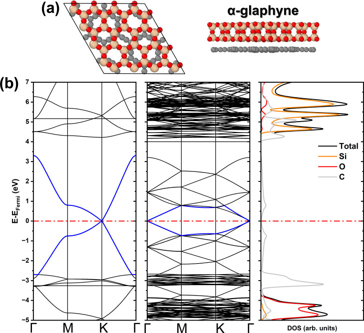
(a) Top and side views of the α-glaphyne unit cell. (b) Electronic band structure of α-graphyne and α-glaphyne, with the corresponding density of states of α-glaphyne.
(a) Top and side views of the β-glaphyne unit cell. (b) Electronic band structure of β-graphyne and β-glaphyne, accompanied by the density of states of β-glaphyne.
(a) Top and side views of the γ-glaphyne unit cell. (b) Electronic band structure of γ-graphyne and γ-glaphyne, with the corresponding density of states of γ-glaphyne.
The α-graphyne presents a zero band gap, featuring a Dirac cone at the K-point (see Figureb). In contrast, α-glaphyne retains a Dirac cone structure, now centered at the Γ-point, while maintaining an almost zero band gap (∼10 meV). The DOS analysis indicates that, although the presence of the SiO_2_ layer alters the band structure, the states near the Fermi level remain predominantly composed of the carbon 2p orbitals. This suggests that the presence of the silica monolayer induces only minor perturbations in the electronic structure of α-graphyne. Nevertheless, silicon contributes significantly to the conduction band through its 3d orbitals, while the valence band remains mainly composed of states from carbon and oxygen atoms.
In the case of β-graphyne, Figureb shows that the system exhibits a Dirac cone along the Γ–M path, maintaining a zero band gap, consistent with previously reported theoretical results. ?,? In contrast, β-glaphyne exhibits a direct band gap at the Γ-point, with a value of 1.16 eV, while preserving the overall band structure, albeit with slight band flattening, which leads to reduced electron mobility. This gap opening in the SiO_2_/β-graphyne heterostructure follows the same qualitative trend reported for glaphene,? where the interaction with the SiO_2_ layer substantially alters the electronic character of an initially gapless carbon lattice. DOS calculations indicate that the primary contributors to the band gap are the C 2p orbitals in the valence band. Nevertheless, the conduction band is mainly composed of C 2p orbitals, with additional contributions from Si 3d orbitals. Oxygen contributes only to deeper energy levels within the conduction band. Despite substantial modifications in the electronic structure, the system retains the symmetry of its pristine configuration. The band gap opening may result from the strain induced in the C–C bonds due to the lattice mismatch. However, although this strain did not lead to structural deformation and the overall symmetry was preserved, the single C–C bonds experienced more significant stretching. Additionally, interlayer interactions play a crucial role, further contributing to the observed variation in the band gap.
To further validate the electronic-structure description provided by DFTB, we benchmarked the β-glaphyne system using density functional theory (DFT) calculations in the CRYSTAL software? with PBE, BLYP, HSE06, B3LYP, and B3LYP-D3 functionals. The lattice parameter obtained with DFTB (matsci-0–3), a = 10.27 Å, differs by less than 1% from the DFT values, which range from 10.17 to 10.23 Å, and the C–C and CC bond lengths predicted by DFTB (1.58, 1.25, and 1.58 Å) remain within approximately 5% of the corresponding DFT intervals of 1.54–1.58, 1.25–1.27, and 1.51–1.58 Å. The Si–O bond obtained with DFTB (1.60 Å) deviates by at most about 2% from the DFT range of 1.58–1.60 Å, and the SiO_2_/β-graphyne separation (3.22 Å) lies within roughly 6% of the DFT values, which span 3.05–3.11 Å. Regarding the electronic properties, the semilocal functionals PBE and BLYP predict β-glaphyne to be metallic, while the hybrid functionals open finite band gaps in the range 0.63–1.14 eV (HSE06, B3LYP, and B3LYP-D3). Despite these quantitative differences, the band structures obtained with the hybrid functionals and with DFTB exhibit the same overall dispersion and consistently show a direct gap at the Γ point induced by the SiO_2_ overlayer. Within the hybrid-DFT interval, DFTB predicts a gap of 1.16 eV, in very close agreement with the B3LYP-D3 result, a hybrid functional known for its high accuracy in carbon- and silicon-based systems, indicating that DFTB captures both the correct qualitative trend of band gap opening upon silica deposition and a quantitatively consistent estimate relative to the most accurate electronic-structure treatment employed here. For completeness, the B3LYP-D3 band structure of β-glaphyne is presented in Figure S3 of the Supporting Information. These results align with benchmark studies of self-consistent-charge (SCC) DFTB for 2D materials, ?,? which demonstrate that DFTB reliably reproduces structural parameters, band topology, and band gap values.
Finally, for the γ-graphyne, a direct band gap of 1.47 eV is observed at the M-point, in good agreement with previous studies. ?,? As shown in Figureb, the presence of SiO_2_ induces a slight modification in the band structure, characterized by a band flattening and an increase in the band gap to 1.51 eV, still located at the M-point. DOS calculations reveal a composition similar to that of α-glaphyne, with the 2p orbitals of carbon atoms dominating the states near the Fermi level. Contributions from silicon and oxygen appear only in deeper energy levels, suggesting that their influence remains confined to lower-lying electronic states.
Another crucial property to examine is the spatial distribution of the Frontier orbitals, namely the highest occupied crystalline orbital (HOCO) and the lowest unoccupied crystalline orbital (LUCO), which are presented in Figure. For α-glaphyne (Figurea,b) and γ-glaphyne (Figuree,f), both HOCO and LUCO exhibit a relatively delocalized character, with HOCO showing slightly greater localization. In contrast, β-glaphyne displays highly localized and well-defined Frontier orbitals, which may favor improved electron transport. This behavior can be attributed to its stacking configuration, in which the pores of the SiO_2_ and β-graphyne layers are periodically aligned. Additionally, Figurec,d reveal a slight charge redistribution from the graphyne layer to the SiO_2_, further reinforcing the system’s structural integrity and indicating that the electronic characteristics of the pristine graphynes are largely preserved.
Highest occupied crystalline orbital (HOCO) and lowest unoccupied crystalline orbital (LUCO) isosurfaces for glaphynes. Panels (a,b) show the top and side views of α-glaphyne, (c,d) correspond to β-glaphyne, and (e,f) to γ-glaphyne, respectively. Blue and yellow isosurfaces represent positive and negative charge densities.
In order to unveil and compare the behavior of glaphynes with that of glaphenes, Figure presents the phonon density of states (DOS) of α-, β-, and γ-glaphyne. This analysis aims to better understand the vibrational behavior and, in particular, to verify the presence of Si–O–C bonding features, similar to those reported by Iyengar et al.? Interestingly, although the three glaphyne structures share common characteristics, such as the presence of both low- and high-frequency vibrational modes, distinct differences emerge in the distribution and intensity of the phonon peaks.
Phonon density of states of α-, β-, and γ-glaphyne. The red dotted region highlights the Si–O–C frequency range.
The α-glaphyne exhibits well-defined and intense peaks across several frequency regions, with a notable concentration in the high-frequency range (900–1200 cm^–1^). In contrast, β-glaphyne shows a more dispersed peak distribution, with generally lower intensities, yet retains a well-marked high-frequency region with a slightly different spectral profile. Meanwhile, γ-glaphyne presents behavior similar to β-glaphyne in the low-frequency range, with less pronounced peaks than α-glaphyne, while still displaying distinct vibrational features across the spectrum. The similarities between α- and γ-glaphynes may be attributed to their larger unit cells and the periodic alignment of the pores in the stacking configuration.
A key spectral region in the phonon DOS lies between approximately 950 and 1150 cm^–1^, which is primarily associated with Si–O–C bonding. Vibrational modes in this range are typically attributed to Si–O and C–O bond stretching, as well as coupled modes involving all three elements. In α-glaphyne, pronounced peaks appear near 980 and 1080 cm^–1^, indicating strong Si–O–C bonding. The intensity and position of these peaks serve as signatures of the formation and structural stability of such bonds, resembling those observed in glaphene.?
Notably, while the phonon DOS profiles of glaphynes and glaphene share some similarities, the G-band and the D′-like shoulder appear shifted toward higher frequencies. This displacement may be attributed to the parametrization adopted in this study, which, as with most parametrizations, is tuned for general-purpose simulations and may introduce deviations in specific vibrational modes. When considered alongside the Mulliken population analysis, the phonon data strongly suggest that β-glaphyne exhibits the most pronounced Si–O–C bonding character. This is evidenced by a distinct peak at 1132 cm^–1^ and the emergence of a secondary shoulder in the same region, reinforcing the relevance of the Si–O–C bond formation to the structural and electronic properties of the system. For comparison, in the case of glaphene, a characteristic frequency associated with the same interaction is observed around 1050 cm^–1^.?
Conclusions
Using the DFTB methodology, we proposed a class of materials referred to as glaphynes, similar to glaphenes. The comparative analysis of α-, β-, and γ-glaphyne reveals distinct electronic modifications induced by the growth of SiO_2_, with potential implications for functional material applications. While α-glaphyne retains a nearly zero band gap and exhibits a redistribution of electronic states near the Fermi level, β-glaphyne undergoes a significant band gap opening of 1.16 eV, accompanied by band flattening that may reduce electron mobility. In contrast, γ-glaphyne maintains a direct band gap, which increases slightly to 1.51 eV upon SiO_2_ incorporation, suggesting nonpronounced quantum confinement effects.
These findings demonstrate that structural modifications achieved through heterostructure engineering enable the precise tuning of electronic properties, thereby influencing charge transport and band characteristics. The band gap variations and orbital rearrangements observed in these systems highlight the potential of glaphyne-based heterostructures for use in nanoelectronic devices, semiconducting applications, and surface-mediated catalysis, where control over band alignment and electronic coupling is essential.
In summary, we have observed that in the case of glaphynes, the electronic proximity effect can indeed open the electronic band gap, but not for all cases, even with the formation of Si–O–C bonds. These results, combined with the distinct electronic responses observed for the three phases, suggest that the SiO_2_/graphyne interface offers a versatile platform for modulating band alignment and tailoring electronic behavior. Such tunability may be relevant for future device-oriented investigations, particularly in contexts where controlled gap opening and interfacial coupling play a central role in transport, optical, or catalytic functionalities.
Simulation Method
Computational simulations were carried out using the SCC-DFTB approximation,? as implemented in the DFTB+ code.? DFTB+ allows quantum simulations of electronic and structural properties for relatively large systems, offering computational efficiency comparable to traditional tight-binding approaches while achieving accuracy similar to DFT in specific cases. ?−? ? ? ? The methodology relies on a second-order expansion of the Kohn–Sham total energy, as extensively discussed in the literature. ?,? In this study, we employed the matsci-0–3 parametrization,? developed for materials science applications, including graphynes and silicon dioxide nanostructures. Atomic structure visualizations were generated using OVITO,? and orbital representations were obtained with the VESTA code.?
Geometry optimizations were performed using the conjugate gradient algorithm implemented in DFTB+, with convergence criteria set to 10^–8^ a.u. for SCC interactions and 10^–5^ a.u. for atomic forces. The first Brillouin zone was sampled using a 10 × 10 × 1 Monkhorst–Pack grid,? where the 6-fold sampling was applied along the periodic direction of the structures. Dispersion interactions were included through the Lennard–Jones potential,? combined with the universal force field (UFF) parametrization.? Isosurface plots were generated using an isovalue resolution of 0.001 Å^–3^.
The creation of glaphyne heterostructures began with the computational modeling of 2D silica, and the α-, β-, and γ-graphyne structures. This preliminary stage aimed to extract key physical properties and obtain optimized geometries in agreement with reference data. Subsequently, supercells were generated for each graphyne type and the silica structure. The supercell sizes were selected in order to minimize the lattice mismatch between graphyne and silica, thus minimizing strain accumulation at the periodic boundaries. Figure illustrates the created graphyne supercells (in gray), the 2D silica unit cell (in orange), and the resulting glaphyne configurations.
Structural representation of two-dimensional SiO2, including α-, β-, and γ-graphynes (glaphynes). The graphynes are depicted using their respective supercells, while the glaphynes are illustrated through their unit cells, which are highlighted by the orange hexagonal outline.
Supplementary Material
The reference list from the paper itself. Each links out to its DOI / PubMed record.
- 1Novoselov K. S.Geim A. K.Morozov S. V.Jiang D.-E.Zhang Y.Dubonos S. V.Grigorieva I. V.Firsov A. A.Electric field effect in atomically thin carbon films Science 200430666666910.1126/science.110289615499015 · doi ↗ · pubmed ↗
- 2Lee C.Wei X.Kysar J. W.Hone J.Measurement of the elastic properties and intrinsic strength of monolayer graphene Science 200832138538810.1126/science.115799618635798 · doi ↗ · pubmed ↗
- 3Balandin A. A.Thermal properties of graphene and nanostructured carbon materials Nat. Mater.20111056958110.1038/nmat 306421778997 · doi ↗ · pubmed ↗
- 4Baughman R.Eckhardt H.Kertesz M.Structure-property predictions for new planar forms of carbon: Layered phases containing sp 2 and sp atoms J. Chem. Phys.1987876687669910.1063/1.453405 · doi ↗
- 5Li X.Li B.-H.He Y.-B.Kang F.-Y.A review of graphynes: Properties, applications and synthesis New Carbon Mater.20203561962910.1016/s 1872-5805(20)60518-2 · doi ↗
- 6Cranford S. W.Brommer D. B.Buehler M. J.Extended graphynes: simple scaling laws for stiffness, strength and fracture Nanoscale 201247797780910.1039/c 2nr 31644 g 23142928 · doi ↗ · pubmed ↗
- 7Kang J.Wei Z.Li J.Graphyne and Its Family: Recent Theoretical Advances ACS Appl. Mater. Interfaces 2019112692270610.1021/acsami.8b 0333829663794 · doi ↗ · pubmed ↗
- 8Puigdollers A. R.Alonso G.Gamallo P.First-principles study of structural, elastic and electronic properties of α-, β- and γ-graphyne Carbon 20169687988710.1016/j.carbon.2015.10.043 · doi ↗