Dietary Fiber Regulation of Gut Microbiota and Bile Acid Metabolism in Animals: Implications for Animal Nutrition
Jinhua Lai, Jürgen Zentek, Łukasz Marcin Grześkowiak

TL;DR
This review explores how dietary fiber affects gut bacteria and bile acid metabolism in animals, highlighting differences between ruminants and non-ruminants for better livestock management.
Contribution
The paper provides a comprehensive review of the bidirectional interactions between dietary fiber, gut microbiota, and bile acid metabolism in animals.
Findings
Dietary fiber influences gut microbiota and bile acid metabolism differently in ruminant and non-ruminant animals.
Short-chain fatty acids produced by fiber fermentation affect bile acid metabolism through microbial processes.
Inappropriate dietary fiber supplementation can negatively impact gastrointestinal function and health.
Abstract
Dietary fiber plays a crucial role in animal nutrition and gut health, and its digestion and metabolic pathways differ fundamentally between non-ruminant and ruminant animals. This review summarizes the interactions among dietary fiber, gut microbiota, and bile acids in both non-ruminant and ruminant animals, which vary depending on fiber type, source, and dosage. By highlighting these interactions, this review provides foundational information for livestock management, helping farmers and caretakers use dietary fiber more effectively. It also offers preliminary insights into the mechanisms of the dietary fiber–microbiota–bile acid axis, which can guide future research and improve animal health and productivity. Dietary fiber (DF) is a fundamental component of animal nutrition and has been widely studied for its nutritional and physiological functions in animals. While existing studies…
Genes, proteins, chemicals, diseases, species, mutations and cell lines named across the full text — each resolved to its canonical identifier and authoritative record.
Click any figure to enlarge with its caption.
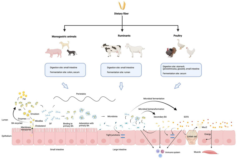 Figure 1
- —China Scholarship Council
Peer Reviews
No public reviews on file for this paper yet. If you reviewed it on a platform where reviews are public (OpenReview, ICLR, NeurIPS, ICML), you can paste yours below so the community can read it here.
Videos
No videos yet. Explain this paper in a talk, walkthrough, or lecture? Add one.
Taxonomy
TopicsRuminant Nutrition and Digestive Physiology · Gut microbiota and health · Animal health and immunology
1. Introduction
Dietary fiber (DF) constitutes an important component of animal nutrition. Numerous studies have demonstrated that DF benefits gut health, metabolism, and immune function in livestock [1]. However, other studies have shown that excessive intake of DF may lead to digestive discomfort, diarrhea, and poor nutrient absorption, resulting in lower performance and even posing health issues [2,3]. Moreover, these phenomena manifest differently among various livestock species. At present, a diverse array of DF is incorporated into livestock diets, encompassing synthetic monosaccharide- or oligosaccharide-based compounds (e.g., polydextrose, fructooligosaccharides), purified plant-derived polysaccharides (e.g., cellulose, lignin, pectin), and structurally complex fiber matrices originating from whole plant sources such as vegetables, fruits, and cereal grains [4]. The complexity and diversity of DF in terms of its sources, types, composition, and physicochemical properties, combined with the presence of other nutrients (e.g., trace elements and vitamins) in natural DF sources, causes significant challenges in investigating the effects of DF on the gut health of livestock.
The animal gastrointestinal tract hosts trillions of microorganisms, including bacteria, archaea, fungi and viruses. These microorganisms establish complex, taxonomically diverse, and metabolically dynamic ecosystems collectively termed the microbiota [5]. The gut microbiota is essential for host development, nutrition, metabolism, physiology, and immunity. It supports these processes by metabolizing nutrients ingested by the host to produce transport proteins, enzymes, and receptor-interacting molecules [6]. The composition of gastrointestinal microbiota varies among different livestock species and is influenced by multiple factors, including animal genetics, location in the gastrointestinal tract, age, sex, diet, and environmental conditions [1]. DF is directly fermented by microorganisms in the gut, significantly impacting the composition of the gut microbiota [7]. Furthermore, the gut microbiota is recognized as a “metabolic organ”, deriving nutrients and energy from DF and producing various essential metabolites, such as short-chain fatty acids (SCFAs) or participating in biotransformation of BAs, which can regulate host metabolism by binding to homologous receptors and mediating related signaling pathways [8].
Bile acids (BAs) are a group of amphipathic steroid acids synthesized from cholesterol in all livestock animals [9]. After synthesis, primary BAs are transported to the intestine, where they are converted into secondary BAs by specific gut bacteria. As signaling molecules, BAs can bind to receptors and perform various physiological functions, including regulation of lipid, glucose, steroid, xenobiotic, and energy metabolism, as well as modulation of the host immune function [6]. There is a bidirectional interaction between BAs and the gut microbiota: the gut microbiota participates in the transformation and synthesis of BAs, while BAs influence the composition of the gut microbiota.
DF not only influences BA metabolism indirectly through microbial activity but can also act directly on BAs via binding and physicochemical interactions. Conversely, BAs can affect the composition of the microbiota, thereby influencing the metabolism of DF within the host. However, the crosstalk among DF, microbiota, and BAs is complex.
DF is a common feed ingredient in livestock diets and plays a critical role in animal health. Based on digestive physiology and feeding characteristics, animals can be categorized into non-ruminant animals and ruminants. Although gastrointestinal structure, digestive capacity, and BA composition differ among species, dietary DF act through similar metabolic pathways. This allows meaningful cross-species synthesis while acknowledging species-specific differences.
Therefore, the objective of this narrative review is to elucidate the effects of DF on different animal species within the framework of the DF–microbiota–BA metabolic axis.
2. Dietary Fiber
DF has a long history as part of carbohydrate fraction within food. But because of the complex structure and chemical compounds of DF, it is difficult to give a precise definition. The word “dietary fiber” was first proposed by Hipsley in 1953, who defined DF as the indigestible ingredient of plant cell wall [10]. However, the definition of DF has changed over many years [11,12,13]. In 2001, the American Association of Cereal Chemists (AACC) described DF as the edible part of plant or analogous carbohydrates that are resistant to digestion and absorption in the human small intestine with complete or partial fermentation in the large intestine [14]. This is a basic concept also useful in animal nutrition research. DF is generally considered to include polysaccharides, oligosaccharides, resistant starch, lignin, and associated plant substances [14,15].
2.1. Physicochemical Properties
DF is one of the plant-based feed sources for animals, providing energy for their growth and development while potentially imposing a digestive burden. The effect of DF depends on its source, type, physicochemical properties, and inclusion level, as well as the animal species and physiological status [16]. Some common DF sources and their composition added to livestock feed are listed in Table 1. Fiber fractions are presented according to established feed analysis systems, which describe soluble/insoluble fiber fractions and structural plant cell wall components and do not directly correspond to the physiological DF definition proposed by AACC.
DF is divided into different types and functions based on their origin, different locations in plant cell walls, chemical structures and components [4]. Moreover, this also determines that different DF have varied physical and chemical properties. The physicochemical characteristics of DF include particle size and bulk volume, surface area characteristics, hydration properties, adsorption or binding of ions and organic molecules, solubility, viscosity and fermentability [1,23].
DF can be classified based on its composition, oligosaccharide/polysaccharide type, physicochemical properties, and physiological role in digestion. However, these classification methods do not fully cover all fiber categories, and generally the most accepted classification of DF is based on its solubility and fermentability. The chemical structure of DF and its interaction with water molecules determine its solubility type. Based on solubility, DF is divided into two categories: soluble dietary fiber (SDF) and insoluble dietary fiber (IDF). The SDF mainly includes fructo-oligosaccharides (FOS), galacto-oligosaccharides (GOS), inulin, β-glucan, pectins, gums, psyllium, and others [24]. The insoluble fraction of DF includes cellulose, partial hemicellulose, and lignin, forming a linear and ordered crystal structure. The different solubility of DF has different effects on the digestion process of nutrients and microbial fermentation metabolism in the body. It has been reported that SDF is generally easily fermentable, whereas IDF is less fermentable by the gut microbiota [25]. In addition, the physicochemical characteristics affect the digestion of DF in animals and produce different physiological effects, for example, feed transit time in gut, fecal production and excretion, digestion and absorption of nutrients, bioavailability of trace elements, composition and metabolism of microorganisms and properties of BAs [11,26]. Lignin, a complex non-carbohydrate phenolic polymer, is particularly recalcitrant due to its highly branched and cross-linked aromatic structure. Unlike polysaccharide-based fibers, lignin is virtually non-fermentable in most non-ruminant animals and can physically encase other fiber components, reducing their digestibility and microbial accessibility [27].
2.2. Summary of Common Dietary Fiber and Its Components
Table 1 provides an overview of commonly used DF sources and ingredients in animal diets, highlighting their respective fiber fractions. SDF generally contribute to gut viscosity and fermentability, whereas IDF are associated with gut motility and fecal bulk. acid detergent fiber (ADF) and neutral detergent fiber (NDF) are measures of plant cell wall components: ADF reflects cellulose and lignin, while NDF includes hemicellulose in addition to ADF. Acid detergent lignin (ADL) indicates the lignin content, which can limit overall fiber fermentability.
The data illustrate considerable variation among fiber sources. For example, sugar beet pulp is notable for its high SDF content, making it a highly fermentable fiber source. In contrast, oat hulls and sorghum hulls exhibit high IDF and NDF levels, indicating a predominance of poorly fermentable structural carbohydrates. By contrast, cereal grains like corn, wheat, and barley contain lower overall fiber levels, with relatively low lignin contents.
2.3. Dietary Fiber Digestion and Fermentation Characteristics in Different Animals
The specificity of digestion of nutrients in different gut parts leads to spatial heterogeneity in the colonization of microorganisms in the intestine [18]. As one of the important sources of nutrients for animals, DF has different digestive characteristics in different animal species.
In non-ruminant animals, DF largely escapes enzymatic digestion in the small intestine and undergoes microbial fermentation predominantly in the hindgut. However, certain fiber-degrading bacteria, such as Ruminococcus, Butyrivibrio, and Prevotella, colonize the stomach and proximal small intestine, partial digestion of DF occurs in the small intestine in pigs [28]. It should be noted that excessive or inappropriate DF supplementation may increase intestinal viscosity, reduce hindgut fermentation, decrease nutrient digestibility, and impair growth performance in pigs, with the magnitude of these effects depending on fiber source and production stage [29].
Adding SDF to the diet of chickens and other poultry can increase the intestinal viscosity, prolong the chyme passage through the gastrointestinal tract, create a hypoxic environment in the intestine, and reduce the growth of pathogenic bacteria [30]. In poultry, SDF can be fermented in the gizzard and duodenum, where the majority of the feed bolus is mixed with enzymes and mechanically ground in the gizzard [30]. Due to the lack of enzymes for degrading non-starch polysaccharides in poultry, the fermentation and utilization of IDF are limited [30]. Therefore, the crude fiber content in commercial diets is typically limited to 2–3%. However, adding a small amount (3–5%) of specific IDF such as cellulose to the poultry feed can improve gizzard function and nutrient utilization, but excessive supplementation may be counterproductive and act as an antinutritional factor, that affects the digestion and metabolism of nutrients by digestive enzymes [31].
In cats and dogs, most of the DF is fermented in the large intestine [32]. However, cats are carnivores with limited cecum and colon length, and certain DF such as cellulose and wheat bran are not extensively fermented [33,34]. Following microbial fermentation, DF serves as an energy source for microbes, supports microbial proliferation and influences chyme retention time in the gastrointestinal tract [32]. It can also contribute to the competitive exclusion of pathogens, stimulate the host’s production of antimicrobial compounds and modulate microbial colonization patterns in the intestines of cats and dogs [32]. However, inappropriate DF type or excessive inclusion, especially in feline diets, may reduce protein and fat digestibility and cause abnormal fecal characteristics or gastrointestinal discomfort [32,35].
Ruminant animals, in contrast to non-ruminants in which DF is mainly fermented in the hindgut after escaping small intestinal digestion, possess a specialized foregut fermentation system, where DF is extensively degraded before entering the lower gastrointestinal tract [36]. However, young ruminants have immature rumens at birth and rely mainly on the abomasum and small intestine for digestion, resulting in limited fiber fermentation. With increasing solid feed intake, rumen microbial communities gradually develop. Adult ruminants such as cattle and sheep have well-developed rumens, which contain a large number of DF-degrading microorganisms [36]. Roughage rich in DF is fermented by rumen microorganisms to produce organic acids and some primary fatty acids, which are absorbed delivering energy for the rumen epithelium and the intermediary metabolism [36]. The physical effectiveness and inclusion level of DF are crucial for maintaining rumen homeostasis. Insufficient fiber supply or imbalance in structural fiber proportion may impair rumination and salivary secretion, decrease ruminal pH, and increase the risk of ruminal metabolic disorders [37].
3. Dietary Fiber–Microbiota Interaction in Different Animals
Table 2 summarizes representative studies investigating how different DF sources modulate gut microbiota composition and, where reported, BA profiles across animal species. The table highlights variations in microbial responses to distinct DF types and provides an overview of associated changes in BA metabolism.
4. Microbial Regulation of Bile Acid Metabolism Across Animal Species
In the intestine, BAs are biotransformed by microbial-derived enzymes. BAs are synthesized by hepatocytes in the liver and stored in the gallbladder. After feed ingestion, they are released into the duodenum to emulsify fat. In the gut, intestinal bacteria metabolize and transform BAs through several processes such as deconjugation, dehydroxylation, oxidation, epimerization, and reconjugation, creating a diverse pool of BAs [6]. The diversity of the BA pool is shaped by the combined enzymatic activities of both the host and microbiota, with variations influenced by the composition of intestinal bacterial species, which differ across host species [92].
Microorganisms can influence gut health by regulating their metabolic products in the intestine, such as by secondary BAs. Studies have shown that secondary BA produced by the gut microbiota can stimulate the expression of P-glycoprotein in intestinal epithelial cells, thereby alleviating inflammation, maintaining colonic epithelial integrity, and mitigating colitis [93]. Additionally, research showed that a physiological level of two secondary BAs, such as deoxycholic acid and lithocholic acid, transformed by clostridia in the intestine, can inhibit the proliferation of harmful bacteria, such as Clostridioides difficile [94,95]. Furthermore, bacterial BA metabolism can promote the generation of peripheral regulatory T cells, contributing to immune regulation [96].
4.1. Types, Function and Physiological Role of Bile Acids
The composition of the BA pool differs among various livestock species. BAs can be detected in numerous biological fluids (such as bile, blood, urine, and follicular fluid), as well as in feces and liver tissue, with varying concentrations of primary BAs in different components. Based on origin, BAs are mainly classified into primary and secondary bile acids. Primary BAs are steroid molecules synthesized in the liver from cholesterol precursors such as cholic acid (CA) and chenodeoxycholic acid (CDCA) through various enzymatic reactions [97]. Secondary BAs are formed in the intestine from conjugated primary BAs through the action of the gut microbiota, undergoing 7α-dehydroxylation and 7α-dehydrogenation. The main secondary BAs include deoxycholic acid (DCA), lithocholic acid (LCA), ursodeoxycholic acid (UDCA), and hyodeoxycholic acid (HDCA) [98]. Primary and secondary BAs form their respective bile salts after conjugation with glycine or taurine. Conjugated BAs are more potent at fat emulsification, helping in fat digestion and energy provision to the host. The conjugated BAs mainly include: glycocholic acid (GCA), glycodeoxycholic acid (GDCA), taurocholic acid (TCA), taurodeoxycholic acid (TDCA), glycochenodeoxycholic acid (GCDCA), taurochenodeoxycholic acid (TCDCA), glycolithocholic acid (GLCA), and taurolithocholic acid (TLCA). Secondary BAs undergo further transformations, including epimerization, sulfation, glucuronidation, and conjugation with N-acetylglucosamine, resulting in a range of further modified bile acids, such as hyocholic acid (HCA), tauroursodeoxycholic acid (TUDCA), and sulfolithocholate (SLC). Additionally, BAs can be classified into two categories based on their structure: free BAs and conjugated BAs. Based on molecular groups, they can be further divided into hydrophilic BAs and hydrophobic BAs [9].
BAs are metabolic and immune signaling molecules that regulate various physiological processes in the body, such as glucose, lipid, steroid, and energy metabolism, by activating different BA receptors [6]. They also possess digestive-promoting, antioxidant, and antimicrobial properties, such as detoxifying endogenous or exogenous harmful compounds like bilirubin, bacterial lipid metabolites (endotoxins), and some inflammatory mediators [99].
4.2. Bile Acid Production and Metabolism
The synthesis of BAs is the primary pathway for cholesterol metabolism in animals. BAs are synthesized from cholesterol through oxidation catalyzed by cytochrome P450 (CYPs), followed by the synthesis of BA-CoA synthetase (BACS) and the conjugation catalyzed by BA-CoA: amino acid N-acyltransferase (BAAT) [6]. Primary BAs can be synthesized through four pathways: the classical pathway, the alternative pathway, the Yamasaki pathway, and the 25-hydroxylation pathway [100]. The classical and alternative pathways are the main routes for BA synthesis. In the classical pathway, cholesterol is hydroxylated by cholesterol 7α-hydroxylase (CYP7A1), which is the rate-limiting enzyme in the BA synthesis process. This is followed by conversion to CA through the actions of microsomal sterol 12α-hydroxylase (CYP8B1) and mitochondrial sterol 27-hydroxylase (CYP27A1). In the absence of CYP8B1, cholesterol is converted to CDCA. The alternative pathway begins with the activation of cholesterol by CYP27A1, converting it into 27α-hydroxycholesterol, and then 27α-hydroxycholesterol is hydroxylated by the rate-limiting enzyme 7α-hydroxylase (CYP7B1) to form CDCA [101].
The metabolism of BAs in the body is mediated via the enterohepatic circulation. In this process, hepatocytes synthesize BAs and secrete them into the bile canaliculi through active transport mechanisms involving key proteins such as the bile salt export pump (BSEP) and multidrug resistance-associated protein 2 (MRP2). The bile in the canaliculi then flows into the bile ducts surrounding the liver lobules and enters the gallbladder or directly into the duodenum. Following feed ingestion, cholecystokinin (CCK) is secreted by enteroendocrine I-cells in the duodenal and jejunal mucosa, stimulating gallbladder contraction and the release of bile into the small intestine to emulsify dietary fats. In the terminal ileum, approximately 95% of BAs are reabsorbed through the apical sodium-dependent BA transporter (ASBT) expressed on the apical membrane of enterocytes. BAs are co-transported with sodium ions through the ASBT into the enterocytes. Subsequently, BAs are transported across the basolateral membrane of enterocytes into the portal circulation via the organic solute transporters alpha and beta (OSTα/OSTβ). BAs are subsequently reabsorbed into hepatocytes through the sodium/taurocholate co-transporting polypeptide (NTCP) and organic anion-transporting polypeptides (OATP) located on the basolateral membrane of liver cells [92,101]. Additionally, other BA transporters in hepatocytes, such as MRP2 on the canalicular membrane and MRP3/MRP4 on the basolateral membrane, can transport BAs into the systemic circulation [102].
Multiple signals are involved in regulating BA metabolism, which affect intestinal immunity and health. Farnesoid X receptor (FXR) is a natural nuclear receptor for BA, highly expressed in the liver, intestine, and kidneys. FXR regulates BA metabolism by inhibiting the expression of rate-limiting enzymes in BA synthesis through three pathways. The first pathway involves BA activating FXR in the liver, which directly suppresses the expression of CYP7A1 [103]. The second pathway involves FXR inducing the expression of small heterodimer partner (SHP), which inactivates liver receptor homolog-1 (LRH-1), a signaling molecule necessary for the expression of CYP7A1, thereby inhibiting its expression [104]. The third pathway involves FXR promoting the binding of fibroblast growth factor 15/19 (FGF15/19) to fibroblast growth factor receptor 4 (FGFR4), which also inhibits the expression of CYP7A1. FGF19 is primarily expressed in the epithelial cells of the distal ileum, and the FXR-FGF19 pathway serves as the main BA sensing axis in intestinal epithelial cells [105].
Takeda G-protein-coupled receptor 5 (TGR5) is a BA receptor located on the cell surface, primarily expressed in the gallbladder and gastrointestinal tract. It plays an important role in regulating gallbladder relaxation and contraction, as well as in BA metabolism and intestinal function [106]. Upon binding with BAs on the surface of intestinal epithelial cells, TGR5 activates adenylate cyclase (AC), cyclic adenosine monophosphate (cAMP), and calcium signaling, leading to the release of glucagon-like peptides-1/2 (GLP-1/2) into the portal vein [107,108]. This represents another important BA sensing pathway in intestinal epithelial cells.
Other BA receptors in the intestine, such as pregnane X receptor (PXR), vitamin D receptor (VDR), and retinoic acid-related orphan receptor gamma t (RORγt), are mainly present in intestinal epithelial cells. Their expression and function are influenced by the intestinal microbiota [102,109]. VDR is a nuclear receptor that primarily regulates intestinal calcium absorption. Studies have shown that VDR plays a role in maintaining intestinal epithelial cell homeostasis. Increased expression of VDR in the mouse intestine can reduce epithelial cell apoptosis and maintain the intestinal mucosal barrier [110]. However, in mice with VDR knockout (KO) in intestinal epithelial cells, the expression of inflammatory factors, including tumor necrosis factor-alpha (TNF-α), monocyte chemoattractant protein-1 (MCP-1), and interleukin-1 beta (IL-1β) is elevated [111]. Moreover, Carnobacterium maltaromaticum, in combination with other microbes, can promote an increase in vitamin D levels and its metabolites in the colon of mice with colorectal cancer (CRC), as well as increase VDR activity in the mucosa, inhibiting CRC [109]. This suggests that VDR may play a potential role in maintaining epithelial cell homeostasis, preserving the intestinal mucosal barrier, and regulating immunity [112].
PXR is an important BA nuclear receptor that senses foreign compounds [113]. Research has shown that in mice receiving microbiota transplantation from ulcerative colitis patients, the expression of VDR and PXR in the colon is downregulated, enhancing inflammation [114]. Additionally, the microbial metabolite indole 3-propionic acid (IPA), as a ligand for PXR, can downregulate TNF-α expression in the intestine. When PXR is absent in the mouse intestine, intestinal barrier damage occurs, and the Toll-like receptor (TLR) signaling pathway is upregulated [115]. PXR is involved in regulating intestinal barrier function, downregulating inflammatory factor expression, and plays a key role in protecting the liver and intestine as well as regulating immunity.
RORγt is a BA nuclear receptor associated with multiple inflammation and immunity-related processes [116]. Studies have found that BA, after being metabolized by the microbiota, can regulate the expression of the transcription factor RORγt in an important population of colonic Foxp3+ regulatory T cells (Tregs), thereby enhancing mucosal immune tolerance and reducing susceptibility to inflammatory bowel disease—an effect mediated in part through other BA receptors such as the vitamin D receptor (VDR) [117]. Further studies have shown that innate lymphoid cells type 3 (ILC3) promote microbiota-specific RORγt+ Tregs and prevent their expansion into inflammatory Th17 cells, thereby defending against aberrant immune responses and inflammation in the microbiota and establishing intestinal tolerance [118].
4.3. Microbial Metabolites Regulate Bile Acid Metabolism and Immunity via Bile Acid-Related Signaling Molecules
SCFAs, as the main products of DF microbial fermentation in the intestine, participate in BA metabolism and immune regulation by modulating multiple BA-related signaling pathways. From the perspective of bile acid metabolism, SCFAs suppress BA synthesis by inhibiting Cyp7a1 through the intestinal FXR-FGF15 axis, thereby improving BA homeostasis [119]. Acetate and propionate activate the G protein-coupled receptors FFAR2 (GPR43) and FFAR3 (GPR41) on intestinal epithelial and enteroendocrine cells, promoting GLP-1 secretion and indirectly regulating bile acid metabolism by modulating intestinal function, energy homeostasis, and bile secretion [120]. Butyrate inhibits histone deacetylase (HDAC) activity and enhances histone acetylation, which increases PXR transcription and promotes PXR-mediated expression of the glucose transporter GLUT2 and P-glycoprotein (ABCB1), accelerating cholesterol metabolism and transport [121]. From an immunological perspective, butyrate activates intestinal VDR expression and related signaling pathways, enhances intestinal epithelial barrier integrity, and exerts anti-inflammatory effects [122]. In addition, butyrate suppresses Th17 cell development by inhibiting RORγt expression and increases IL-10 production in T cells, which may be involved in colitis development [123].
4.4. Microbes Influence Bile Acid Metabolism in the Intestine of Different Animals
Table 3 summarizes representative studies demonstrating the direct involvement of gut microorganisms in BA metabolism across different animal species. The table highlights the role of microbial interventions in regulating BA transformations, including deconjugation, dehydroxylation, and conversion between primary and secondary BA. These findings indicate that the gut microbiota plays a central role in shaping BA pools through enzymatic and metabolic activities.
Free primary BAs in the liver are further modified by the gut microbiota during the BA enterohepatic circulation, and there are various physiological processes of mutual influence between gut bacteria and BAs. However, these physiological processes primarily depend on the expression of functional bacterial proteins related to BA metabolism, including enzymes associated with BA microbial transformation and BA transport proteins [135]. The gut microbiota can promote the secretion of intestinal enzymes, thereby influencing BA synthesis [136]. BSH is mainly found in genera such as Clostridium, Bacteroides, Bifidobacterium, Lactobacillus, Enterococcus, and Listeria. BSH dissociates and binds BAs by hydrolyzing the amide bond between the glycine and taurine parts that are attached to the steroid core of bile salts, which is the critical first step in microbial transformation of BAs [102,103,137,138]. 7α/β-dehydroxylases are mainly found in anaerobic microorganisms in the gut, such as Clostridium and Eubacterium, where they catalyze the 7α- or 7β-dehydroxylation of glycine- or taurine-conjugated BAs like CA and CDCA, converting them into secondary BAs such as DCA and LCA [102]. 3α-/3β-/7α,β-/12α,β-hydroxy steroid dehydrogenases (HSDHs) have been detected in Clostridium species, and these enzymes play a crucial role in BA metabolism by oxidizing or reducing hydroxyl groups at various positions on the BA molecule, thereby altering the structure and activity of BAs [102]. Furthermore, Clostridium species also express the proton-dependent BA transporter BaiG, which mediates the entry of free primary BAs into the bacteria [139]. Additionally, microbes can influence BA metabolism by mediating related BA receptor signaling [140].
As shown in Table 3, in non-ruminant animals, microbial interventions, including fecal microbiota transplantation and targeted probiotic supplementation, have been shown to alter bile acid composition and modulate microbial enzymatic activities related to bile acid metabolism in pigs and poultry. Studies have demonstrated that supplementing piglet diets with Bacillus subtilis fermented liquid enhanced the growth of bile salt hydrolase-active and 7α-dehydroxylase-active bacteria in the intestine, disrupting the normal production of unconjugated BAs [126]. In dogs and cats, Peptacetobacter (Clostridium) hiranonis and certain members of Oscillospirales have been confirmed as key bacterial species responsible for converting primary BAs into secondary BAs. These bacteria contain functional bai operons and influence the composition and function of BA through specific enzymes such as BSH [130,131].
In ruminants, rumen microorganisms and microbial additives participate in bile acid metabolism through diverse enzymatic pathways and microbial enrichment.
4.5. Bile Acids Influence the Composition of Microbiota in Different Animals
Table 4 summarizes representative studies investigating how BA interventions, including exogenous BAs, BA extracts, and BA receptor agonists, influence gut microbiota and host physiological responses in different animal species. The table highlights BA-mediated antimicrobial effects, receptor signaling pathways, and shifts in microbial community structure associated with altered BA pools. These findings reveal the bidirectional interaction between bile acids and the gut microbiota within the DF–microbiota–BA axis.
BAs influence gut microbial composition through multiple interconnected mechanisms. BAs exert direct antimicrobial effects by disrupting bacterial membranes and inhibiting the growth of bile-sensitive taxa, thereby selectively shaping microbial communities [8]. BAs can modulate the intestinal environment and nutrient availability, indirectly affecting the competitive fitness of different microbial populations [8]. In addition, BA signaling via host receptors, such as FXR and TGR5, regulates intestinal immune responses and barrier function, creating ecological niches that favor specific bacterial taxa [8,154]. The effects of BAs on gut microbiota are highly dependent on BA species, dosage, dietary context, and host physiology. Together, these mechanisms contribute to species-specific and diet-dependent remodeling of gut microbial communities.
As shown in Table 4, in non-ruminant animals, supplementation with specific bile acids or bile acid-related compounds generally modulated the abundance of dominant bacterial taxa, including Prevotella, Lactobacillus, and Bifidobacterium, and affected overall microbial diversity. However, a study also reported no significant effects of dietary bile acid supplementation on gut microbiota in pigs [144], which may be related to differences in bile acid type and dosage. In poultry, bile acid supplementation under different dietary conditions produced distinct effects. For example, under low-fat diets, supplementation with HCA, HDCA, and CDCA increased the abundance of Akkermansia in the cecum, whereas under high-fat diets, bile acid supplementation reduced the relative abundance of Bacteroides and increased Bifidobacterium, Escherichia coli, and Lactobacillus [149]. In broilers, bile acid supplementation was frequently associated with reduced colonization of pathogenic bacteria, such as Clostridium perfringens and Campylobacter jejuni, and enrichment of beneficial genera [146,147,148].
In ruminants, BA supplementation altered the relative abundance of BA- and DF-responsive bacteria, including decreased Prevotella and Treponema and increased Akkermansia. In addition, microbiota-derived BA, such as ursodeoxycholic acid, have been shown to inhibit antibiotic-resistant bacteria, including ESBL-producing Escherichia coli, in dairy calves [100].
5. The Mechanisms by Which Dietary Fiber Affects Bile Acid Metabolism
DF affects BA metabolism in livestock animals through various mechanisms. DF has the ability to adsorb BAs, influencing their reabsorption in the animal body and increasing the excretion of BAs in feces [155]. Additionally, DF can alter the passage rate, thus influencing the excretion of BAs from the gut. Furthermore, DF can modify the microbial composition in the animal intestine, affecting the enzymatic activity of bacteria involved in BA conversion, thereby influencing the microbial transformation of BAs [156,157].
5.1. Binding of Dietary Fiber and Bile Acids
DF can directly influence the structure and composition of BAs by binding to them. The binding of DF to primary BAs is an effective method to reduce cholesterol levels in the blood, while binding to secondary BAs can reduce the toxic effects of these metabolites on colon cells. Studies have shown that different bile salts have varying binding affinities for pectin, with the order being GCA > GDCA > CA > DCA > CDCA > GCDCA. As for cellulose, its binding affinity to bile salts is in the order of GDCA > CA > GCA > DCA > CDCA > GCDCA [100].
After mixing citrus pectin with BA, an endothermic reaction occurs, and the concentration of BA decreased. This result suggests that there is some interaction between BA and pectin [158]. Current research indicates several possible mechanisms of interaction between SDF and BA. It may be due to the nonpolar surface of BA binding to the nonpolar methyl groups on the pectin chains [159]. Both BAs and pectin have hydrophobic groups, which may mediate adsorption through hydrophobic interactions [160]. The interaction between BAs and DF, but also the viscous chyme matrix formed by DF, can encapsulate bile salt micelles [161,162].
IDF has a lower BA-binding capacity and primarily plays an adsorptive role for BA [163]. Some studies have suggested that the interaction between DF and bile salts depends on the physicochemical properties of the fiber, including particle size and surface area, as well as the quantity and type of substituents, rather than the molecular weight of the fiber [164]. However, the adsorption capacity of lupin cotyledons, which is rich in both cellulose and pectin, for primary BAs is not related to the protein and DF content within them [160]. Therefore, the specific mechanisms of interaction between DF and BAs still need to be further elucidated.
5.2. Dietary Fiber Affects Bile Acid Metabolism Through the Microbiota in the Intestine
DF can influence the composition of gut microbiota, and certain microorganisms are involved in the conversion of secondary BAs. Therefore, DF can indirectly affect BA metabolism by altering the microbial composition. Table 2 presents examples of studies demonstrating how DF feeding in livestock leads to changes in specific microorganisms and BAs. Furthermore, studies in mice have shown that inulin fiber promoted microbiota-derived BAs and triggered inflammation [165]. Fructooligosaccharides have also been found to regulate the gut microbiome by enriching bacteria involved in the production of 6α-hydroxy BA, thereby activating the TGR5-GLP1R axis [157]. However, there is limited research on how DF influences BA metabolism through gut microbiota in livestock animals, and further studies are needed to clarify it.
6. Implications for Dietary Fiber Application Based on Microbial and Bile Acid Alterations
While studies on the DF–microbiota–BA axis improve our understanding of underlying biological mechanisms, such findings alone are not adequate to establish practical dietary fiber recommendations for animal nutrition without integrating performance and health-related outcomes.
In non-ruminant animals, mostly studied in pigs, supplementation with different types and levels of DF consistently induced alterations in gut microbial structure. Most studies indicated that low to moderate DF inclusion (approximately 0.05–15%) was associated with increased abundances of bacteria related to gut health, including Lactobacillus, Bifidobacterium, Prevotella, and Faecalibacterium. Meanwhile, reductions in Helicobacter, Desulfovibrio, Proteus, and some Clostridium taxa were also observed, suggesting a potential inhibitory effect on unfavorable microbial populations. Regarding BA metabolism, only a limited number of studies in pigs evaluated related parameters. These studies reported changes in BA composition, such as decreased CA and GDCA and increased UDCA and THCA, as well as shifts in BA proportions. However, current evidence mainly reflects alterations in BA profiles, and their physiological implications remain unclear.
In poultry, low-dose supplementation with functional fibers or oligosaccharides (e.g., FOS, MOS, and AXOS, approximately 0.05–0.5%) was commonly associated with increased abundances of Lactobacillus and Bifidobacterium, along with reductions in potential pathogens such as Escherichia coli, Salmonella, Campylobacter, and Clostridium perfringens. However, in a few high-fiber treatments (approximately 6–8% DDG and cereal by-products), increased abundances of Escherichia coli and Campylobacter were observed, indicating potential adverse effects at higher inclusion levels. Some studies also reported changes in BA composition, including alterations in CA, TCA, and DCA and reductions in LCA. These findings mainly reflect BA redistribution, and their functional relevance requires further investigation.
Studies in dogs showed that supplementation with fermentable or mixed DF (approximately 0.5–8%) was generally associated with increased abundances of Bifidobacterium, Lactobacillus, and Faecalibacterium, along with decreased Fusobacterium. Under some alternative formulations or high-protein/high-fiber conditions, reductions in Bifidobacterium or Prevotella were also reported. Regarding BA, some studies reported decreases in CA and DCA and increases in UDCA following DF or high-fiber diets, whereas high-dose prebiotic supplementation was occasionally associated with increased DCA and LCA, indicating dose-dependent effects. Most studies focused on DF inclusion levels between 1% and 8%. Higher levels (e.g., 25% grain-based diets) were mainly applied under specific experimental conditions, and their long-term safety remains uncertain. Data on cats remain limited.
In ruminants, DF is mainly supplied in the form of roughage or fibrous by-products. Moderate to relatively high DF supplementation (e.g., 10% alfalfa, 15–30% FDG, or high-NDF diets) was generally associated with increased abundances of fiber-degrading bacteria, including Ruminococcus, Prevotella, and Butyrivibrio, reflecting enhanced ruminal fiber fermentation. Some studies also reported decreases in Clostridium, Blautia, or Bacteroides, suggesting that changes in fiber structure and proportion may affect specific commensal populations. DF supplementation was associated with alterations in BA composition, including decreased CA, CDCA, and TCA and increased DCA and TDCA, mainly reflecting BA redistribution. However, direct functional evidence remains limited.
Based on the data collected in this review, most included studies mainly focused on changes in gut microbiota composition and BA profiles, whereas comprehensive health and production indicators, such as nutrient digestibility, growth performance, immune status, and metabolic responses, were not consistently evaluated. Therefore, in practical feeding and diet formulation, DF inclusion levels should be adjusted dynamically according to species, physiological stage, and fiber characteristics, as well as potential synergistic effects with other nutrients, and evaluated comprehensively using production performance and health status.
7. Conclusions
As summarized in Figure 1, this review comprehensively synthesizes the current understanding of DF in animal nutrition, focusing on its complex interplay with the gut microbiota and BA metabolism. DF is a fundamental component of animal diets, profoundly influencing gut health, overall metabolism, and immune function across various species. A significant insight from this review is the bidirectional relationship between BAs and the gut microbiota; the microbiota actively participates in BA transformation, and conversely, BAs influence the composition of the gut microbiota. Furthermore, DF not only impacts BA metabolism indirectly through microbial activity but also exhibits direct interactions with BAs. Despite extensive research, the precise and intricate mechanisms governing the crosstalk among DF, gut microbiota, and BA metabolism remain to be fully elucidated. Therefore, future research should aim to unravel these complex molecular and physiological pathways in greater detail.
The reference list from the paper itself. Each links out to its DOI / PubMed record.
- 1Holscher H.D. Dietary fiber and prebiotics and the gastrointestinal microbiota Gut Microbes 2017817218410.1080/19490976.2017.129075628165863 PMC 5390821 · doi ↗ · pubmed ↗
- 2Wang W. Dernst A. Martin B. Lorenzi L. Cadefau-Fabregat M. Phulphagar K. Wagener A. Budden C. Stair N. Wagner T. Butyrate and propionate are microbial danger signals that activate the NLRP 3 inflammasome in human macrophages upon TLR stimulation Cell Rep.20244311473610.1016/j.celrep.2024.11473639277863 · doi ↗ · pubmed ↗
- 3Zimmermann C. Wagner A.E. Impact of Food-Derived Bioactive Compounds on Intestinal Immunity Biomolecules 202111190110.3390/biom 1112190134944544 PMC 8699755 · doi ↗ · pubmed ↗
- 4Gill S.K. Rossi M. Bajka B. Whelan K. Dietary fibre in gastrointestinal health and disease Nat. Rev. Gastroenterol. Hepatol.20211810111610.1038/s 41575-020-00375-433208922 · doi ↗ · pubmed ↗
- 5Wan T. Wang Y. He K. Zhu S. Microbial sensing in the intestine Protein Cell 20231482486010.1093/procel/pwad 02837191444 PMC 10636641 · doi ↗ · pubmed ↗
- 6Cai J. Rimal B. Jiang C. Chiang J.Y.L. Patterson A.D. Bile acid metabolism and signaling, the microbiota, and metabolic disease Pharmacol. Ther.202223710823810.1016/j.pharmthera.2022.10823835792223 · doi ↗ · pubmed ↗
- 7So D. Whelan K. Rossi M. Morrison M. Holtmann G. Kelly J.T. Shanahan E.R. Staudacher H.M. Campbell K.L. Dietary fiber intervention on gut microbiota composition in healthy adults: A systematic review and meta-analysis Am. J. Clin. Nutr.201810796598310.1093/ajcn/nqy 04129757343 · doi ↗ · pubmed ↗
- 8Wahlstrom A. Sayin S.I. Marschall H.U. Backhed F. Intestinal Crosstalk between Bile Acids and Microbiota and Its Impact on Host Metabolism Cell Metab.201624415010.1016/j.cmet.2016.05.00527320064 · doi ↗ · pubmed ↗