An Analysis of Mandibular Characteristics According to Biological Sex Using Three-Dimensional Computed Tomography Scans in Koreans: A Retrospective and Observatoinal Study
Byeongjun Kim, Junghyun Lee, Donghyun Lee, Kuylhee Kim, Jiwon Jeong, Soyeon Jung

TL;DR
This study uses 3D CT scans to identify mandibular differences between biological sexes in Koreans, aiding facial feminization surgery planning.
Contribution
Provides population-specific mandibular morphological data for Korean individuals to support gender-affirming surgical planning.
Findings
Males had significantly larger mandibular angles, lengths, and widths compared to females.
Females predominantly showed pointed chins and straight inferior mandibular borders, while males had round/square chins and rocker-shaped borders.
Sexual dimorphism in the Korean population includes angularity, transverse width, and chin morphology differences.
Abstract
Background and Objectives: With the increasing demand for gender-affirming procedures, facial feminization surgery (FFS) has become an essential component in the management of patients with gender dysphoria. In this study, ‘male’ and ‘female’ refer to biological sex as recorded in the medical record; gender identity was not assessed. The mandible is widely recognized as one of the most sexually dimorphic facial bones and plays a critical role in defining masculine and feminine facial contours. However, quantitative mandibular data directly applicable to surgical planning for FFS, particularly in Asian populations, remain limited. The purpose of this study was to analyze gender differences in mandibular morphology using three-dimensional (3D) computed tomography (CT) images and to provide clinically relevant anatomic data applicable to mandibular contouring in FFS. Materials and Methods:…
Genes, proteins, chemicals, diseases, species, mutations and cell lines named across the full text — each resolved to its canonical identifier and authoritative record.
Click any figure to enlarge with its caption.
 Figure 1
 Figure 2
 Figure 3
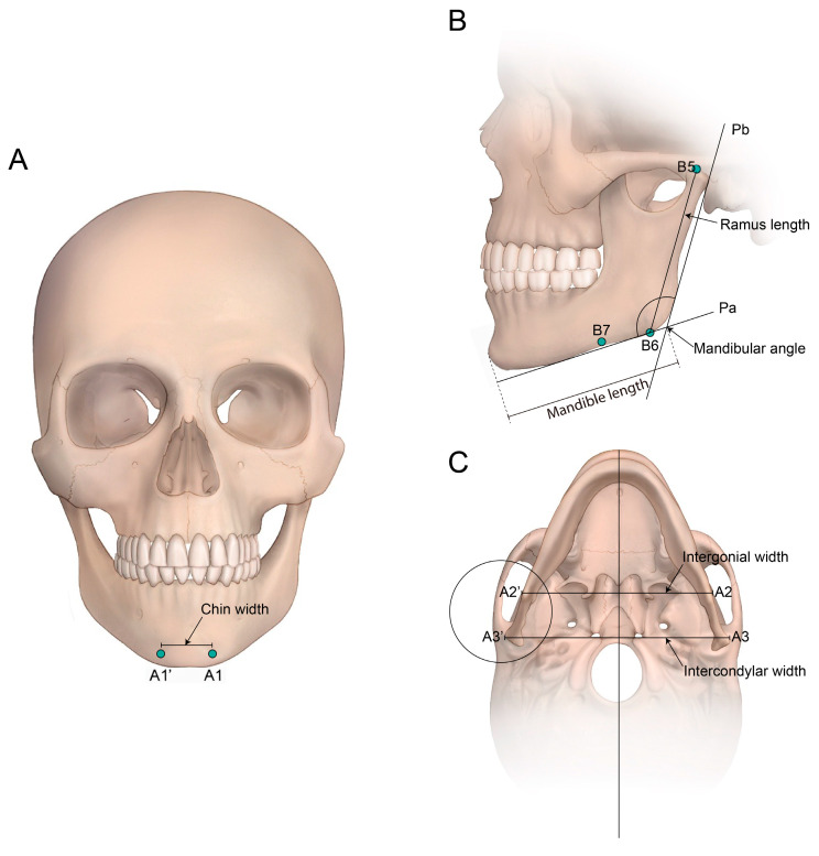 Figure 4
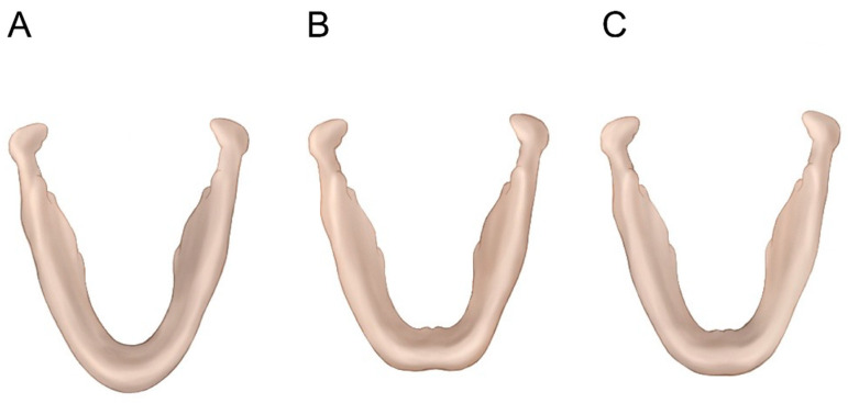 Figure 5
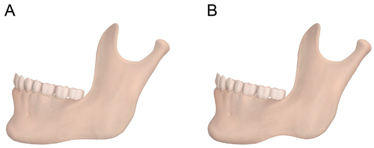 Figure 6
Peer Reviews
No public reviews on file for this paper yet. If you reviewed it on a platform where reviews are public (OpenReview, ICLR, NeurIPS, ICML), you can paste yours below so the community can read it here.
Videos
No videos yet. Explain this paper in a talk, walkthrough, or lecture? Add one.
Taxonomy
TopicsDental Radiography and Imaging · Facial Rejuvenation and Surgery Techniques · Orthodontics and Dentofacial Orthopedics