Quantitative Characterization of Microfiltration Membrane Fouling Using Optical Coherence Tomography with Optimized Image Analysis
Song Lee, Hyongrak Cho, Yongjun Choi, Juyoung Andrea Lee, Sangho Lee

TL;DR
This paper introduces a new method using optical coherence tomography and image analysis to measure and track membrane fouling in real time, improving diagnostics and system control.
Contribution
A reproducible OCT image-analysis workflow with automated segmentation and benchmarked thresholding algorithms for quantitative membrane fouling diagnostics.
Findings
The Triangle–Moments thresholding combination was identified as the most robust for OCT image analysis.
OCT-derived thickness measurements closely matched SEM results for humic-acid fouling.
The method revealed flux loss periods with minimal thickness growth, indicating permeability and compaction changes.
Abstract
Membrane fouling reduces permeate flux and treatment efficiency, yet most diagnostic methods are destructive and require offline analysis. Optical coherence tomography (OCT) enables in situ, real-time visualization; however, quantitative image extraction of thin foulant layers is often limited by manual processing and subjective thresholding. Here, we develop a reproducible OCT image-analysis workflow that combines band-pass filtering, Gaussian smoothing, and unsharp masking with a dual-threshold subtraction strategy for automated fouling-layer segmentation. Seventeen global thresholding algorithms in ImageJ (289 threshold pairs) were benchmarked against SEM-measured cake thickness, identifying Triangle–Moments as the most robust combination. For humic-acid fouling, the OCT-derived endpoint thickness (14.23 ± 1.18 µm) closely agreed with SEM (15.29 ± 1.54 µm). The method was then…
Genes, proteins, chemicals, diseases, species, mutations and cell lines named across the full text — each resolved to its canonical identifier and authoritative record.
Click any figure to enlarge with its caption.
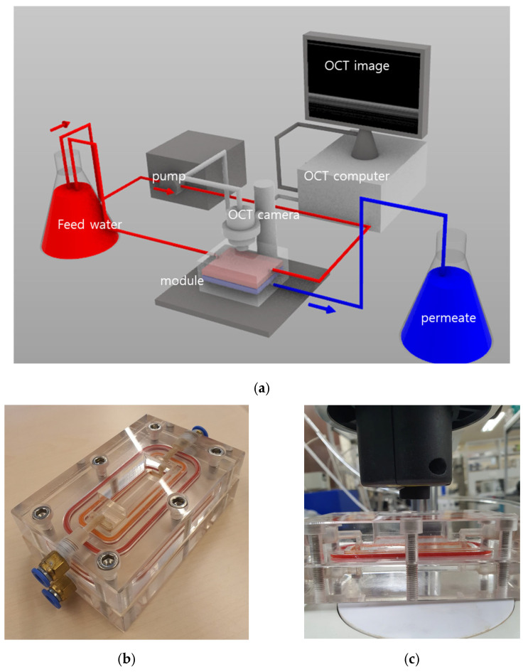 Figure 1
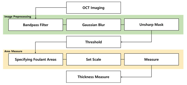 Figure 2
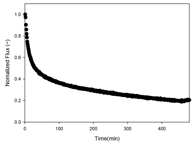 Figure 3
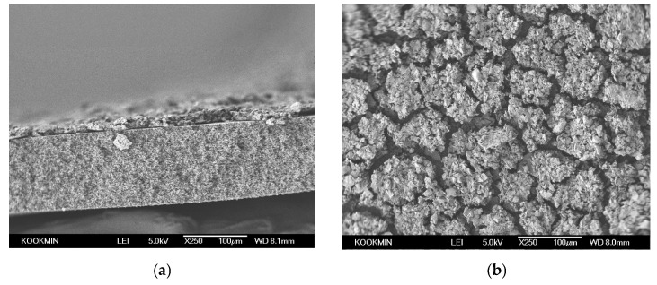 Figure 4
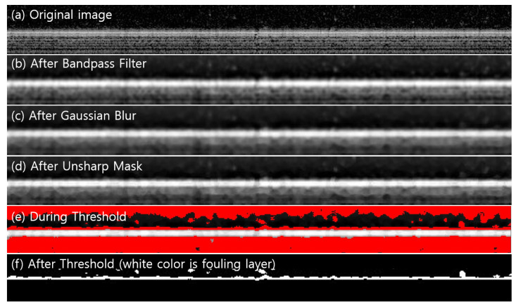 Figure 5
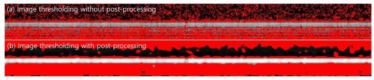 Figure 6
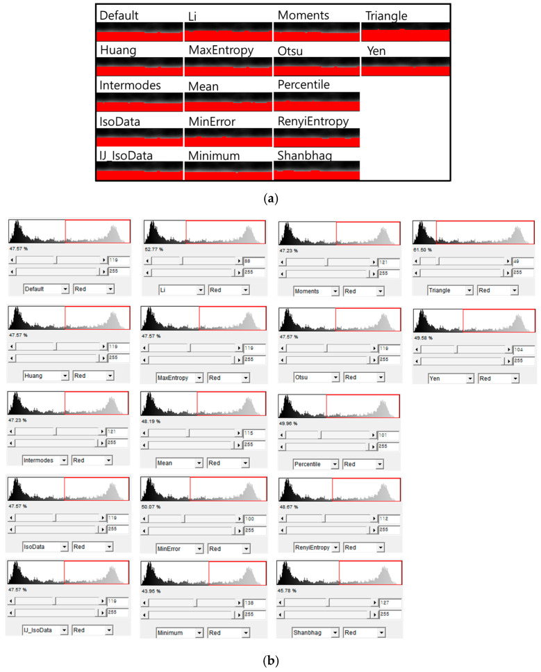 Figure 7
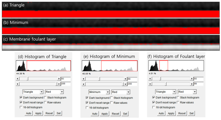 Figure 8
 Figure 9
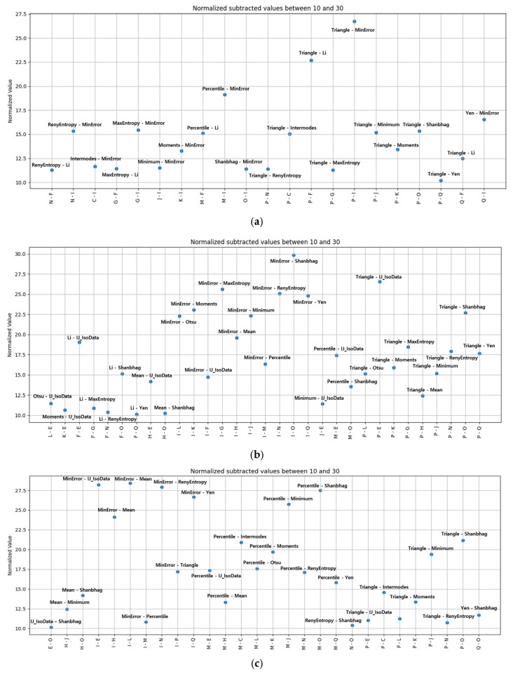 Figure 10
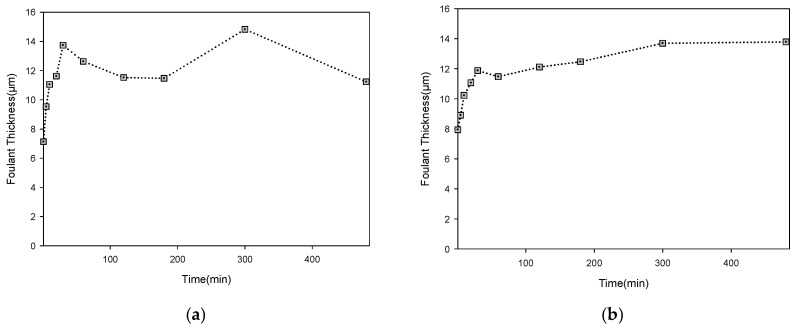 Figure 11
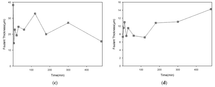 Figure 12
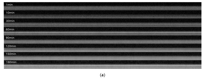 Figure 13
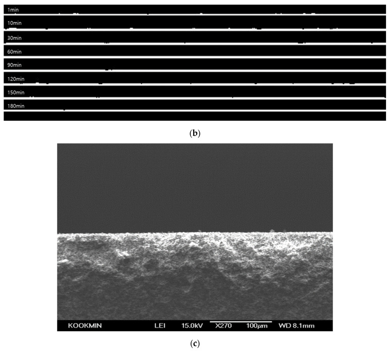 Figure 14
 Figure 15
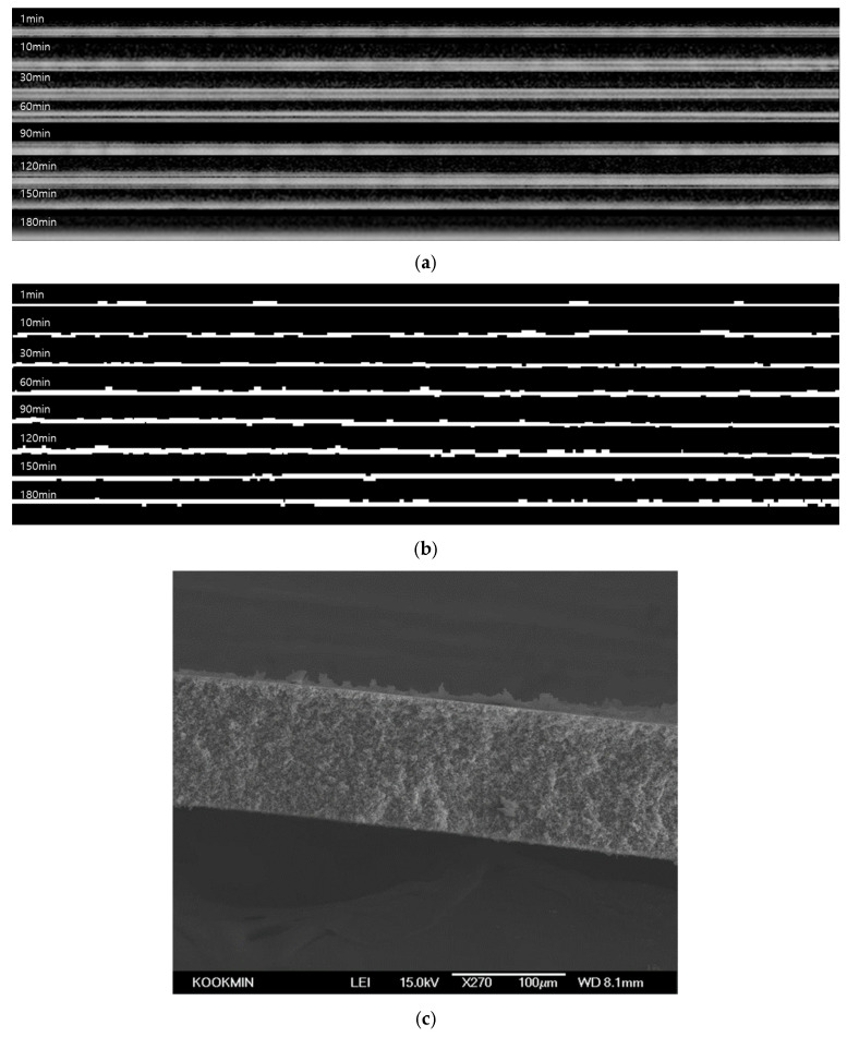 Figure 16
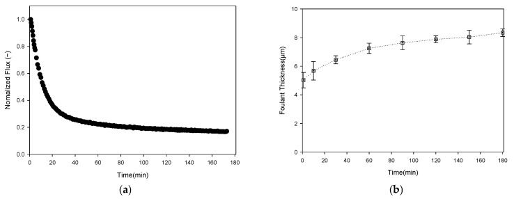 Figure 17
Peer Reviews
No public reviews on file for this paper yet. If you reviewed it on a platform where reviews are public (OpenReview, ICLR, NeurIPS, ICML), you can paste yours below so the community can read it here.
Videos
No videos yet. Explain this paper in a talk, walkthrough, or lecture? Add one.
Taxonomy
TopicsMembrane Separation Technologies · Wound Healing and Treatments · Nanocomposite Films for Food Packaging