Social stress worsens colitis through β-adrenergic–driven oxidative stress in intestinal mucosal compartments
Maria Elisa Caetano-Silva, Miranda E. Hilt, Ivan Valishev, Casey Lim, Mikaela Kasperek, Akriti Shrestha, Helen Fu, Eleanor Eck, Robert McCusker, Heather Armstrong, Brett Loman, Michael T. Bailey, Jacob M. Allen

TL;DR
This study shows that social stress worsens gut inflammation through β-adrenergic and oxidative stress pathways, offering new therapeutic targets for stress-related inflammatory bowel disease.
Contribution
The study identifies β-adrenergic signaling and NADPH oxidase as key mediators of stress-induced gut inflammation, providing novel therapeutic insights.
Findings
Social stress increases oxidative stress markers like Duox2 and Nos2 in intestinal epithelial cells.
β-adrenergic blockade with propranolol reverses stress-induced inflammation and microbial dysbiosis.
NADPH oxidase inhibition with apocynin reduces stress-related gut inflammation and disease severity.
Abstract
Psychological stress is a known risk factor for inflammatory bowel disease (IBD), but the mechanisms linking stress to worsened disease remain unclear. Because distinct stress paradigms activate different neuroimmune circuits, it is critical to investigate model-specific effects. We examined how social stress primes the gut for heightened inflammation and whether this is mediated by specific neuroendocrine pathways, including α2-/β-adrenergic (sympathetic) or glucocorticoid/ corticotropin-releasing hormone receptor (CRHR1) (HPA axis) signaling. Mice were exposed to social disruption (SDR) stress and pre-treated with pharmacological antagonists targeting α2-adrenergic receptors (idazoxan), β-adrenergic receptor (β-AR) (propranolol), glucocorticoid receptor (mifepristone), or CRHR1 (antalarmin). Intestinal epithelial cell (IEC) gene expression and microbiota composition were assessed…
Genes, proteins, chemicals, diseases, species, mutations and cell lines named across the full text — each resolved to its canonical identifier and authoritative record.
Click any figure to enlarge with its caption.
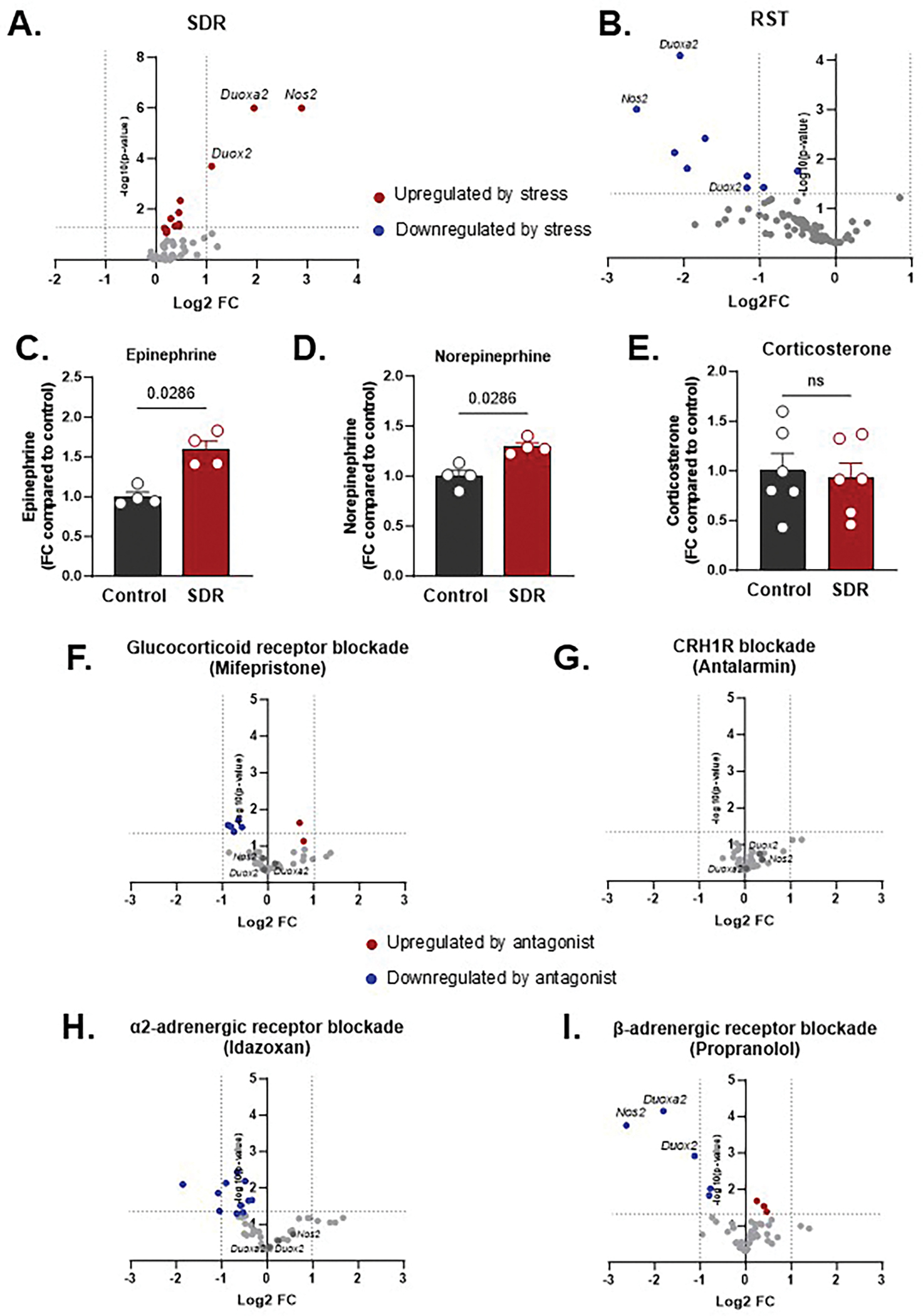 Figure 1
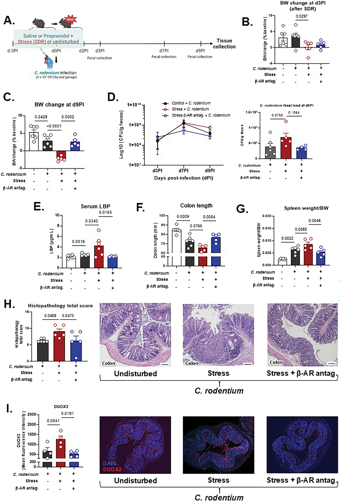 Figure 2
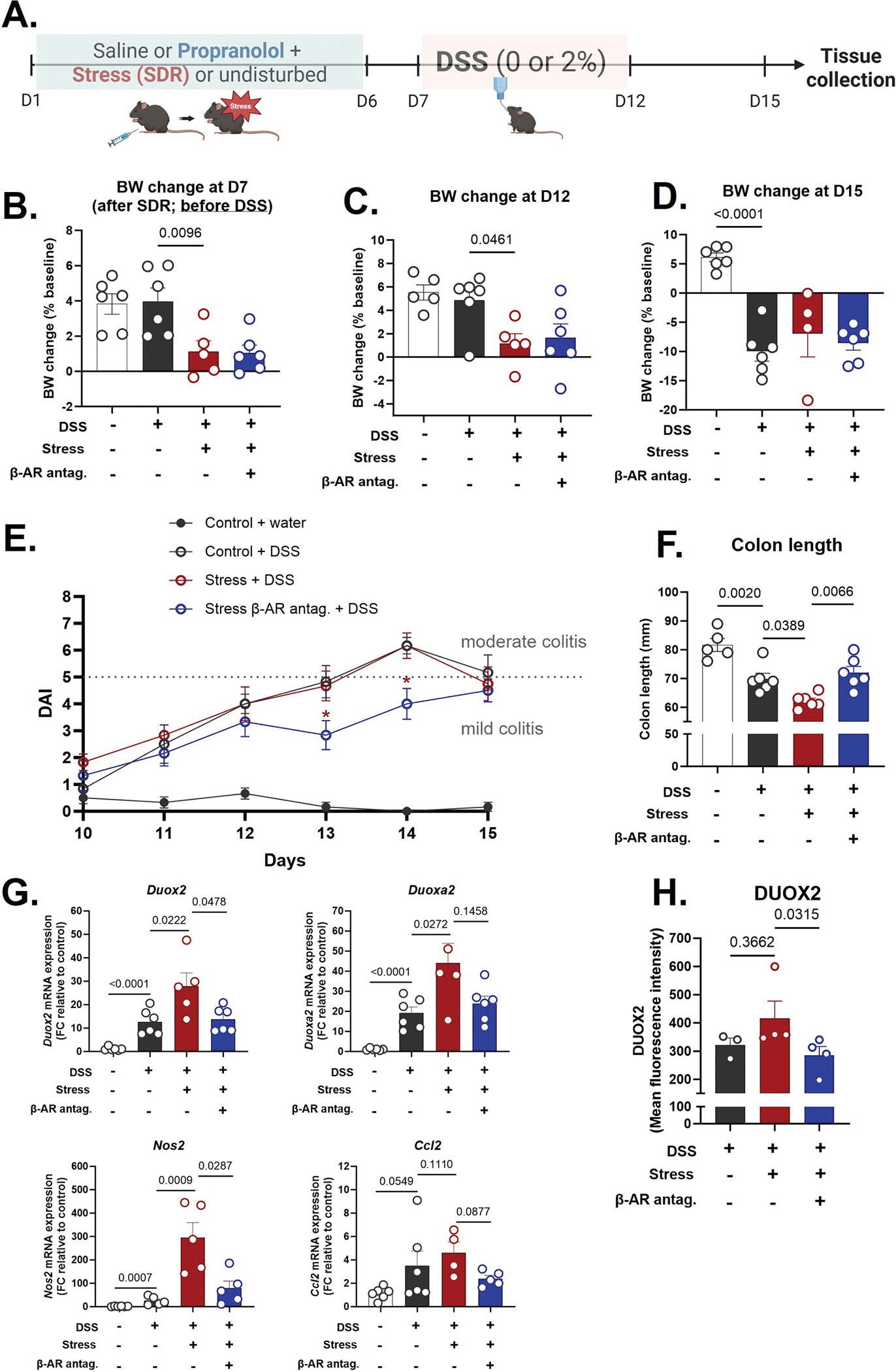 Figure 3
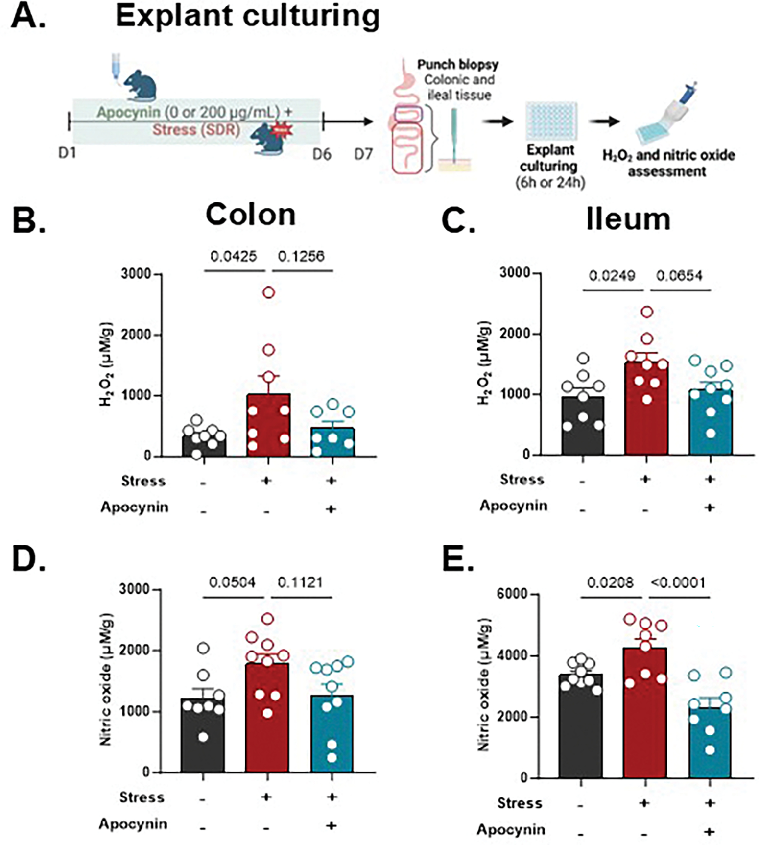 Figure 4
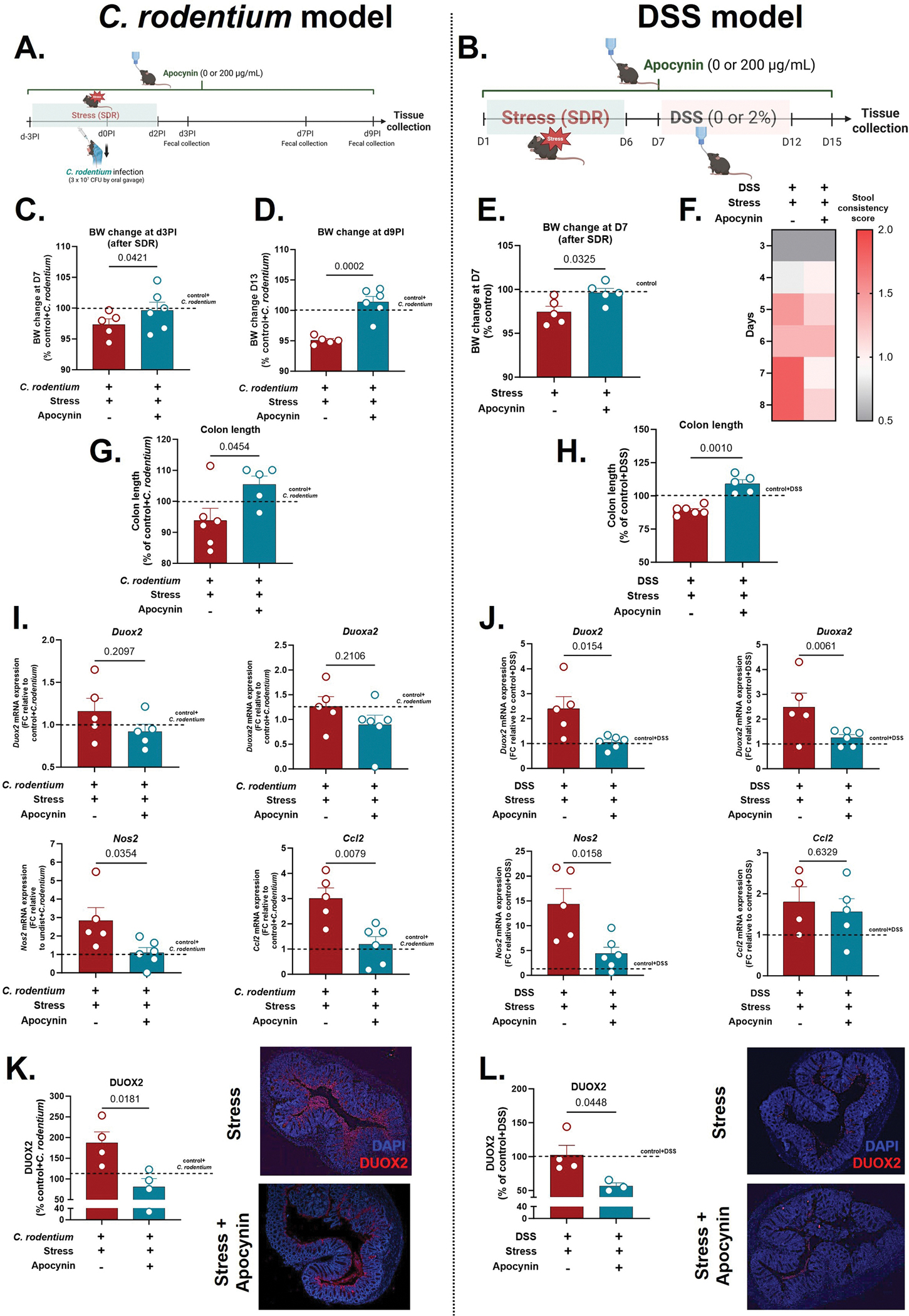 Figure 5
Peer Reviews
No public reviews on file for this paper yet. If you reviewed it on a platform where reviews are public (OpenReview, ICLR, NeurIPS, ICML), you can paste yours below so the community can read it here.
Videos
No videos yet. Explain this paper in a talk, walkthrough, or lecture? Add one.
Taxonomy
TopicsStress Responses and Cortisol · Gut microbiota and health · Gastrointestinal motility and disorders
Introduction
Inflammatory bowel diseases (IBD), including Crohn’s disease and ulcerative colitis, have increased in prevalence in recent decades and represent a significant global health burden (Agrawal and Jess, 2022; Caron et al., 2024). IBD pathogenesis is multifactorial, arising from complex gene-environment interactions, with growing evidence implicating disrupted brain-gut-microbiota communication as a central contributor (Craig et al., 2022). Among environmental risk factors, psychological stress has emerged as a robust and clinically relevant trigger, with perceived stress, trauma, and adverse life events linked to increased disease onset, relapse, and symptom severity (Bitton et al., 2008; Black et al., 2022).
Physiological responses to stress involve activation of two key neuroendocrine pathways: the sympathetic nervous system (SNS), which rapidly releases catecholamines, and the hypothalamic–pituitary–adrenal (HPA) axis, which promotes glucocorticoid secretion (Godoy et al., 2018). Both pathways influence intestinal physiology, but emerging data suggest they may play distinct roles depending on the nature of the stressor. For example, recent work by Schneider et al. (2023) implicates glucocorticoids as primary mediators of stress-induced colitis, yet the generalizability of this mechanism across stress paradigms remains unclear. Social stress models, such as social disruption (SDR), may preferentially engage SNS signaling and provoke unique host responses that differ from those elicited by restraint or early-life stress (Hanke et al., 2012; Lauten et al., 2025; Wohleb et al., 2011; Yadav et al., 2023). Thus, model-specific interrogation of stress signaling is essential to define the biological mechanisms linking stress to IBD.
Intestinal epithelial cells (IECs), which form the frontline of host-microbiota interaction, are increasingly recognized as central players in IBD pathogenesis (Malik et al., 2023). Beyond acting as a physical barrier, IECs secrete cytokines, antimicrobial peptides, and reactive oxygen and nitrogen species (ROS/RNS) to maintain mucosal homeostasis (Castrillón-Betancur et al., 2023; Soderholm and Pedicord, 2019). ROS/RNS generated by NADPH oxidases—particularly DUOX2—and inducible nitric oxide synthase (iNOS), help defend against pathogens but can also disrupt the mucosal barrier when dysregulated. Indeed, DUOX2 and iNOS are consistently elevated in IBD and correlate with disease severity in human cohorts (Burgueño et al., 2021; Haberman et al., 2014; Urbauer et al., 2024).
Our previous work shows that SDR stress upregulates ROS- and RNS-generating pathways in IECs, sensitizing the epithelium to microbial insults (Allen et al., 2022). Yet, the neuroendocrine mechanisms that elevate epithelial ROS tone and how they influence susceptibility to IBD remain poorly understood. In this study, we investigated the role of SNS-driven versus HPA-driven signaling in mediating social stress-induced modifications to mucosal compartments. Using pharmacologic blockade during SDR stress, we identified β-adrenergic signaling as a key driver of DUOX2 and NOS2 upregulation in IECs. Targeting this pathway in both infectious and chemically induced colitis models revealed that inhibiting β-adrenergic receptors (β-AR) or ROS signaling through NADPH oxidases provides effective protection against stress-aggravated colitis. These findings suggest that stress-induced colitis susceptibility may arise through distinct hormonal mechanisms depending on the stress context, with catecholaminergic and ROS pathways playing a dominant role in the SDR model.
Results
Distinct stress paradigms engage divergent neuroimmune pathways to shape ROS/RNS epithelial response and the gut microbiome
2.1.
To investigate how psychological stress impacts intestinal epithelial and microbial homeostasis, we utilized two established murine stress models: SDR and restraint stress (RST). SDR is characterized by daily exposure to an aggressive intruder for 2 h over 6 days, whereas RST involves placing mice in ventilated restrainers for 2 h per day over 6 days. While prior work by the Thaiss group (Schneider et al., 2023) showed that RST worsens colitis via corticosterone-driven mechanisms, our preliminary findings suggested that SDR engages distinct neuroimmune pathways. We therefore hypothesized that SDR and RST differentially regulate IEC responses through divergent upstream mechanisms.
Transcriptomic profiling of IECs (EPCAM+, CD45−) revealed that SDR significantly upregulated oxidative stress–related genes compared to unstressed controls, including Duox2, Duoxa2, and Nos2 (Fig. 1A). In contrast, RST suppressed expression of these genes (Fig. 1B). These findings suggest opposing effects on mucosal redox signaling and implicate ROS/RNS as central to the epithelial stress response in this model. In addition, SDR elevated colonic tissue levels of epinephrine from 1.11 to 1.78 ng/g (a 61 % increase) and norepinephrine from 162.1 to 210.0 ng/mg (a 30 % increase) compared to unstressed controls (Fig. 1C–D), with no change in corticosterone (Fig. 1E), indicating a dominant role for local SNS activation over systemic HPA signaling during SDR. In contrast, catecholamine levels in colonic tissue from RST mice did not differ from unstressed controls (Suppl. Fig. S1A–B). Instead, RST was associated with a significant elevation of corticosterone in serum (Suppl. Fig. S1C), supporting predominant activation of the HPA axis rather than local sympathetic signaling under this stress paradigm.
To further define the upstream mediators of SDR, mice were pre-treated with antagonists targeting glucocorticoid receptors (mifepristone), CRH1R (antalarmin), α2-adrenergic receptors (idazoxan), or β-adrenergic receptors (propranolol) (Fig. 1F–I). Only propranolol suppressed SDR-induced expression of Duox2, Duoxa2, and Nos2 (Fig. 1I), while HPA axis blockade had minimal effects (Fig. 1F–G), implicating β-adrenergic signaling as the primary driver of SDR-induced oxidative stress gene transcription. SDR also impaired barrier integrity, as evidenced by serum levels of lipopolysaccharide-binding protein (LBP), a marker of microbial translocation, which was reversed by propranolol (βAR-blocker) (Suppl. Fig. S2A).
Given growing evidence that stress alters the gut microbiome (Allen et al., 2022; Beurel, 2024; Hylander et al., 2023; Karl et al., 2018) and that microbial changes can influence intestinal inflammation, we next investigated which neuroendocrine signaling pathways contribute to microbial dysbiosis. SDR induced significant microbial restructuring, altering Bray-Curtis β-diversity and reducing α-diversity across multiple cohorts (Suppl. Fig. S2B–F). Among all antagonists tested, only β-AR blockade with propranolol significantly restored microbial α-diversity (Suppl. Fig. S2F), while antagonism of α2-adrenergic, CRH, or glucocorticoid receptors had negligible effects (Suppl. Fig. S2C–E), further highlighting the central role of β-adrenergic signaling in stress-induced microbial disruption. Taxonomic composition analysis revealed pronounced shifts in microbial communities following SDR exposure, with numerous taxa significantly upregulated or downregulated compared to non-stressed controls (Suppl. Fig. S2G). However, mice subjected to SDR and treated with propranolol exhibited markedly fewer taxonomic changes relative to controls (Suppl. Fig. S2H), indicating that blockade of β-adrenergic signaling blunts stress-induced microbial alterations. Together, these findings suggest that SDR drives microbial dysbiosis primarily through β-adrenergic signaling pathways, likely in conjunction with epithelial ROS production.
β-adrenergic receptor blockade mitigates stress-exacerbated infectious colitis
2.2.
Given the strong effects of β-adrenergic signaling on mucosal ROS and inflammatory pathways, we next tested whether blocking this pathway could prevent stress-induced exacerbation of colitis. We first used Citrobacter rodentium, a murine pathogen that recapitulates key features of human enteropathogenic Escherichia coli (EPEC) and enterohaemorrhagic E. coli (EHEC) infections, as well as modeling some versions of infection-driven IBD (Bouladoux et al., 2017). Mice underwent six consecutive days of SDR stress with or without the β-adrenergic antagonist propranolol. On day 4 of the SDR paradigm, mice were inoculated with C. rodentium (Fig. 2A), a time point previously identified in preliminary experiments as corresponding to peak stress-induced immune alterations.
By d3PI, three days post-infection and before C. rodentium-induced weight loss began, stressed mice had already gained less weight than non-stressed controls (p < 0.05; Fig. 2B), indicating early effects of stress on body weight (BW) regulation. By d9PI, infection decreased BW, but stressed, infected mice exhibited significantly greater BW loss compared to infected-only controls (p < 0.0001). Propranolol effectively rescued this weight loss (p < 0.001; Fig. 2C). Notably, these changes occurred without differences in food intake across groups (Suppl. Fig. S3A), suggesting a metabolic effect of stress rather than reduced consumption.
Fecal C. rodentium load tended to be higher in stressed animals by d9PI (p < 0.10), while propranolol-treated mice showed lower pathogen load across the course of infection (Fig. 2D). To evaluate epithelial barrier integrity, we quantified serum levels of LBP, a marker of microbial translocation. C. rodentium-infected mice showed a trend towards elevated circulating LBP levels, an effect that was further amplified by stress, yet reversed by propranolol treatment (p < 0.05; Fig. 2E).
Stress also worsened traditional colitis readouts induced by C. rodentium: colon length was significantly reduced in stressed, infected mice compared to unstressed counterparts, and this was ameliorated by β-AR blockade (Fig. 2F). Splenomegaly, a marker of systemic immune activation, was elevated in C. rodentium infected mice and further so in C. rodentium infection plus stress mice, which was reversed by β-AR blockade (Fig. 2G). Consistent with these findings, histopathological assessment revealed that stress increased the total histological inflammation score (which includes epithelial hyperplasia, goblet cell depletion, lamina propria inflammation, area affected, and presence of severe features) by approximately 50 % relative to infection alone, whereas propranolol decreased the total score compared to stressed counterparts (p < 0.05) (Fig. 2H).
C. rodentium infection triggered robust upregulation of oxidative stress-related genes: Duox2 (8-fold), Duoxa2 (25-fold), and Nos2 (98-fold) compared to uninfected controls (p < 0.001). Stress further increased Nos2 expression in infected mice by a further ~ 3-fold (p < 0.05). In contrast, Duox2 and Duoxa2 transcript levels were not significantly impacted by stress or propranolol during C. rodentium infection (Suppl. Fig. 4B), while DUOX2 protein levels increased with stress and were reduced by propranolol (p < 0.05; Fig. 2I). Interestingly, Ccl2, a chemokine driving monocyte recruitment, was increased 3-fold by infection and further increased up to 11-fold by stress, with propranolol restoring expression to baseline (p < 0.05; Suppl. Fig. 4B).Because propranolol is a nonselective β-adrenergic receptor blocker, we next asked whether β2-specific antagonism would reproduce its protective effects. To address this, we tested zenidolol, a selective β2-adrenergic receptor antagonist, in stressed mice with C. rodentium-induced colitis (Suppl. Fig. S5A). Zenidolol treatment prevented stress-exacerbated disease outcomes, improving body-weight recovery, preserving colon length, and reducing total histopathology score compared to stressed vehicle-treated controls (Suppl. Fig. S5B–E). These findings indicate that β_2_-adrenergic signaling plays a key role in mediating the detrimental effects of stress on colitis severity.
β-AR signaling also drives stress-aggravated DSS-induced colitis
2.3.
We next asked whether β-adrenergic signaling contributes to stress-induced worsening of chemically-induced colitis. In a separate cohort, mice underwent SDR stress followed by induction of colitis with 2 % DSS in drinking water for 5 days (Fig. 3A). By Day 7 (before DSS administration), stressed mice had already gained significantly less weight than controls (Fig. 3B). During DSS-induced colitis (Day 12), stressed mice showed significantly lower weight gain than non-stressed DSS-treated controls (p < 0.05), indicating that stress exacerbated disease-associated weight loss (Fig. 3C). However, by the end of the experiment (Day 15), this difference was no longer apparent, possibly due to the robust BW reduction induced by DSS across all groups (Fig. 3D). Treatments did not result in consistent reductions in food intake (Suppl. Fig. S3B). While propranolol treatment did not prevent DSS-induced weight loss or mitigate the transient exacerbation caused by prior stress, several other clinical markers of disease were significantly responsive to β-AR blockade. Disease Activity Index (DAI), which includes BW loss, stool consistency and blood in stool/rectal bleeding, was significantly elevated in stress + DSS mice by Day 10, compared to control mice, while addition of propranolol displayed reduced DAI beginning at day 13 compared to DSS alone and stress + DSS mice (Fig. 3E). By Day 14, stress + DSS mice exhibited moderate to severe colitis (DAI scores of 5–7), whereas their propranolol-treated counterparts showed markedly lower DAI scores (≤4), indicating attenuated disease severity (Fig. 3E). Colon shortening caused by DSS was worsened by stress, an effect that was significantly attenuated with propranolol treatment (p < 0.05; Fig. 3F).
DSS increased Duox2, Duoxa2, and Nos2 expression by 8- to 32-fold, and stress further amplified this ROS/RNS response by 2- to 16-fold over DSS alone. Propranolol significantly reduced this stress-induced enhancement (p < 0.05; Fig. 3G), providing further support that β-AR signaling mediates social stress driven mucosal ROS signaling. At the protein level, DUOX2 showed a similar pattern, with propranolol reducing its expression (Fig. 3H).
Apocynin attenuates stress-induced exacerbation of intestinal ROS and RNS
2.4.
We previously showed that SDR increases epithelial expression of the NADPH oxidase DUOX2 in the colon, implicating ROS signaling in stress-related mucosal dysfunction (Allen et al., 2022). To determine whether this pathway plays a direct role in stress-induced colitis severity, we targeted NADPH oxidases, the primary enzymes responsible for mucosal ROS production. We used apocynin, a well-characterized inhibitor that blocks the assembly of the NADPH oxidase complex and prevents downstream ROS generation, including hydrogen peroxide (H_2_O_2_). First, mice received apocynin via drinking water during the SDR stress paradigm to establish effects on gut-specific ROS levels (Fig. 4A). In ex vivo explant cultures, stress increased H_2_O_2_ and nitric oxide (NO) levels in both colon and ileum, while apocynin attenuated these effects, most notably in the ileum (Fig. 4B–E). This regional difference suggests that apocynin more potently blunts stress-induced ROS and RNS signaling in the small intestine, potentially reflecting site-specific sensitivity or differences in redox regulation along the gut. Although apocynin does not directly inhibit iNOS, the reduction in NO levels may reflect an upstream role for ROS in facilitating iNOS expression or activity, highlighting potential redox crosstalk between ROS and RNS pathways.
NADPH oxidase inhibition limits social stress-exacerbated colitis
2.5.
In light of our findings that apocynin attenuates stress-induced exacerbation of gut ROS production, we next asked if these effects could also limit disease outcomes associated with heightened ROS activity. To test this, we treated stressed mice with apocynin in both C. rodentium and DSS colitis models (Fig. 5A, B). In both models, apocynin rescued stress-induced BW loss, with higher BW already evident immediately post-stress, even before colitis onset (Fig. 5C–E). Apocynin treatment also alleviated colitis severity as evidenced by improved stool consistency scores (Fig. 5F) and increased colon length (Fig. 5G, H) compared to stress-colitis controls. In the C. rodentium model, apocynin further reduced total histopathology score compared to stressed mice (p < 0.05) (Suppl. Fig. S6). Consistent with these phenotypic improvements, apocynin significantly suppressed key stress-amplified inflammatory and oxidative transcripts, reducing Nos2 and Ccl2 expression in the infection model (Fig. 5I) and downregulating Duox2, Duoxa2, and Nos2 in DSS model (Fig. 5J), compared to C.rodentium + stress and DSS-stress controls, respectively. At the protein level, DUOX2 expression was also decreased by apocynin treatment in both models (Fig. 5K–L), consistent with its inhibitory effect on NADPH oxidase activity. Together, these transcriptional and protein data align with apocynin’s effects on explant ROS/RNS production, reinforcing its impact in mitigating stress-induced redox signaling. Furthermore, given our previous findings that Ccl2 expression is essential for multiple stress-induced changes in infection-driven colitis, including increased colonic macrophage accumulation, heightened inflammatory gene expression, and greater bacterial translocation to the spleen (Mackos et al., 2016), these results support the notion that stress-induced ROS signaling primes the gut for heightened immune infiltration and worsened disease severity.
To further delineate the temporal requirement for NADPH oxidase inhibition, we next restricted apocynin administration to the SDR period (Day 1–6) only, without continuing treatment during colitis induction (Suppl. Fig. S7A, B). Remarkably, this limited regimen was sufficient to recapitulate the protective effects observed with continuous apocynin exposure, including attenuated stress-exacerbated body weight loss (Suppl. Fig. S7C, D) and increased colon length in both C. rodentium and DSS models (Suppl. Fig. S7E, F), as well as reduced disease activity index (DAI) in the DSS model (Suppl. Fig. S7G). These findings indicate that NADPH oxidase activation during the stress phase alone is a key driver of subsequent colitis vulnerability.
Because apocynin can also act as a general ROS scavenger, we tested N-acetylcysteine (NAC), a known ROS scavenger, at concentrations previously reported to ameliorate C. rodentium colitis (Bonetti et al., 2024) (Suppl. Fig. S8A). In our study, NAC was administered only during the SDR paradigm, matching the transient apocynin treatment window (Suppl. Fig. S7A). Under these conditions, NAC treatment failed to recapitulate the protective effects of apocynin and did not prevent stress-exacerbated body weight loss or colon shortening (Suppl. Fig. S8B–D).
Together, these results demonstrate that NADPH oxidase activity is a critical amplifier of stress-induced gut dysfunction, and that its inhibition reverses both oxidative stress priming and downstream colitis severity.
Discussion
Psychological stress is a known risk factor for human IBD relapse and symptom severity (Black et al., 2022) and exacerbates colitis in preclinical models (Chen et al., 2021; Gao et al., 2018; Mackos et al., 2016), but the underlying mechanisms remain poorly understood. Building on our previous work showing that social stress upregulates ROS- and RNS-generating pathways in IECs, priming the gut for exaggerated inflammatory responses (Allen et al., 2022), we now identify β-adrenergic receptor (β-AR) signaling and oxidative stress as critical factors linking social stress to enhanced colitis severity in a mouse model.
We first found that β-AR blockade reversed stress-induced transcriptional changes in IECs, including upregulation of ROS generating enzymes Duox2, Duoxa2, and Nos2. These effects were not seen with glucocorticoid or corticotrophin releasing hormone (CRH) receptor antagonism, highlighting a predominant role for catecholaminergic signaling in epithelial priming (Schneider et al., 2023; Tena-Garitaonaindia et al., 2022; Zheng et al., 2013). We further found that social stress–induced changes to the gut microbiome were β-AR–dependent, providing the first mechanistic link between sympathetic signaling and stress-driven microbial remodeling and offering a framework to interpret extensive evidence of stress-associated microbiome shifts in animals (Allen et al., 2022; Geng et al., 2020; Hylander et al., 2023; Shevchenko et al., 2023; Yadav et al., 2023) and humans (Almand et al., 2022; Delgadillo et al., 2025). We anticipate that SDR and RST produce distinct effects on gut physiology because they differentially engage upstream SNS and HPA-axis signaling pathways. SDR combines psychological and physical stressors through repeated social defeat, producing robust, prolonged neuroendocrine and immune activation and preferentially engaging sympathetic pathways and catecholamine release (Hanke et al., 2012; Lauten et al., 2025; Wohleb et al., 2011; Yadav et al., 2023). In contrast, RST represents a purely psychological stressor without physical confrontation and is characterized by strong activation of the HPA axis and elevated glucocorticoid levels (Schneider et al., 2023). These distinctions likely underlie the divergent mucosal and gut microbiota we observed and emphasize the importance of considering stress paradigm-specific features on the gut and other tissues.
We observed social stress exposure exacerbated colitis severity in both C. rodentium and DSS models, and these effects were blunted by either a pan–β-adrenergic blocker or the β2-selective antagonist zenidolol. β-blockade also reduced stress-induced expression of epithelial ROS/RNS enzymes, particularly DUOX2, suggesting that catecholamine signaling promotes a β-adrenergic–dependent ROS priming in intestinal epithelial cells that heightens susceptibility to subsequent inflammatory challenge. While β-blockers have previously been shown to limit social stress-induced immune activation in other tissues (Hanke et al., 2012; Wohleb et al., 2013), this is, to our knowledge, the first evidence demonstrating β-adrenergic blockade attenuates social stress-induced colitis exacerbation.
Beyond epithelial regulation, additional mechanisms may contribute. Propranolol is lipophilic and can penetrate the blood–brain barrier, raising the possibility that both central and peripheral adrenergic pathways shape gut responses to stress. Moreover, vascular regulation remains an important alternative mechanism: stress-induced redistribution of blood flow- potentially involving α1-mediated gut vasoconstriction and β2-mediated skeletal muscle vasodilation could be altered by β-blockade and influence mucosal inflammation. Future studies dissecting epithelial, vascular, and central adrenergic contributions will be essential for defining how sympathetic signaling governs stress-exacerbated gut pathology.
Our findings point to colonic ROS signaling as a key pathway linking stress to heightened colitis susceptibility. The observation that NADPH oxidase inhibitor apocynin robustly attenuated stress-driven disease severity, both in infectious and chemical colitis models reinforces this conclusion. We also found that apocynin administered only during stress, and not throughout colitis, was sufficient to confer lasting protection. Moreover, apocynin attenuated body weight loss and ROS/RNS production in the gut before colitis consent, suggesting that mucosal ROS signaling contributes to the early inflammatory priming induced by social stress.
Apocynin can also act as general ROS scavenger, with myeloperoxidase (MPO)-dependent activation required for its classical NADPH oxidase–blocking effects (Heumüller et al., 2008; Kovacevic et al., 2020). However, protection from stress-exacerbated colitis by apocynin is unlikely to be explained solely by nonspecific antioxidant activity, as N-acetylcysteine (NAC), tested at concentrations previously reported to ameliorate C. rodentium colitis (Bonetti et al., 2024) but matching the transient apocynin treatment window, failed to prevent stress-exacerbated weight loss or colon shortening.
We found that in vivo apocynin treatment during infectious colitis suppressed stress-induced upregulation in colonic DUOX2 expression, which we previously identified as a mediator of stress-induced epithelial and microbiome changes (Allen et al., 2022). Because DUOX2 is not a direct apocynin target, we anticipate apocynin modulates upstream inflammatory or adrenergic cues that normally drive DUOX2 induction at the mucosal interface. Regardless, the ability of apocynin to suppress colitis severity and DUOX2 expression aligns with recent work showing that epithelial DUOX2 deficiency protects against colitis, whereas DUOX2 overexpression drives microbiome shifts that promote disease progression (Castrillón-Betancur et al., 2023). Overall, strategies that modulate overexpression of DUOX2 and other NADPH oxidases – like NOX enzymes – may represent a promising therapeutic approach for IBD. However, both hyperactive and hypoactive ROS signaling at the mucosal interface have been implicated in intestinal inflammatory diseases (Stenke et al., 2019), underscoring the need to disentangle the nuanced roles of ROS in IBD and to clarify when ROS inhibition is beneficial versus detrimental.
In summary, we found that social disruption stress enhances epithelial ROS signaling and colitis severity through β-adrenergic receptor pathways. While various stress paradigms can impact gut physiology, social disruption stress uniquely mimics psychosocial stressors that heavily engage sympathetic outputs, including post-traumatic stress disorders in humans (Lauten et al., 2025; Lauten et al., 2024). Our findings show that pharmacological β-AR and NADPH oxidase inhibition were successful in attenuating colitis severity induced by social stress, revealing potential therapeutic avenues for mitigating stress-exacerbated inflammation in IBD in humans.
Material and methods
Animals
4.1.
Adult C57BL/6 mice (Charles Rivers Laboratories, Wilmington, MA) 6–8 weeks of age were used for experiments. Mice were given ad libitum access to water and standard chow for the duration of experiments, excluding the daily two-hour stress time, during which chow and water were removed for all animals. A 12-hour light/dark cycle was used in the room housing mice (lights on at 5 am). Separate cohorts were used for each antagonist experiment (n = 6). All experimental procedures were conducted at the Animal Care Facility at the University of Illinois at Urbana-Champaign with the approval of the Institutional Animal Care and Use Committee (UIUC-Protocol #23225).
Social disruption stress (SDR)
4.2.
Male mice were exposed to either a social disruption (SDR) stressor or a home cage control for six consecutive days. All SDR experimental groups included only male mice, as this model relies on social defeat and hierarchy formation, which occur robustly in males but not females. Including females would risk mating or uncontrolled aggression, compromising the stress paradigm.
The SDR stressor used was an aggressive CD-1 retired breeder male mouse (Charles Rivers Laboratories, Wilmington, MA) which was added to a cage with three younger and smaller experimental mice at 4 pm (1 h prior to lights off, i.e. active cycle) for to 2 h per day. Social avoidance (aka. defeat) of experimental mice was verified if they displayed the classical defeat posture (forelimbs raised, upright posture against the sides of the cage). Mouse sacrifice followed by tissue collection and cell isolation occurred on day seven after the beginning of the six-day SDR protocol.
Restraint stress model (RST)
4.3.
Male and female mice were subjected to restraint stress for 2 h per day across six consecutive days, in parallel with the SDR protocol. Beginning 3 h after lights on, mice were placed in well-ventilated 50 mL conical restraint tubes that restricted movement but allowed normal respiration. Tubes were appropriately sized to prevent injury and stress-induced hypothermia and were sanitized between uses. Mice were continuously monitored throughout each restraint session, then returned to their home cages immediately afterward. Control animals remained undisturbed in their home cages. Sacrifice and tissue collection occurred on day seven, approximately 24 h after the final restraint session.
Stress-hormone pharmacological blockade
4.4.
For each stress/antagonist experiment, mice were randomized to one of three groups: 1) No stress + vehicle; 2) SDR + vehicle; and 3) SDR + antagonist (n = 9/group). Antagonist or corresponding vehicle control was administered via intraperitoneal (i.p.) injection (25-gauge needle) daily for six days right before the beginning of 2-hour SDR exposure. Pharmacological antagonists were used to inhibit the major stress hormone receptors: α2-adrenergic receptor (Idazoxan), β-adrenergic receptor (Propranolol), CRH1 receptor (Antalarmin), and glucocorticoid receptor (RU-486/Mifepristone). All antagonists were purchased from Sigma Aldrich (St. Louis, MO, USA). Propranolol hydrochloride (Cat #P0884) and Idazoxan hydrochloride (Cat #16138) were dissolved in saline solution (0.9 % NaCl) and administered at a dosage of 10 and 2 mg/kg, respectively. Antalarmin hydrochloride (Cat #A8727) and RU-486 (Mifepristone; Cat #M8046) were dissolved in 10 % Tween 80 and 10 % DMSO in sterile PBS and administered at a dosage of 20 mg/kg or 50 mg/kg, respectively.
Intestinal epithelial cells (IEC) (CD45−; EpCAM + ) isolation
4.5.
Colons were removed from mice immediately after sacrifice. Luminal contents were first gently collected and separated for microbiome analysis; the tissues were flushed with cold PBS using a gavage needle. Tissues were then opened longitudinally, thoroughly washed with PBS, and cut into 2–3 mm pieces. The tissue pieces were placed in a 50-ml conical tube containing 20 mL pre-digestion solution (1x HBSS, 5 mM EDTA, 1 mM DTT, 5 % FBS with Antibiotic/Antimycotic solution (Sigma Aldrich, St. Louis, MO)) and rotated for 20 min at 37 °C. After vortexing for 10 s, the tissue homogenate was filtered with a 100 μM mesh filter with the resulting pass through (containing colonic cells) placed on ice. The remaining colon pieces were placed in 20 mL of fresh pre-digestion buffer and rotated again. These steps were repeated 3 consecutive times with pass through stored on ice after each rotation to ensure adequate IEC removal from lamina propria. Next, single cell suspensions were diluted to 10^8^ cells/mL in MACs buffer (0.5 % BSA 2 mM EDTA). After a wash step, live cells were incubated with CD45 magnetic beads (20 μL per 10^7^ cells) (Cat# 130–052–301, Miltenyi Biotec, Auburn, CA, USA), for 10 min before being passed through Miltenyi MS columns (Cat #130–042–201) per manufacturer’s instructions. The eluted CD45 – cell fraction (10^7^–10^8^ cells) was then incubated with EpCAM + beads (20 μL per 10^7^ cells) (Cat #130–105–958) for 10 min before again passing through MS columns per manufacturer instructions. Resulting cells (CD45−; EpCAM + ) were collected and named IECs (intestinal epithelial cells). Cells were stored at −80 °C until further analysis.
Catecholamine and corticosterone assessment in colon tissue
4.6.
Colonic levels of epinephrine, norepinephrine, and corticosterone were measured using ELISA kits (Adrenaline: Cat #BA E-5100R, Noradrenaline: Cat #BA E-5200R; Version 14.2; Rocky Mountain Diagnostics, CO, USA) according to the manufacturer’s instructions. Colon segments were flash-frozen in liquid nitrogen immediately after collection and stored at −80 °C until processing. Tissues were weighed and homogenized in cold extraction buffer (0.01 N HCl, 1 mM EDTA, 4 mM metabisulfite) using tissue homogenizer. Homogenates were centrifuged at 15,000 × g for 15 min at 4 °C, and supernatants were used for quantification in both assays, epinephrine and norepinephrine.
Colonic corticosterone levels were determined using an ELISA kit (Cat #ab108821; Abcam, Waltham, MA, USA). Colon segments were weighed and homogenized in 1 % Triton-X (1 mL/g tissue) using a bead beater. Homogenates were centrifuged at 14,000 × g for 20 min at 4 °C, and supernatants were collected for analysis.
All samples were run at least in triplicate, and concentrations were normalized to tissue weight. Data was expressed as fold-change compared to the unstressed control.
Gene expression by Fluidigm
4.7.
Colon tissue RNA was isolated using the Zymo RNA extraction kit (Cat# R2062, Zymo Research Corporation, Irvine, CA, USA), followed by cDNA synthesis with a High-Capacity cDNA Reverse Transcription Kit with RNA inhibitor (Cat# 4374967, Thermo Fisher Scientific, Waltham, MA USA). Real-Time PCR Fluidigm analysis (96 × 96 chip) was conducted by the University of Illinois at Urbana-Champaign Functional Genomics Unit of the W.M. Keck Center. Data acquisition was performed using the Fluidigm Real-Time PCR Analysis 3.0.2 software (Fluidigm, San Francisco, CA, USA). Relative expression was determined using the delta-delta cycle threshold method (ddCt) with Undisturbed mice as control, and Eukaryotic elongation factor 2 (Eef2) served as the housekeeping gene. Values were log2 transformed before statistical analysis. Table S1 shows the Fwd and Rv primers used (Integrated DNA Technologies, Coralville, IA).
16S rRNA microbiome sequencing analysis
4.8.
Proximal colon contents were removed from animals’ colons immediately after sacrifice, snap frozen in liquid nitrogen, and stored at −80 °C. For sample preparation, proximal colon contents were incubated for 45 min at 37 °C in lysozyme buffer (22 mg/ml lysozyme, 20 mM TrisHCl, 2 mM EDTA, 1.2 % Triton-x, pH 8.0) before homogenization for 150 s with 0.1 mm zirconia beads. Next, samples were incubated at 95 °C for 5 min with InhibitEX Buffer, followed by incubation at 70 °C for 10 min with Proteinase K and lysis Buffer AL. QIAamp Fast DNA Stool Mini Kit (Cat # 51604, Qiagen, Hilden Germany) was utilized to extract DNA (~10 mg) from the homogenized content. All conditions followed manufacturer’s instructions, with slight modifications as previously described by Allen et al. (2022). dsDNA Broad Range Assay Kit was used to quantify DNA with Qubit 2.0 Fluorometer (Life Technologies, Carlsbad, CA). Illumina MiSeq was used to obtain amplified PCR libraries sequencing done from both ends of the 250-nucleotide region of V3-V4 16S rRNA hypervariable region. Amplicon processing and downstream taxonomic assignment using the ribosomal RNA database SILVA v138 was performed using the DADA2 and QIIME 2.0 platforms. EMPeror tool was used to visualize the microbial diversity (β-diversity, Unweighted Unifrac) in 3-dimensional PCoA plots.
Apocynin and N-acetylcysteine (NAC) treatments
4.9.
To assess the role of ROS and NADPH oxidases, Apocynin (Cat #178385, Sigma Aldrich, St. Louis, MO, USA) was administered via drinking water (200 μg/mL) alongside the SDR paradigm for ex vivo culturing, and also used in both colitis models with the SDR paradigm. A stock solution (40 mg/mL) was prepared in 100 % ethanol and subsequently diluted in tap water. Fresh solutions were prepared every 2–3 days, and fluid intake was monitored to ensure adequate consumption. For the DSS model, apocynin was diluted in a 2 % DSS solution instead of tap water, allowing for simultaneous delivery of both compounds. Apocynin was administered either throughout the entire experiment or transiently, limited to the period of SDR paradigm.
As a control for non-NADPH antioxidant activity, N-acetylcysteine (NAC; CAS: 616–91–1; MedChem Express LCC, cat #HY-B0215) was administered via drinking water at 40 mM, a concentration previously reported to ameliorate C. rodentium colitis Bonetti et al. (2024). However, in our study, NAC was provided only during the SDR paradigm, to match the transient apocynin treatment window. NAC was freshly prepared every 2–3 days.
Colonic and ileal tissue-derived explant culturing and ROS/RNS assessment
4.10.
After sacrifice of mice fed Apocynin in drinking water and submitted to 6-day SDR course, colon and ileum tissues were collected and cleaned of residual fat and Peyer’s patches. Contents were gently removed, and the tissues flushed with cold PBS using a gavage needle. After opening the tissues longitudinally with scissors, they were rinsed and put in a tube containing cold 2 % FBS HBSS for further processing within an hour. Biopsies were collected from the mid-colon and mid-ileum using a sterile disposable biopsy Tru-Punch 6 mm (Sklar, West Chester, PA, USA). Ex vivo biopsy cultures were performed following Armstrong et al. (2023) with modifications. Briefly, biopsies were carefully placed onto sterile SURGIFOAM^®^ absorbable gelatin sponge (10 × 10 × 10 mm cube; Cat# 1974, Ethicon, Inc. Somerville, NJ, USA) positioned in 48-well plates. The sponges were soaked but not submerged in supplemented DMEM/F12 containing 10 % FBS. Explant tissues were then incubated at 37 °C 5 % CO_2_. The supernatants were collected at 6 h for hydrogen peroxide analysis using the Amplex Red assay (Cat #A22188) with Amplex Ultra Red reagent (Cat # A36006; Thermo Fisher Scientific, Eugene, OR, USA), and at 24 h for nitric oxide analysis using the Griess Reagent assay (Cat #G7921).
C. rodentium induced colitis
4.10.1.
Citrobacter rodentium strain DBS120 (pCRP1::Tn5) was grown in Difco Lennox broth (LB) and prepared as previously described (Mackos et al., 2013). Mice were infected via oral gavage with 100 μL PBS containing 3 × 10^7^ CFU on Day 4, during the ongoing stress paradigm. Mice were deprived of food and water for 2 h before and after challenge to aid in colonization. Fecal samples were collected at baseline (Day 0) and on Days 3, 7, 9 post-infection (dPI) to assess bacterial colonization after growth on MacConkey agar supplemented with kanamycin (40 μg/mL) based on prior work (Mackos et al., 2013). Body weight, food and fluid intake were daily monitored. Mice were euthanized on Day 9 dPI, a time point previously determined as the peak of disease severity in a similar C. rodentium-induced colitis protocol using this mouse strain. The entire colon (from the cecum to the anus) was excised and measured using digital calipers as an indicator of disease severity. Spleens were also collected and weighed, and the spleen weight-to-body weight ratio was calculated as an additional indicator of systemic inflammation. Distal colon sections were fixed overnight in 10 % neutral-buffered formalin, paraffin-embedded, and stained with hematoxylin and eosin (H&E) for histopathological analysis, based on Bouladoux et al. (2017) scoring system. The total histopathology score included the following parameters: epithelial hyperplasia, goblet cell depletion, lamina propria inflammation, area affected, and presence of severe features.
Chemically-induced colitis (DSS)
4.10.2.
The methodology for the DSS colitis model and monitoring was adapted from previous work (Caetano-Silva et al., 2024). Dextran sulfate sodium (DSS; molecular weight 36,000–50,000; MP Biomedicals, Illkirch, France) was administered via drinking water at concentrations of 0 % or 2 % for five days. Colitis induction with DSS began after the 6-day SDR paradigm was completed. A single DSS batch was used across all experiments to ensure consistency. Daily assessments included body weight, food and water intake, stool consistency, and stool blood presence. Starting on the third day of treatment, stool consistency was evaluated using a modified scoring system based on Wirtz et al. (2017), with the following scale: 0 – Normal; 1 – Soft; 2 – Very soft/wet; 3 – Watery diarrhea. Blood in stool was detected using the Hemoccult test (HemoCue America Beckman Coulter^™^ Hemoccult^™^ SENSA^™^ Fecal Occult Blood Slide Test System) and scored as follows: 0 – Negative; 1 – Positive; 2 – Positive with visible traces of blood in stool; 3 – Positive with gross bleeding. Mice were sacrificed on Day 8 after DSS start, a time point previously determined as the peak of disease severity in a similar DSS-induced colitis protocol using this mouse strain (Cook et al., 2013). As in the C. rodentium model, colon length and the spleen weight-to-body weight ratio were assessed as indicators of colitis severity and systemic inflammation.
β2-adrenergic receptor blockade with zenidolol
4.11.
To selectively inhibit β2-adrenergic receptor (β2-AR) signaling, mice subjected to the SDR paradigm and C. rodentium infection were treated with zenidolol (ICI-118551) hydrochloride (cat. #S8114, Selleckchem, Houston, TX, USA). Zenidolol was dissolved in DMSO and further diluted in PBS, yielding a final DMSO concentration of 1.5 % in the working solution. Mice received intraperitoneal injections of zenidolol at 10 mg/kg once daily throughout the stress paradigm. Vehicle-treated control groups were administered DMSO/PBS on the same schedule. Disease parameters were assessed at the experimental endpoint.
DUOX2 immunofluorescence staining
4.12.
Colonic tissue sections were fixed in 10 % neutral-buffered formalin, paraffin-embedded, and sectioned for analysis. Sections from both C. rodentium and DSS experiments were processed for immunofluorescence staining to assess DUOX2 protein expression. Slides were deparaffinized in Clear-Rite 3 (two 10-min incubations) followed by sequential rehydration in 100 %, 95 %, 75 %, 50 %, and 25 % ethanol (5 min each) and rinsed in PBS. Antigen retrieval was performed in citrate buffer (pH 6.0) at 90 °C for 15 min, followed by cooling to room temperature. After permeabilization and blocking in 1 % BSA with 3 % donkey serum and 0.3 % Triton X-100, sections were incubated overnight at 4 °C with primary antibody against DUOX2 (primary pan-DUOX antiserum; 1:1000; provided by Xavier De Deken’s laboratory). Normal rabbit IgG (R&D systems, cat. #AB105C) was used as an isotype control.
Slides were washed in PBS-T and incubated for 1 h at room temperature with donkey anti-rabbit IgG Alexa Fluor 568 secondary antibody (Invitrogen, cat. #A10042; 1:500–1:2000) diluted in permeabilization buffer. After washing in PBS, autofluorescence was quenched using 0.5 × TrueBlack (Biotium) for 30 s, followed by a brief rinse in distilled water to remove excess reagent. Nuclear staining was performed by incubating slides for 5 min in NucBlue/PBS solution (ThermoFisher, cat. #R37606; 2 drops/mL PBS), followed by a PBS wash. Sections were mounted using ProLong Gold Antifade Mountant (ThermoFisher, cat. #P10144), allowed to cure for 24 h, and sealed with clear nail polish prior to imaging. Images were acquired on a Nikon Ti2 microscope equipped with a Crest X-Light V3 confocal scanner using a 20x/0.8NA PLAN APO λD air objective. DAPI (405/444.5 nm) and Cy3 (592.5 emission) channels were collected using a 70 μm pinhole and 1 s exposure time. All image acquisition settings were kept consistent across samples. Images were analyzed using an NIS-Elements General Analysis 3 algorithm designed by the Nationwide Children’s Hospital Microscopy Core.
Statistical analysis
4.13.
Data are presented as mean ± SEM. Statistical analyses were performed using GraphPad Prism v8.0.1 (GraphPad Software, San Diego, CA), with statistical significance set at p < 0.05. Details of the specific statistical tests used are provided in each figure caption.
Microbiome analyses were conducted using the MicrobiomeAnalyst platform (www.microbiomeanalyst.ca) (Lu et al., 2023). Taxonomic classification was based on the SILVA database. Sequencing data were normalized to the minimum library size and transformed using the centered log-ratio (CLR) method. Multiple comparisons were corrected using the Benjamini-Hochberg false discovery rate (FDR). Beta-diversity was evaluated using Bray–Curtis dissimilarity and visualized via principal coordinates analysis (PCoA); significance was tested using PERMANOVA. Alpha-diversity was measured using the Chao1 index. Differential abundance at the genus level was analyzed using multiple linear regression with covariate adjustment and FDR correction. Taxonomic differences between control and stress groups, with or without propranolol treatment, were visualized using heat tree plots (Foster et al., 2017).
Supplementary Material
Supplementary Data
The reference list from the paper itself. Each links out to its DOI / PubMed record.
- 1Agrawal M, Jess T, 2022. Implications of the changing epidemiology of inflammatory bowel disease in a changing world. United Eur. Gastroenterol. J. 10 (10), 1113–1120. 10.1002/ueg 2.12317.PMC 975230836251359 · doi ↗ · pubmed ↗
- 2Allen JM, Mackos AR, Jaggers RM, Brewster PC, Webb M, Lin C-H, Ladaika C, Davies R, White P, Loman BR, Bailey MT, 2022. Psychological stress disrupts intestinal epithelial cell function and mucosal integrity through microbe and host-directed processes. Gut Microbes 14 (1), 2035661. 10.1080/19490976.2022.2035661.35184677 PMC 8865257 · doi ↗ · pubmed ↗
- 3Almand AT, Anderson AP, Hitt BD, Sitko JC, Joy RM, Easter BD, Almand EA, 2022. The influence of perceived stress on the human microbiome. BMC. Res. Notes 15 (1), 193. 10.1186/s 13104-022-06066-4.35659718 PMC 9164568 · doi ↗ · pubmed ↗
- 4Armstrong HK, Bording-Jorgensen M, Santer DM, Zhang Z, Valcheva R, Rieger AM, Sung-Ho Kim J, Dijk SI, Mahmood R, Ogungbola O, Jovel J, Moreau F, Gorman H, Dickner R, Jerasi J, Mander IK, Lafleur D, Cheng C, Petrova A, Wine E, 2023. Unfermented β-fructan fibers fuel inflammation in select inflammatory bowel disease patients. Gastroenterology 164 (2), 228–240. 10.1053/j.gastro.2022.09.034.36183751 · doi ↗ · pubmed ↗
- 5Beurel E, 2024. Stress in the microbiome-immune crosstalk. Gut Microbes 16 (1), 2327409. 10.1080/19490976.2024.2327409.38488630 PMC 10950285 · doi ↗ · pubmed ↗
- 6Bitton A, Dobkin PL, Edwardes MD, Sewitch MJ, Meddings JB, Rawal S, Cohen A, Vermeire S, Dufresne L, Franchimont D, 2008. Predicting relapse in Crohn’s disease: a biopsychosocial model. Gut 57 (10), 1386–1392.18390994 10.1136/gut.2007.134817 · doi ↗ · pubmed ↗
- 7Black J, Sweeney L, Yuan Y, Singh H, Norton C, Czuber-Dochan W, 2022. Systematic review: the role of psychological stress in inflammatory bowel disease. Aliment. Pharmacol. Ther. 56 (8), 1235–1249.36082403 10.1111/apt.17202 PMC 9825851 · doi ↗ · pubmed ↗
- 8Bonetti L, Horkova V, Grusdat M, Longworth J, Guerra L, Kurniawan H, Franchina DG, Soriano-Baguet L, Binsfeld C, Verschueren C, Spath S, Ewen A, Koncina E, Gérardy J-J, Kobayashi T, Dostert C, Farinelle S, Härm J, Fan Y-T, Brenner D, 2024. A Th 17 cell-intrinsic glutathione/mitochondrial-IL-22 axis protects against intestinal inflammation. Cell Metab. 10.1016/j.cmet.2024.06.010.38986617 · doi ↗ · pubmed ↗