MOF-Derived Co3O4 Dodecahedrons with Abundant Active Co3+ for CH4 Gas Sensing at Room Temperature
Xueqi Wang, Yu Hong, Guohui Wu, Yujie Hou, Shengnan Zhao, Binbin Dong, Jianchun Fan, Jun Yu

TL;DR
Researchers developed a room-temperature methane sensor using Co3O4 dodecahedrons derived from MOFs, which improves efficiency and reliability for detecting methane leaks.
Contribution
The novel contribution is the fabrication of Co3O4-350 with high Co3+ content and large surface area for efficient room-temperature methane sensing.
Findings
Co3O4-350 calcined at 350°C showed a response of Rg/Ra = 1.53 to 2000 ppm CH4 at room temperature.
Pulse heating in MEMS sensors reduced response and recovery times to 26 s and 21 s, respectively.
Abstract
Gas sensors based on metal oxide semiconductors (MOS) have attracted significant attention in monitoring of methane emission and leakage monitoring due to their high sensitivity, fast response time, simple structure and low cost. However, the high power consumption caused by long-term high-temperature operation of MOS sensors restricts their application in mobile and portable devices. In this study, MOF-derived Co3O4 dodecahedrons for low-concentration methane detection at room temperature was prepared using Zeolitic Imidazolate Framework-67 (ZIF-67) as a template and with various calcination temperatures. Among them, the Co3O4-350 calcined at 350 °C exhibited the optimal CH4 sensing performance at room temperature, with a response of Rg/Ra = 1.53 to 2000 ppm CH4. This enhanced gas sensing performance is attributed to the highest Co3+ proportions and the largest specific surface area in…
Genes, proteins, chemicals, diseases, species, mutations and cell lines named across the full text — each resolved to its canonical identifier and authoritative record.
Click any figure to enlarge with its caption.
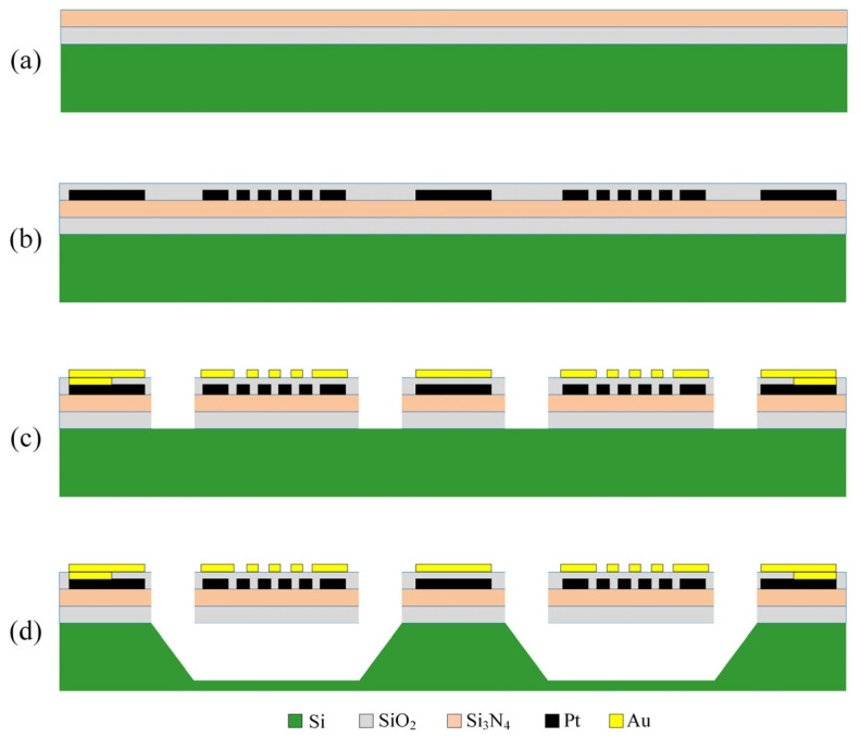 Figure 2
Peer Reviews
No public reviews on file for this paper yet. If you reviewed it on a platform where reviews are public (OpenReview, ICLR, NeurIPS, ICML), you can paste yours below so the community can read it here.
Videos
No videos yet. Explain this paper in a talk, walkthrough, or lecture? Add one.
Taxonomy
TopicsGas Sensing Nanomaterials and Sensors · 2D Materials and Applications · Metal-Organic Frameworks: Synthesis and Applications
1. Introduction
Methane (CH_4_), the simplest organic compound widely found in nature, is extensively used in industrial production (such as petroleum and natural gas production) and daily life due to its combustibility [1,2,3]. Real-time monitoring of CH_4_ is essential for the safe processing of industrial and daily activities. Compared with optical and thermoelectric sensors, metal oxide semiconductor (MOS)-based CH_4_ sensors possess advantages such as low cost, simple structure, and highly tunable properties, attracting widespread attention [4,5,6]. However, the working mechanism of conventional MOS gas sensors necessitates operation at elevated temperatures (100–450 °C) [7,8,9], thereby restricting their practical use.
Metal–organic frameworks (MOFs) are porous materials constructed from metal ions or clusters coordinated with organic linkers [10,11]. Their large specific surface area, tunable porosity, and structural diversity make them ideal self-sacrifice templates for generating metal oxides. Recent studies have confirmed that MOS materials synthesized using MOFs templates can retain their original structure and exhibit remarkably enhanced gas sensing performance [12,13,14,15]. For instance, Song et al. created ZIF-67-derived hollow Co_3_O_4_ nanospheres that achieved a room-temperature response of 3.5 to 100 ppm NH_3_ and possessed high humidity resistance, enabling low-concentration NH_3_ detection in exhaled breath [16]. Fan et al. synthesized ZIF-67-derived hollow Co_3_O_4_ nanocages vertically wrapped by ultrathin NiO cilia, demonstrating an exceptionally high response (47.4) to 100 ppm NO_2_ at room temperature, along with an ultrafast response/recovery time (1.3/9.6 s) [17]. Li et al. reported that a high-performance room-temperature H_2_ sensor based on MOF-derived porous Pd@SnO_2_ composite exhibits an exceptional response (R_a_/R_g_ = 25.4 to 50 ppm), fast response/recovery, and excellent long-term stability, offering a promising strategy for low-power H_2_ detection [18]. Chu et al. synthesized a MOF-derived ZnCo_2_O_4_/Co_3_O_4_ nanocomposite that enabled highly sensitive and selective room temperature detection of NH_3_ down to 0.5 ppm [19]. Therefore, using MOFs as self-sacrifice templates is a feasible and effective strategy for synthesizing MOS materials with improved room-temperature gas sensing performance. A previous study [20] has shown that highly active MOS materials possess efficient C-H bond activation and adsorption capabilities for CH_4_ at room temperature. Thus, effective detection of CH_4_ gas under room temperature conditions could be achieved through the modification of appropriate MOS gas sensing materials.
In this study, ZIF-67 was employed as a self-sacrifice template to investigate the CH_4_ sensing characteristics of Co_3_O_4_ samples obtained at different calcination temperatures. It was found that the calcination temperature can alter the Co^3+^/Co^2+^ ratio and the specific surface area in the Co_3_O_4_ samples. The Co_3_O_4_-350 with the highest Co^3+^/Co^2+^ ratio and the larger specific surface area exhibited the optimal CH_4_ sensing performance and a low detection limit at room temperature. Furthermore, a pulse heating mode was employed to further enhance the gas sensing performance of Co_3_O_4_-350, which significantly improved both the response and recovery times to 26 s and 21 s, respectively.
2. Materials and Methods
2.1. Materials
Cobalt nitrate hexahydrate (Co(NO_3_)2·6H_2_O), ethanol (C_2_H_5_OH), and methanol (CH_3_OH) were purchased from Xilong Scientific Co., Ltd., Guangzhou China, and 2-Methylimidazole (C_4_H_6_N_2_) was purchased from Shanghai Aladdin Biochemical Technology Co., Ltd., Shanghai China. All the chemicals and reagents were of chemical purity and used as received.
2.2. Synthesis of ZIF-67 and Co3O4
The preparation process is shown in Figure 1. To synthesize ZIF-67, 2 mmol of Co(NO_3_)2·6H_2_O and 16 mmol of C_4_H_6_N_2_ were separately dissolved in methanol solution during magnetic stirring. Then, the solution of Co(NO_3_)2·6H_2_O in methanol was added to the solution of C_4_H_6_N_2_ and stirred thoroughly for 30 min. The mixed solution was aged at room temperature for 24 h, then the purple ZIF-67 precursors were collected by centrifugation and washed three times to remove C_4_H_6_N_2_ with methanol. Finally, the purple ZIF-67 precursors were dried at 60 °C overnight. The thermogravimetric (TGA) curve of ZIF-67 in air is shown in the lower left of Figure 1. ZIF-67 begins to lose weight at around 280 °C and stabilizes at approximately 330 °C. Therefore, the prepared ZIF-67 precursors were heated in an air atmosphere at a rate of 3 °C/min to 300 °C, 350 °C, 400 °C, and 450 °C, respectively, and maintained at each temperature for 2 h. The resulting products were denoted as Co_3_O_4_-300, Co_3_O_4_-350, Co_3_O_4_-400 and Co_3_O_4_-450.
2.3. Characterization
The crystal structure was analyzed by X-ray diffraction (XRD, D8 Advance, Bruker, Germany) using Cu Kα radiation (λ = 0.15418 nm) in the range of 5–90°. To investigate the morphology and microstructure of the samples, a field-emission scanning electron microscope (FESEM, JSM-7900F, Tokyo, Japan) and a transmission electron microscope (TEM, JEM-F200, Tokyo, Japan) were employed. The XPS patterns were measured on an X-ray photoelectron spectroscope (XPS, Thermal Scientific K-Alpha, Waltham, MA, USA). Additionally, the specific surface area was measured through N_2_ gas adsorption using Brunauer–Emmett–Teller (Micromeritics ASAP 2460 3.01, Micromeritics Instrument Corporation, Norcross, GA, USA) surface analysis techniques. A thermogravimetric (TGA) test was performed using a thermogravimetric analyzer (TGA, HITACHI STA200, Tokyo, Japan).
2.4. Fabrication and Measurement of MEMS Gas Sensor
In this study, a low-power, high-reliability micro-heater plate (MHP) based on MEMS technology was fabricated. A schematic diagram of the fabrication process is illustrated in Figure 2, with the steps detailed as follows:
(1) An N-type (100) silicon wafer was selected and subjected to high-temperature thermal oxidation to grow a silicon dioxide (SiO_2_) layer, which serves as an electrical insulation layer, as shown in Figure 2a.
(2) A low-stress silicon nitride (Si_3_N_4_) layer was subsequently deposited via Plasma-Enhanced Chemical Vapor Deposition (PECVD) to function as a mechanical support layer, as depicted in Figure 2a.
(3) A platinum (Pt) thin-film resistor for heating and temperature sensing was fabricated using magnetron sputtering and a lift-off process, as presented in Figure 2b.
(4) A silicon oxide layer was then deposited by PECVD to act as a passivation/insulation layer, as shown in Figure 2b.
(5) Photolithography and dry etching were employed to define holes and the sacrificial release windows. The multilayer stack of silicon oxide and silicon nitride within these release windows was completely etched away to expose the underlying silicon substrate, as illustrated in Figure 2c.
(6) Interdigitated gold (Au) gas sensing electrodes were formed using magnetron sputtering and a lift-off process, as presented in Figure 2c.
(7) Finally, the exposed silicon substrate was etched through the release windows using a Tetramethylammonium Hydroxide (TMAH) solution, resulting in a suspended bridge-type micro-heater plate structure, as shown in Figure 2d.
Schematic diagram of the processing steps for a MEMS MHP array chip: (a) steps 1–2, (b) steps 3–4, (c) steps 5–6, (d) step 7.
SiO_2_/Si_3_N_4_/ SiO_2_ layer thickness is 800 nm/500 nm/800 nm, Pt and Au layer thickness are both 100 nm, heater track and electrode finger width are both 5 μm, electrode finger gap is 5 μm, and the shape of release window is as shown in Figure 3c. Given the reproducibility of micro-scale components [21], MHPs can achieve large-scale mass production.
High-precision Electrohydrodynamic (EHD) printing technology was employed to deposit the as-synthesized Co_3_O_4_ sensitive materials onto the MEMS MHP array chip, thereby forming the MEMS gas sensor. First, a certain amount of as-synthesized Co_3_O_4_ powders were dispersed in a specific quantity of ethanol, followed by thorough grinding to form an ink with a mass fraction of 20%. Next, the ink was printed onto the MHP using commercial EHD printing equipment (RD-EHD-200, Ruidu Photoelectric Technology Co., Ltd., Shanghai, China). The inner diameter of the printing needle is 200 μm, and the distance between the needle and the MHP is 50 μm. The printed film on the MHP has a thickness ranging from 500 to 2000 nm. The top view of the non-printed MHP array chip and the printed MHP array chip are shown in Figure 3a,b. The sensor was dried at 60 °C in a drying oven for 6 h to remove the residual ethanol. Finally, the MEMS gas sensor was aged at 200 °C before the gas sensing test.
A gas sensing test system was developed to evaluate the gas sensing performance of the MEMS MOS sensor (Figure 4). The system is composed of a computer, computer-controlled mass flow controllers (MFCs), gas cylinders (Dalian Guangming Special Gas Products Co., Ltd., Dalian China), and data acquisition instrumentation (Keithley DAQ 6510, Solon, OH, USA). During the experiments, the gas concentration was adjusted to five levels: 200, 400, 800, and 2000 ppm. The total gas flow rate was maintained at 500sccm throughout the testing process to avoid the influence of flow rate variations on the sensor response, when the gas is switched, stability is achieved within 0.7 s. Following the introduction of each target concentration gas, the test chamber of the sensor was purged using synthetic air as the cleaning gas. Throughout the entire testing process, the humidity in the test chamber was maintained at 3%. During the humidity influence tests, humidity levels were regulated by adjusting the mix of dry and wet air. Sensor’s response was defined as R_g_/R_a_, where R_a_ is the sensor’s resistance in air and R_g_ in the target gas. Response and recovery times were defined as the durations required for the sensor’s resistance to reach 70% of its total change during gas adsorption and desorption.
3. Morphology and Structural Characterization
The structure and crystal phase of the obtained samples were investigated by XRD (Figure 5). The XRD pattern of the as-synthesized ZIF-67 sample is shown in Figure 5a, which matched well with those reported in the literature, confirming the formation of pure ZIF-67 crystals [22,23,24]. All samples obtained at four different calcination temperatures exhibited consistent strong diffraction peaks corresponding to the spinel structure of Co_3_O_4_. As shown in Figure 5b, the diffraction peaks located at around 19.1, 31.16, 36.9, 38.54, 44.74, 55.72, 59.4, and 65.2° were recorded, matching with the lattice planes of (111), (220), (311), (222), (400), (422), (511), and (440) of Co_3_O_4_ (PDF# 42-1467). No CoO or Co was detected in the samples, indicating its high purity.
The morphology of the ZIF-67 precursor and the resulting Co_3_O_4_ samples obtained by calcination at different temperatures was investigated using scanning electron microscopy (SEM). As can be seen in Figure 6a, the as-synthesized ZIF-67 exhibited a rhombic dodecahedron morphology with a smooth surface. After high-temperature calcination, ZIF-67-derived Co_3_O_4_-300 and Co_3_O_4_-350 retain the original dodecahedral structure of the precursor, with their surfaces becoming rougher (Figure 6b,c). A histogram of the particle size distribution of Co_3_O_4_-350 was plotted in Figure 6c; the particle diameter of Co_3_O_4_-350 is approximately 177 nm. As the calcinating temperature increases, the structure of Co_3_O_4_ collapses. It can be observed that when the calcinating temperature rises to 400 °C or higher, the dodecahedral structure is destroyed, leaving only collapsed block-like structures (Figure 6d,e). This is due to the excessively high calcinating temperature, which causes small gas molecules to rapidly escape, leading to swift framework contraction and eventual collapse. Transmission Electron Microscopy (TEM) is a supplementary technique to SEM that can be used to further investigate the nanostructure of Co_3_O_4_ metal oxides. Figure 7a,b clearly reveals a rhombic dodecahedron morphology with a length of approximately 200 nm, which is consistent with the SEM results. Figure 7c,d more clearly shows collapsed block-like structures of Co_3_O_4_-400 and Co_3_O_4_-450. Figure 7e shows the high-resolution transmission electron microscopy (HRTEM) image, where clear lattice fringes can be observed. The measured interplanar spacing d = 0.465 nm corresponds to the (111) plane of the spinel-structured Co_3_O_4_. The fast Fourier transform (FFT) of the corresponding region is displayed in the lower right corner, which can be indexed to the diffraction pattern of the cubic phase Co_3_O_4_ along the zone axis.
XPS measurement was used to determine the chemical composition and valence states of Co_3_O_4_ samples obtained at different calcination temperatures. Figure 8a presented the survey spectrum of all the Co_3_O_4_ samples, demonstrating the presence of O and Co elements. Figure 8b shows that the Co 2p curves of all Co_3_O_4_ samples are similar, exhibiting two distinct spin–orbit peaks at approximately 780 eV and 795 eV, which correspond to Co 2p_3/2_ and Co 2p_1/2_, respectively. Among them, the Co 2p_3/2_ peak can be deconvoluted into two peaks located at binding energies of approximately 779.3779.7 eV and 780.8781.2 eV, which correspond to Co^3+^ and Co^2+^, respectively [25]. In the four Co_3_O_4_ samples, the ratios of Co^3+^/Co^2+^ were determined to be 0.37, 2.77, 0.42, and 0.93, respectively, by calculating the integrated areas of the fitted curves. The results demonstrate that different calcination temperatures significantly influence the Co^3+^/Co^2+^ ratios in ZIF-67-derived Co_3_O_4_. It is well known that Co^3+^ serves as the active center for CH_4_ adsorption [26]. The Co_3_O_4_-350 sample exhibits the highest Co^3+^/Co^2+^ ratios, which may contribute to enhanced CH_4_ gas sensing performance.
Figure 8c displays the O 1s spectra of four samples. According to different binding energies, the O 1s spectra can be deconvoluted into three peaks: the peak located at a binding energy of 529.7 ± 0.2 eV corresponds to lattice oxygen (O_L_), the peak at 531.2 ± 0.2 eV is attributed to oxygen vacancy (O_V_), and the peak at 532.7 ± 0.2 eV is assigned to adsorbed oxygen (O_C_) [27]. Among all the samples, the adsorbed oxygen content for Co_3_O_4_-300, Co_3_O_4_-350, Co_3_O_4_-400, and Co_3_O_4_-450 was measured to be 24.51%, 47.01%, 30.72%, and 36.05%, respectively, by calculating the integrated areas of the fitted curves. A higher adsorbed oxygen content may be more conducive to the complete participation of CH_4_ in the reaction.
To investigate the specific surface areas of the Co_3_O_4_ samples, N_2_ adsorption–desorption tests were performed. Figure 9 displays the N_2_ adsorption–desorption isotherms for all these samples. All isotherms exhibit a classical type IV pattern, which are typical of mesoporous materials [28]. These plentiful surface mesopores are beneficial for gas diffusion in the sensing material [29]. As shown in Table 1, the specific surface areas of the samples were: Co_3_O_4_-300, 4.56 m^2^/g; Co_3_O_4_-350, 38.82 m^2^/g; Co_3_O_4_-400, 32.90 m^2^/g; and Co_3_O_4_-450, 24.30 m^2^/g. The pore volumes of the samples were Co_3_O_4_-300, 0.02 cm^3^g^−1^; Co_3_O_4_-350, 0.29 cm^3^g^−1^; Co_3_O_4_-400, 0.16 cm^3^g^−1^; and Co_3_O_4_-450, 0.11 cm^3^g^−1^. And the average pore sizes of all the samples were 21.98, 29.95, 20.09 and 17.95 nm, respectively. The results indicate that the samples obtained at different calcination temperatures exhibit distinct specific surface areas, pore volumes, and average pore sizes. Apparently, Co_3_O_4_-350 exhibited a larger specific surface area. This enhanced feature provides abundant active sites and efficient electron transfer channels for the gas sensing reaction, which may lead to the improvement of sensing performance.
4. Gas Sensing Performance
First, the as-prepared MEMS gas sensors based on Co_3_O_4_-300, Co_3_O_4_-350, Co_3_O_4_-400, Co_3_O_4_-450 were used to test CH_4_ sensing response with a concentration of 400 ppm at room temperature. As shown in Figure 10a, the increase in resistance of all the sensors upon exposure to CH_4_ at room temperature is consistent with the characteristics of p-type semiconductors. Due to incomplete recovery of the sensor’s resistance baseline upon re-exposure to air, it was heated to 285 °C to restore the baseline. Figure 10b illustrates the relationship between the Co_3_O_4_ sensors’ response values and the calcination temperature; the Co_3_O_4_-350 sensor exhibited the highest response to 400 ppm CH_4_ at room temperature, with a value of 1.316. Figure 10c shows the response values of the Co_3_O_4_-350 sensor to 400 ppm CH_4_ and its resistance baseline at operating temperatures ranging from RT (25 °C) to 150 °C; the results indicate that the optimal operating temperature for the Co_3_O_4_-350 sensor is room temperature, and the resistance baseline decreased with increasing operating temperature, which is consistent with the characteristics of MOS materials. Figure 10d displays the dynamic response curve of the Co_3_O_4_-350 sensor to CH_4_ concentrations ranging from 200 to 2000 ppm at its optimal operating temperature. It can be observed that the sensor’s response intensifies with the rising CH_4_ concentration. Among them, the response to 2000 ppm CH_4_ is 1.53. To investigate the selectivity of the Co_3_O_4_-350 sensor, Figure 10e compares its response to different concentrations of different gases, such as 10 ppm H_2_S, 20 ppm C_6_H_6_, 50 ppm H_2_, 100 ppm CO, 400 ppm C_2_H_6_O, and 2000 ppm CH_4_. These gases are all common in petroleum and natural gas production. The results show that the response of Co_3_O_4_-350 to CH_4_ is higher than that to other gases.
The baseline of the Co_3_O_4_-350 sensor does not recover when detecting CH_4_ at room temperature, which limits its application. Our previous work utilizing pulse heating to enable room-temperature H_2_S sensing materials to respond at room temperature and recover by high-temperature cleaning [30]. This approach resolved the issue of baseline non-recovery in room-temperature gas sensors. Therefore, pulse heating is adopted in this work. The pulse heating waveform is a trapezoidal wave with a period of 10 s, including a high level of 190 °C for 3 s, a low level of 45 °C for 5 s, and the rise/fall time of 1 s. The power consumption is 3.44 mW.
Figure 11a shows the typical response curve of the Co_3_O_4_-350 sensor to CH_4_ concentrations ranging from 200 to 2000 ppm under pulse heating mode. The inset in Figure 11a shows the pulse heating waveform and the sensor’s resistance data under a single pulse. In practical testing, data collection is paused during the high temperature phase, with the collected values held constant from the moment before pausing. Once the high temperature phase ends, data collection is resumed and smoothed. As shown in Figure 11b, a linear fit was performed between the CH_4_ gas concentrations and the response value under pulse heating mode. The fitting formula is y = 1.077 + 0.00086x, with an R^2^ value of 0.991. This indicates that the Co_3_O_4_-350 sensor conforms to the linear law. Figure 11c summarizes the response and recovery times, where the response time and recovery time for 2000 ppm CH_4_ are 26 s and 21 s, respectively, suitable for rapid detection of early-stage CH_4_ gas leaks. Figure 11d shows the response curves of the Co_3_O_4_-350 sensor to 800 ppm CH_4_ under pulsed heating mode during five repeated tests, and the results indicate minor variations, which indicate that the sensor’s repeatability is satisfactory. In general, relative humidity has a negative effect on gas sensor performance [31,32]. Therefore, tests were conducted to evaluate the impact of humidity on the performance of the Co_3_O_4_-350 sensor operating in pulsed heating mode. Figure 11e,f shows that as humidity increases; the baseline resistance rises while the response value decreases. When the humidity increases to approximately 30%, the declining trend of the response slows down. In practical applications, it needs to be combined with a pretreatment water removal device for use.
Table 2 summarizes the previously reported low-temperature CH_4_ gas sensing materials. In several studies [33,34,35,36], it has been reported that gas sensing materials can detect CH_4_ at room temperature or near room temperature. However, the reported ZnO material operating at 60 °C requires activation under ultraviolet (UV) to function [34]. The reported Pd-doped SnO_2_/rGO and In_2_O_3_ materials working at room temperature exhibit long response and recovery times, and pulsed heating mode was not employed in these studies [33,35]. The reported VO_x_ can operate at room temperature and exhibits relatively fast response/recovery times [36], but its response is too small. The superiority of Co_3_O_4_-350 in this study lies in its ability to exhibit a significant response to CH_4_ at room temperature without UV assistance while also achieving relatively fast response and recovery in pulsed heating mode.
The comparison of MHP gas sensors is shown in Table 3. In this work, the MHP has a smaller area than that reported in reference [44], resulting in correspondingly lower heating power consumption. Although the MHP in reference [45] consumes even less power, its smaller size makes it inconvenient for transferring gas-sensitive materials. The MHP in this work has a moderate area, facilitating the deposition of MOF-derived gas-sensitive materials by EHD printing. Furthermore, the MHP in this work is designed to provide heating pulse for the room-temperature gas-sensitive material Co_3_O_4_-350, enabling its desorption at high temperature. In contrast, the MHP reported previously [44,45] employs a heating pulse primarily to reduce the power consumption of gas-sensitive materials operating at high temperatures, indicating a difference in their operational sensing temperatures. Overall, MHP helps gas sensing materials achieve better sensing performance while reducing power consumption.
5. Gas Sensing Mechanism
As is well known, the gas sensing mechanism of metal oxide semiconductor materials can be explained by the surface adsorption oxygen model [46,47,48]. Co_3_O_4_ is a common p-type semiconductor characterized by holes as its primary charge carriers; the sensing behavior is illustrated in Figure 12. When Co_3_O_4_ is exposed to air atmosphere, oxygen molecules absorb onto its surface and capture electrons from the conduction band to form oxygen ions while simultaneously forming a hole accumulation layer (HAL), and because Co_3_O_4_ works at room temperature, the main oxygen ions are O_2_^−^, such as in Formulas (1) and (2). When Co_3_O_4_ is exposed to a CH_4_ atmosphere, CH_4_ molecules react with the chemisorbed cations on the surface and donate electrons to the conduction band of Co_3_O_4_. Then, the holes are neutralized by electrons, resulting in an increase in resistance. The reaction is shown in Formulas (3) and (4).
Co_3_O_4_ is a late-transition-metal oxide. The exposed pairs of coordinated unsaturated (cus)-metal and oxygen atoms on the surface of late-transition-metal oxides exhibit high reactivity toward CH_4_ [20]. The former study [20] demonstrated that cus-metal atoms strongly bind CH_4_, giving rise to an adsorbed molecular state that resembles a CH_4_ σ-complex, and the DFT simulation results indicate that the formation of the CH_4_ σ-complex enhances the adsorption of CH_4_ to the surface and facilitates the cleavage of C-H bonds in CH_4_. Additionally, MOF-derived Co_3_O_4_ possesses a large specific surface area, which may expose more cus-metal and oxygen atoms to its surface. Consequently, MOF-derived Co_3_O_4_ exhibits a detectable gas sensing response at room temperature.
Compared to other samples in this work, the MOF-derived Co_3_O_4_-350 exhibits better CH_4_ detection capability at room temperature. This advantage could in part be attributed to the fact that Co_3_O_4_-350 exhibits the highest Co^3+^/Co^2+^ ratio and the highest adsorbed oxygen content among all samples. The Co^3+^ species possess stronger oxidation capability, which further facilitates the adsorption of CH_4_ gas [26], and a higher adsorbed oxygen content may be more conducive to CH_4_ participating more fully in the reaction. This could contribute to the enhancement of CH_4_ gas sensing performance. In addition, according to the characterization results, Co_3_O_4_-350 exhibited well-preserved precursor structure, achieved by using a proper annealing temperature. This dodecahedral structure not only provides the sample with the largest specific surface area among all the samples, providing more active sites for gas adsorption and desorption, but also effectively reduces the particle agglomeration and enhances the material’s stability.
6. Conclusions
In this study, a series of Co_3_O_4_ nanomaterials for CH_4_ detection were synthesized by calcining ZIF-67 at different temperatures. The results indicate that among all four samples, CO_3_O_4_-350 exhibited the highest response to CH_4_ at room temperature (R_g_/R_a_ = 1.53 to 2000 ppm). Furthermore, under pulsed heating mode, the response/recovery time of CO_3_O_4_-350 was significantly improved, with values of 26 s and 21 s respectively for 2000 ppm CH_4_. Accordingly, the gas sensing performance of ZIF-67-derived Co_3_O_4_ is significantly influenced by the calcination temperature during the synthesis process. The Co_3_O_4_-350 obtained at 350 °C exhibits the optimal low-concentration CH_4_ detection performance at room temperature. This straightforward synthesis strategy offers a novel approach for preparing advanced MOS materials capable of CH_4_ detection at room temperature, which has broad application prospects.
The reference list from the paper itself. Each links out to its DOI / PubMed record.
- 1Chen S.D. Zeng L.X. Li Q. Dai Z.D. Zhang Z. Yi S.L. Membrane-based natural gas dehydration: Techno-economic analysis of membrane process designs with different potential application scenarios Sep. Purif. Technol.202537813456410.1016/j.seppur.2025.134564 · doi ↗
- 2Wang F. Xu Y.F. Zhang P. Liu D.M. Zhang G.D. Rapid and continuous generation of methane hydrates under low pressure promotes the efficient capture of associated petroleum gas (APG)Energy 202533213719710.1016/j.energy.2025.137197 · doi ↗
- 3Kan J.Y. Kang J. Qin J.R. Huang X. Li N. Li Z. Chen G.J. Optimizing CH 4 recovery and CO 2 sequestration in natural gas hydrate exploitation through dynamic adjustment of CO 2/N 2 injection composition: A simulation study Fuel 20254001357910.1016/j.fuel.2025.135790 · doi ↗
- 4Fu L. You S.X. Li G.J. Li X.X. Fan Z.C. Application of Semiconductor Metal Oxide in Chemiresistive Methane Gas Sensor: Recent Developments and Future Perspectives Molecules 202328671010.3390/molecules 2818671037764486 PMC 10536930 · doi ↗ · pubmed ↗
- 5Ke J.W. Xie X.Y. Qiu L. Liu F.Z. Huang S.Y. Zhang Z.Y. Chen X.X. Advances in chemiresistive methane gas sensors based on nanostructured metal oxide semiconductor Mater. Sci. Eng. B 202632311875510.1016/j.mseb.2025.118755 · doi ↗
- 6Li Z.X. Qi T.T. Zhao X.H. Zhang Y. Zhang Z. Wang T.T. Xiao X.Z. Yang D.C. Ce O 2 Hollow Nanospheres Decorated with Pd and Pd O Nanodots for Fast Methane Sensing Adv. Funct. Mater.202636 e 1737810.1002/adfm.202517378 · doi ↗
- 7Zhang Z.L. Qiu P.P. Deng Y.H. Luo W. Recent Advances in Functionalizing Metal Oxide Semiconductors for Highly Sensitive Gas Sensors Small Methods 20259250022810.1002/smtd.20250022840331443 · doi ↗ · pubmed ↗
- 8Kumar A. Mazumder J.T. Joyen K. Favier F. Mirzaei A. Kim J.Y. Kwoka M. Bechelany M. Jha R.K. Kumar M. Defect engineering approaches for metal oxide semiconductor-based chemiresistive gas sensing Coord. Chem. Rev.202554121683610.1016/j.ccr.2025.216836 · doi ↗