Phytohemical Profiling, Bioactivity and Toxicity Evaluation of Elsholtzia cypriani, a Potential Multifunctional Natural Feed Additive
Hongxia Zhang, Xinye Tian, Huiwei Zhou, Ziyi Chen, Mingxiang Li, Yongpeng Ma, Zhizhi Du

TL;DR
This study explores Elsholtzia cypriani as a natural feed additive with antimicrobial, anti-inflammatory, and antioxidant properties, and finds it safe and rich in bioactive compounds.
Contribution
The study reports thirty compounds from Elsholtzia cypriani, including two new ones, and evaluates their bioactivity and safety for potential use as a natural feed additive.
Findings
Elsholtzia cypriani contains thirty compounds, two of which are newly identified.
Key compounds like ethyl caffeate and luteolin show anti-inflammatory, antioxidant, and antibacterial effects.
Acute toxicity tests showed no adverse effects at tested dosages.
Abstract
The overuse of antibiotics in animal husbandry is a primary driver of antimicrobial resistance, creating a pressing need for safe and effective natural alternatives. This study systematically evaluated the potential of the edible aromatic plant Elsholtzia cypriani as a comprehensive alternative by investigating its chemical composition, bioactivities, and preliminary safety. Methods included solvent extraction and systematic chromatographic fractionation from the plant aerial parts, complemented by a series of in vitro assays assessing anti-inflammatory, antioxidant, and antibacterial properties, along with an acute toxicity study. A total of thirty compounds were isolated and their structures were elucidated, including two new and twenty-eight known compounds reported for the first time in this species. Key isolates, such as ethyl caffeate and luteolin, demonstrated significant…
Genes, proteins, chemicals, diseases, species, mutations and cell lines named across the full text — each resolved to its canonical identifier and authoritative record.
Click any figure to enlarge with its caption.
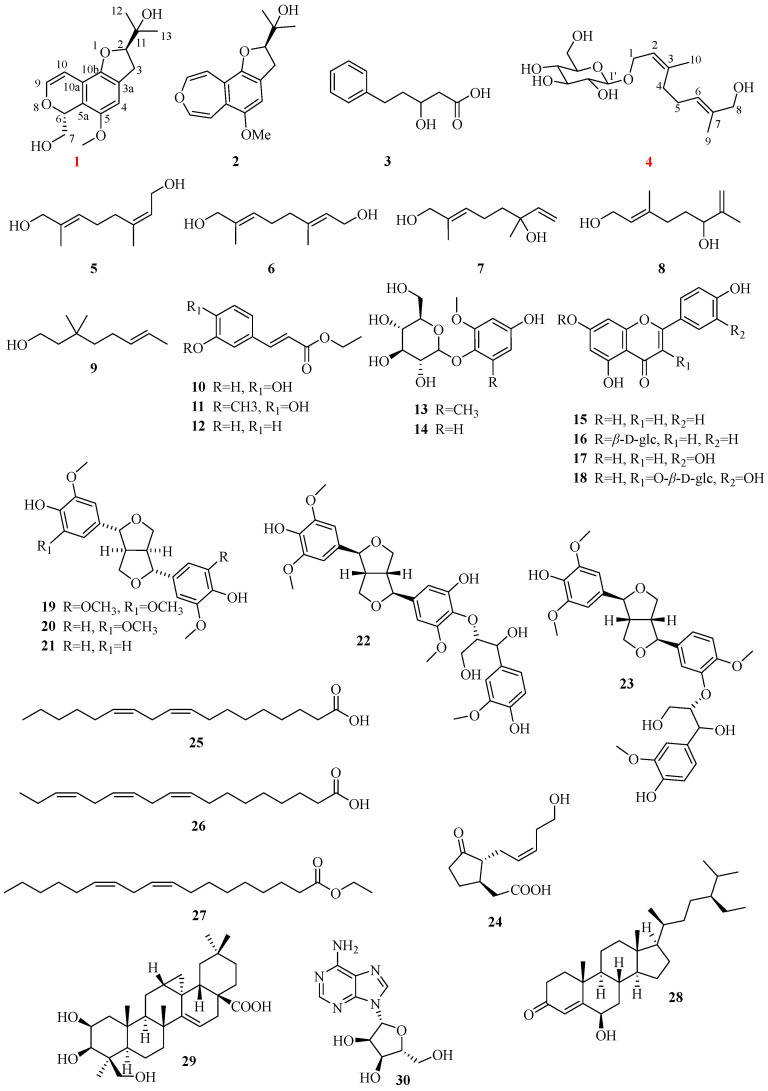 Figure 1
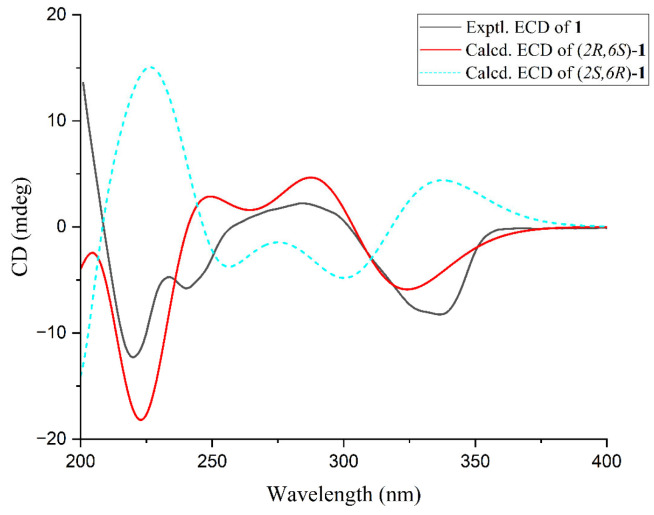 Figure 2
 Figure 3
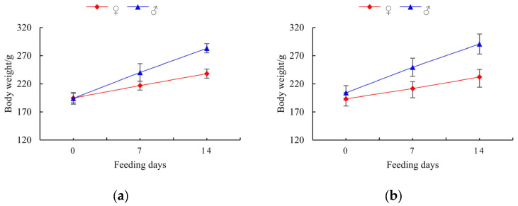 Figure 4
- —Fujian Provincial Department of Science and Technology
- —Yunnan Fundamental Research Project
Peer Reviews
No public reviews on file for this paper yet. If you reviewed it on a platform where reviews are public (OpenReview, ICLR, NeurIPS, ICML), you can paste yours below so the community can read it here.
Videos
No videos yet. Explain this paper in a talk, walkthrough, or lecture? Add one.
Taxonomy
TopicsPhytochemistry and Biological Activities · Ethnobotanical and Medicinal Plants Studies · Phytochemicals and Antioxidant Activities
1. Introduction
The widespread proliferation of bacterial resistance and drug residue issues resulting from the long-term misuse of antibiotics in livestock and poultry farming has become a global public health crisis of significant concern [1,2]. Natural products, as a vital source of bioactive molecules, demonstrate immense potential in addressing global health challenges such as antibiotic resistance [3,4]. Among these, the aromatic plants and their extracts offer advantages such as abundant resources, wide availability, comprehensive functions, and low toxicity with minimal side effects, making them increasingly applied in animal husbandry [5,6]. Systematic research on natural products, including the isolation and structural identification of chemical constituents, comprehensive evaluation of biological activities, and bioexploration of new resources, forms the scientific foundation for fully unlocking this potential [3,7].
Against this research backdrop, plants of the genus Elsholtzia have garnered significant attention due to their rich chemical diversity and traditional medicinal value [8,9,10]. Among them, Elsholtzia cypriani, a medicinal and edible plant distributed across multiple provinces in China, possesses a well-documented history of use. Its fresh leaves or dried inflorescences are commonly employed as flavorings [11,12], while the whole herb is traditionally used to treat conditions such as colds and gastroenteritis [13], suggesting the presence of potential bioactive compounds. Notably, recent progress in an animal study has provided new impetus for in-depth research on this plant. Research has revealed that E. cypriani herbal powder can enhance poultry immune function by activating the body’s antioxidant defenses and regulating the intestinal microbiota [14]. This discovery validates its biological regulatory potential at the application level while highlighting the urgency for systematic natural product research at the fundamental level. There is an urgent need to comprehensively investigate the pharmacological basis of its efficacy, evaluate its direct in vitro anti-inflammatory, antioxidant, and antibacterial activities, as well as establish preliminary safety assessments.
However, existing research has primarily focused on its volatile oil components, which have been demonstrated to possess anti-inflammatory and antibacterial activities [15,16,17]. In contrast, systematic research on its non-volatile constituents remains limited. Although isolated flavonoids and triterpenoids have been reported sporadically, with preliminary indications of antioxidant and antibacterial effects [18,19,20], the plant’s overall chemical profile remains unclear. Its comprehensive in vitro bioactivity spectrum and key bioactive components have yet to be fully elucidated.
This study systematically characterized the chemical profile of E. cypriani, leading to the isolation of thirty compounds, including two previously undescribed ones (1 and 4). These isolates were tested for in vitro anti-inflammatory, antioxidant, and antibacterial activities. Furthermore, an acute toxicity study of the extracts was conducted and reported here for the first time, showing no adverse effects. Collectively, this work supplies essential scientific evidence for developing this plant as a promising, well-defined botanical resource with potential to serve as an effective and safe plant-based alternative to antibiotics.
2. Results and Discussion
2.1. Isolation and Structure Elucidation of Compounds
Thirty compounds were isolated, and their structures were elucidated through comprehensive spectroscopic analysis, including 1D and 2D NMR, MS, IR, UV, and ECD. Notably, compounds 1 and 4 were identified as new compounds. Additionally, twenty-eight compounds (1–15, 18–27, and 28–30) were reported from E. cypriani for the first time. The structures of these compounds are shown in Figure 1.
Cyprianin A (1), the molecular formula, C_16_H_20_O_5_ was deduced by a high-resolution (HR) EI-MS peak at 315.1205 [M + Na]^+^ (calcd for C_16_H_20_O_5_Na^+^, 315.1203) and the ^13^C NMR spectrum, requiring 7 degrees of unsaturation. The ^1^H NMR data (Table 1) revealed the presence of three olefinic proton signals at δH 6.57 (s, 1H, H-4), 6.44 (d, J = 5.8 Hz, 1H, H-9), and 5.79 (d, J = 5.8 Hz, 1H, H-10), two oxygenated methine proton signals at δH 4.55 (t, J = 9.3, 8.7 Hz, 1H, H-2) and 5.51 (dd, J = 9.0, 3.2 Hz, 1H, H-6), two oxygenated methylene proton signals at δH 3.96 (dd, J = 11.8, 9.0 Hz, 1H, H-7) and 3.45 (dd, J = 11.8, 3.2 Hz, 1H, H-7), two methylene proton signals at δH 3.14 (dd, J = 15.8, 8.7 Hz, 1H, H-3) and 3.06 (dd, J = 15.8, 9.3 Hz, 1H, H-3), one methoxy proton signal at δH 3.73 (s, 3H, 5-OMe), and two methyl proton signals at δ_H_1.31 (s, 3H, H-12) and δH 1.18 (s, 3H, H-13). The ^13^C NMR and DEPT spectra indicated the presence of five aromatic quaternary carbon signals at δC 127.1 (C-3a), 149.3 (C-5), 114.3 (C-5a), 112.8 (C-10a), 147.4 (C-10b), and one aliphatic quaternary carbon signal at δC 71.8 (C-11). The NMR data indicated that 1 was structurally similar to perilloxin, a compound isolated from Perilla frutescens [21], with the notable difference being the substitution of a heterocyclic ring in compound 1 for the benzoxepine ring present in perilloxin. In comparison, the ^1^H NMR spectrum of compound 1 showed a decrease of two olefinic proton signals, with a concomitant increase of one oxygenated methylene proton signal at δH 3.96 (dd, J = 11.8, 9.0 Hz, 1H, H-7) and 3.45 (dd, J = 11.8, 3.2 Hz, 1H, H-7) and an oxygenated methine proton signal at δH 5.51 (dd, J = 9.0, 3.2 Hz, 1H, H-6). A six-membered 2H-pyran ring in 1 differs from a seven-membered oxepine ring in perilloxin was deduced by the HMBC, HSQC, and ^1^H-^1^H COSY correlation signals. A six-membered 2H-pyran ring in 1, which is different from the seven-membered oxepine ring in perilloxin, was established based on HMBC, HSQC, and ^1^H-^1^H COSY correlations. The HMBC correlation supported the presence of 6-hydroxymethyl from H-7 (δH 3.96) to C-6 (δC 74.3) and C-5a (δC 114.3), and the presence of 5-methoxy from H-OMe (δH 3.73) to C-5 (δC 149.3) and C-4 (δC 106.4) (Figure 2). The ECD calculation results for (2R,6S)-1 matched well with its experimental ECD spectrum, finally establishing the absolute configuration of 1 (Figure 2). Therefore, compound 1 is identified as: (2R,6S)-2-(6-(hydroxymethyl)-5-methoxy-3,6-dihydro-2H-furo[2,3-f]isochromen-2-yl)propan-2-ol, named as cyprianin A.
(2Z,6E)-8-hydroxygeraniol-β-ᴅ-glucopyranoside (4), its molecular formula C_16_H_28_O_7_, was elucidated according to HR-ESI-MS peak at m/z 355.1727 [M + Na]^+^ (calcd for C_16_H_28_O_7_Na^+^, 355.1737) and the NMR spectrum (detailed spectral data in Table 2), with 3 degrees of unsaturation. The ^1^H NMR data (Table 2) revealed the presence of two methyl proton signals at δH 1.66 (s, 3H, H-9) and 1.78 (s, 3H, H-10), three oxymethylene proton signals at δH 4.35 (dd, J = 11.9, 6.4 Hz, 1H, H-1), 4.22 (dd, J = 11.9, 7.7 Hz, 1H, H-1), 3.92 (s, 2H, H-8), 3.87 (dd, J = 11.9, 2.3 Hz, 1H, H-6′), and 3.68 (dd, J = 11.9, 5.6 Hz, 1H, H-6′), two olefinic proton signals at δH 5.41 (m, 1H, H-2) and 5.42 (m, 1H, H-6), and one anomeric proton signal at δH 4.28 (d, J = 7.8 Hz, 1H, H-1′), which were corroborated by the corresponding ^13^C NMR data at δC 13.8 (q, C-9) and 23.6 (q, C-10), 66.3 (t, C-1), 68.8 (q, C-8), 62.8 (q, C-6′), 122.7 (d, C-2), 125.9 (d, C-6), and 102.9 (d, C-1′). The NMR spectra and MS data suggested that 4 was highly similar to 8-hydroxygeraniol-β-ᴅ-glucopyranoside [22]. However, significant differences in chemical shifts were observed for C-4, C-9, and C-10 between the two compounds. The ROESY correlations between H-1 and H-4, as well as between H-5 and H-9, confirmed the (2Z,6E) configuration of 4. The absolute configuration of ᴅ-glc (tR = 45.868 min) unit was determined by comparing the retention time of its derivative with that of the standard derivative as the method reported in the previous literature [23]. The spectrum of HMBC (Figure 3) showed that this glucosyl was correlated with C1-OH from H-1′ to C-1 (δC 66.3). Thus, compound 4 was identified as (2Z,6E)-8-hydroxygeraniol-β-ᴅ-glucopyranoside.
In addition, the twenty-eight known compounds were identified as follows: perilloxin (2) [21], 3-hydroxy-5-phenylpentanoic acid (3) [24], (2E,6Z)-2,6-dimethyl-2,6-octadiene-1,8-diol (5) [24], (2E,6E)-2,6-dimethyl-2,6-octadiene-1,8-diol (6) [25], (E)-2,6-dimethyl-2,7-octadiene-1,6-diol (7) [25], (E)-3,7-dimethyl-2,7-octadiene-1,6-diol (8) [25], 3,7-dimethyl-6-octene-1,3-diol (9) [25], ethyl caffeate (10) [26], ethyl ferulate (11) [27], trans-p-hydroxycinnamic acid ethyl ester (12) [28], 2,6-dimethoxy-4-hydroxyphenol-1-O-β-ᴅ-glucopyranoside (13) [29], isotachioside (14) [30], apigenin (15) [31], apigenin-7-O-β-ᴅ-glucopyranoside (16) [32], luteolin (17) [33], isoquercitrin (18) [34], (+)-syringaresinol (19) [35], (+)-medioresinol (20) [35], pinoresinol (21) [36], threo-buddlenol C (22) [37], ficusesquilignan B (23) [38], 12-hydroxyjasmonic acid (24) [39], linoleic acid (25) [40], α-linolenic acid (26) [40], ethyl linoleate (27) [41], (24R)-6β-hydroxy-24-ethyl-cholest-4-en-3-one (28) [42], 2β,3β,24β-trihydroxy-12,13-cyclotaraxer-14-en-28 oic acid (29) [43], adenosine (30) [44].
Previous studies on E. cypriani have predominantly focused on its volatile oils [20], which have limited a comprehensive understanding of its systematic value and controllability. This study, for the first time, systematically elucidated the chemical composition of its non-volatile fractions, identifying 30 compounds, including 28 newly reported ones (two of which are novel), spanning various categories such as flavonoids, phenolic acids, and fatty acids, all closely associated with bioactivity. This breakthrough discovery lays a critical foundation for transforming this empirical herbal resource into a standardized botanical material with well-defined constituents and controllable quality. This represents the primary prerequisite for enabling any plant resource to achieve scalable and standardized application as an “antibiotic alternative” in the feed industry [45,46]. The definitive identification of components such as luteolin, apigenin, ethyl caffeate, and various fatty acids provides direct chemical markers for subsequent bioactivity tracing and product quality control.
2.2. Acute Toxicity Test Results
The acute oral toxicity of extracts 95EWE and 50EWE was evaluated by a single oral gavage to SD rats. The total administered dose is 10,000 mg/kg. Following gastric administration, no abnormal symptoms were observed in any of the four experimental groups. During the 14-day observation period, no treatment-related toxic clinical symptoms or deaths were noted, and the average body weights of rats in all groups exhibited a linear increase (Figure 4). Based on these findings, the test sample is preliminarily judged to have an acute oral LD50 > 10,000 mg/kg body weight in SD rats, indicating it is practically non-toxic. These results provide critical preliminary data on its safety as a feed additive, clearing a fundamental hurdle for its practical application and paving the way for subsequent feeding trials and risk assessments. It should be noted that a comprehensive toxicological evaluation will be necessary before any practical application.
2.3. Inhibitory Effect on Nitric Oxide (NO) Production
In vitro NO production inhibition assays were conducted to evaluate the anti-inflammatory activity of the isolated and identified compounds. The results are summarized in Table 3. The compounds all exhibited a cell viability greater than 90% at a concentration of 50 μM. The findings indicate that ethyl caffeate (10), ethyl ferulate (11), apigenin (15), luteolin (17), and linoleic acid (25) exhibited significant NO production inhibition activity at the concentration of 50 μM, with 50% concentration of inhibition (IC_50_) values lower than that of the positive control drug L-NMM. Compound 26, α-linolenic acid, exhibited significant NO production inhibition at the concentration of 25 μM, with an inhibition rate of 72.19 ± 3.16%. The IC_50_ values reported in this study are estimates based on a limited number of concentration points. In subsequent research on the lead compounds, they will be accurately determined through comprehensive dose–response experiments. Excessive nitric oxide serves as a critical inflammatory mediator in infection- and stress-induced intestinal inflammation, capable of compromising the integrity of the intestinal mucosal barrier. The active components identified in this study, such as luteolin and ethyl caffeate, have been reported in the literature as inhibitors of inducible nitric oxide synthase (iNOS) or modulators of the NF-κB signaling pathway. Therefore, their documented activities provide a plausible explanation for the NO inhibition observed in our cellular assay [47]. Consequently, through its polyphenolic constituents—such as phenolic acids and flavonoids—and polyunsaturated fatty acids, E. cypriani can directly interfere with inflammatory signaling at the molecular level, thereby alleviating excessive intestinal inflammatory responses triggered by pathogens or environmental stressors. This host-oriented “anti-inflammatory” modulation synergizes with the “direct antibacterial” effects discussed later: while suppressing pathogenic bacteria, it simultaneously mitigates inflammation and safeguards the physical and immune barriers of the gut. Together, these actions establish an internal environment that is unfavorable for pathogen colonization and proliferation, fundamentally reducing the reliance on antibiotics.
2.4. Antioxidant Assay
While elucidating its anti-inflammatory activity, this study also systematically evaluated the antioxidant capacity of E. cypriani, which serves as another crucial pillar for its role as a comprehensive “antibiotic alternative” resource in modulating host health [48]. Persistent oxidative stress can damage intestinal epithelial cells, disrupt tight junctions, and exacerbate inflammatory responses, thereby creating favorable conditions for pathogen colonization. The antioxidant potentials of extracts and isolated compounds were determined through in vitro DPPH, ABTS, and OH radical scavenging assays (Table 4). Results indicate that both the 95% ethanol extract (95EWE) and 50% ethanol extract (50EWE) of E. cypriani demonstrated strong DPPH and ABTS radical scavenging activities, while exhibiting weaker scavenging capacity against hydroxyl radicals. Ethyl caffeate (10), luteolin (17), isoquercitrin (18), and (+)-medioresinol (19) exhibited good DPPH and ABTS radical scavenging activities at 200 μM, with IC_50_ values of 22.92 ± 2.60 μM and 21.19 ± 2.87 μM, 28.91 ± 3.42 μM and 37.62 ± 5.00 μM, 26.63 ± 2.54 μM for DPPH radical scavenging activities, and 31.39 ± 3.19 μM, 41.54 ± 1.60 μM, and 19.02 ± 1.65 μM for ABTS radical scavenging activities, respectively. Ethyl ferulate (11) also exhibited moderate DPPH and ABTS radical scavenging activities with scavenging rates of 58.92 ± 1.56% and 78.37 ± 2.29%, respectively, and IC_50_ values of 111.9 ± 13.97 μM and 51.58 ± 2.89 μM. Compound 13 exhibited great ABTS radical scavenging activity with a scavenging rate of 85.54 ± 1.64% and an IC_50_ of 37.74 ± 3.26 μM. Hydroxyl radical scavenging experiments indicated that all tested compounds exhibited weak scavenging capacity. Only 10 and 29 demonstrated marginal scavenging abilities, with scavenging rates of 33.89 ± 2.88% and 36.34 ± 1.83%, respectively. It is speculated that the antioxidant activity primarily arises from scavenging DPPH radicals and ABTS radicals.
Oxidative stress serves as a common pathological basis for numerous chronic diseases and aging processes [49]. Although the scavenging effect on hydroxyl radicals is generally weak, the strong scavenging capacity against DPPH and ABTS radicals sufficiently demonstrates that E. cypriani can effectively neutralize common reactive oxygen species generated during immune activation and metabolic processes.
2.5. Antibacterial Activity
The antibacterial potentials of extracts and isolated compounds were evaluated through in vitro antibacterial activity assays against E. coli, with results presented in Table 5. The results indicate that 95EWE exhibits no antibacterial activity against E. coli, while 50EWE demonstrates weak antibacterial activity, with an inhibition rate of 27.527 ± 1.354% at a concentration of 128 μg/mL. Evaluation of the antibacterial activity of isolated compounds revealed that all tested compounds exhibited antibacterial activity. Compounds 3-hydroxy-5-phenylvaleric acid (3), apigenin (15), isoquercitrin (18), 12-hydroxyjasmonic acid (24), linoleic acid (25), and α-linolenic acid (26) demonstrated significant antibacterial activity with inhibition rates exceeding 70%. The new compound cyprianin A (1) exhibited weak antibacterial activity with an inhibition rate of 31.92 ± 9.81%. This study identified compounds from E. cypriani that showed in vitro antibacterial activity against E. coli, complementing its observed anti-inflammatory and antioxidant activities. The research not only confirmed the inhibitory effect of its crude extract (50EWE) against E. coli but, more importantly, identified for the first time a series of structurally diverse monomeric compounds with significant antibacterial activity from this plant. This discovery surpasses previous limitations in understanding its antibacterial components. The active compounds encompass various categories, including organic acids (compounds 3 and 24), flavonoids (compounds 15 and 18), and polyunsaturated fatty acids (compounds 25 and 26). These components may operate through complementary mechanisms: flavonoids (e.g., apigenin) may interfere with bacterial energy metabolism or topoisomerase activity; organic acids can lower intracellular pH or affect membrane function; while fatty acids disrupt cell membrane integrity through their surfactant properties [50]. This library of antibacterial constituents, built upon diverse chemical scaffolds, makes it difficult for pathogens to develop adaptive resistance through single mutations, thereby offering an ideal chemical foundation for developing sustainable plant-derived antibacterial solutions.
3. Materials and Methods
3.1. Instrument and Materials
Thirty compounds were isolated, and their structures were elucidated through comprehensive spectroscopic analysis, including 1D and 2D NMR, MS, IR, UV, and ECD. Notably, compounds 1 and 4 were identified as new compounds. Additionally, twenty-eight compounds (1–15, 18–27, and 28–30) were reported from E. cypriani for the first time. The structures of these compounds are shown in Figure 1.
3.1.1. Plant Materials
The fresh aerial parts of Elsholtzia cypriani (Pavol.) S. Chow ex P. S. Hsu were collected from Zhaotong City in Yunnan province of China and identified by Professor Pu Chunxia in Yunnan University of Chinese Medicine. A voucher specimen (YCX-2023-1) was stored in Yunnan Key Laboratory for Wild Plant Resources, Kunming Institute of Botany, Chinese Academy of Sciences (CAS).
3.1.2. Animals
A total of 40 specific pathogen-free (SPF) SD rats (20 males and 20 females, weight 180~220 g) were purchased from Hunan Slack Jingda Experimental Animal Co., Ltd. in Changsha city of China (license: SCXK (Xiang) 2020-0002). All rats were maintained under standard conditions (20.0–26.0 °C, 40.0–70.0% humidity) with free access to food and water for a 5-day acclimatization period, then the rats were fasted overnight prior to experiments. The 40 SPF SD rats were divided into 4 groups, as shown in Table S1 for the acute toxicity test.
3.1.3. Chemicals, Instruments, and Procedures
The isolation and purification of compounds were performed using D101 macroporous adsorption resin (Tianjin Xingnan Yunneng Polymer Technology Co., Ltd., Tianjin, China), silica gel (200–300 mesh, QingdaoMarine Chemical Co., Ltd., Qingdao, China), and a semi-preparative high-performance liquid chromatography (HPLC) system (Jiangsu Hanbon Science & Technology Co., Ltd., Huaian city, China). RAW264.7 cells were purchased from the Cell Bank of the Chinese Academy of Sciences (Shanghai, China). DMEM medium and fetal bovine serum (FBS) were purchased from VivaCell (Shanghai, China). Ferrous sulfate (FeSO_4_), hydrogen peroxide (H_2_O_2_), and salicylic acid were purchased from Tianjin Damao Chemical Reagent Factory (Tianjin, China). Griess Reagent, lipopolysaccharide (LPS), NG-methyl-L-arginine acetate salt (L-NMMA), trolox, 1,1-diphenyl-2-picrylhydrazyl (DPPH), gentamicin, and 2,2′-azinobis (3-ethyl-benzothiazoline-6-sulfonic acid, ABTS) are purchased from Sigma–Aldrich Chemical Co. (Sigma-Aldrich, Shanghai, China). Mueller-Hinton (MH) broth was purchased from Qingdao Science and Technology Industrial Park, Haibo Biotechnology Co., Ltd. (Qingdao, China).
Nuclear magnetic resonance (NMR) spectra data were obtained using a Bruker AVANCE 500 MHz NMR spectrometer (Bruker, Bremen, Germany). MS data were collected by a Shimadzu ultraperformance liquid chromatograph–quadrupole time of flight–mass spectrometer (Shimadzu, Tokyo, Japan). The ultraviolet (UV) spectra were recorded on a UV-2700 series spectrophotometer (Shimadzu, Tokyo, Japan). Specific optical rotations were measured with a Rudolph Autopol VI polarimeter (Rudolph Research Analytical, Hackettstown, NJ, USA). Infrared (IR) spectra were obtained using a Thermo NICOLET Is10 mid-IR spectrometer (Thermo Fisher Scientific, Waltham, MA, USA).
3.2. Chemical Composition Research
The dried sample of E. cypriani (8.4 kg) was extracted sequentially with petroleum ether, 95% ethanol-water, and 50% ethanol-water for three times (24 h each) to produce petroleum ether extract (PPE, 60 g), 95% ethanol-water extract (95EWE, 290 g), and 50% ethanol-water extract (50EWE, 460 g). Thirty compounds were isolated, and their structures were elucidated through comprehensive spectroscopic analysis, including 1D and 2D NMR, MS, IR, UV, and ECD.
The extract 50EWE (460 g) was fractionated on a macroporous adsorption resin D101 column chromatography (9.5 cm × 97 cm) and eluted with an ethanol-water gradient (0%, 30%, 50%, 70%, 90%). Based on TLC, the eluates were combined to yield seven fractions (Fr. A–G). Fraction D, E, F, and G were subjected to column chromatography over silica gel (200–300 mesh) to divided into some subfractions. After being subjected to repeated silica gel and Sephadex LH-20 column chromatography, and followed by semi-preparative HPLC or recrystallization, these sub-fractions afforded compound 1 (18 mg), 2 (245 mg), 3 (11.4 mg), 4 (5.5 mg), 5 (40 mg), 6 (8 mg), 7 (2.5 mg), 8 (2 mg), 9 (350 mg), 10 (323.2 mg), 11 (18 mg), 12 (120 mg), 13 (57.6 mg), 14 (1.5 mg), 15 (60 mg), 16 (6 mg), 17 (115 mg), 18 (42 mg), 19 (20 mg), 20 (5.6 mg), 21 (6.1 mg), 22 (4.6 mg), 23 (3.1 mg), 24 (35 mg), 25 (40 mg), 26 (55 mg), 27 (13 mg), 29 (14.5 mg), and an additional 30 (1.5 mg). The detailed separation process is shown in the Supplementary Material.
The extract PPE (60 g) was dissolved in petroleum ether and partitioned by a silica gel column chromatography (1.1 kg, 200–300 mesh), eluted with a petroleum ether–ethyl acetate gradient system (P/E, 100:1, 100:2, 100:4, 100:10, 100:20, and finally 0:100). Based on thin-layer chromatography (TLC) analysis, the eluates were combined to yield 22 fractions (Fr. P1–P22). Fraction P18 (1.62 g) was further separated by successive silica gel column chromatography and Sephadex LH-20 chromatography to afford compound 2 (220 mg). Fraction P19 (1.33 g) was purified via sequential silica gel column chromatography, Sephadex LH-20 chromatography, and preparative thin-layer chromatography to yield compound 28 (10 mg).
3.3. Spectroscopic Data
Compound 1: light yellow powder; −197.80 (c 0.109, MeOH); UV (MeOH) λ_max_ (log ε) 333 (0.1325), 285 (0.1325), 223 (0.4137), 207 (0.3007) nm; IR (KBr) ν_max_ 3424, 3083, 2972, 2936, 1721, 1636, 1620, 1591, 1466, 1442, 1399, 1383, 1349, 1335, 1307, 1251, 1216, 1111, 1092, 1076, 1013, 944, 861, 835, 805, 760 cm^−1^. HR-ESI-MS m/z: 315.1205 [M + Na]^+^ (calcd. for C_16_H_20_O_5_Na, 315.1203). ^1^H and ^13^C NMR data, see Table 1.
Compound 4: Colorless oily substance; −18.82 (c 0.34, MeOH); UV (MeOH) λ_max_ (log ε) 229 (0.0374), 202 (0.5800) nm; IR (KBr) ν_max_ 3412, 2912, 2863, 1631, 1448, 1412, 1383, 1316, 1274, 1222, 1200, 1158, 1098, 1074, 1038, 1023, 893, 840, 819, 745, 630, 615, 576, 536, 492 cm^−1^. HR-ESI-MS m/z 355.1727 [M + Na]^+^ (calcd. for C_16_H_28_O_7_Na, 355.1737). ^1^H and ^13^C NMR data, see Table 2.
Spectral data for known compounds are provided in Supplementary Material.
3.4. Acute Toxicity Test
95EWE and 50EWE were dissolved in sterilized water to make a 0.50 g/mL test solution. The animals were given the test solutions by gavage once, within 24 h at the dose of 2.0 mL/100 g of body weight (bw), individually. After administration of the test solutions, the animals were fasted for an additional 4 h. They were then observed for a 14-day period, during which signs of toxicity, mortality, and time to death were recorded. Body weights were measured on days 0, 7, and 14.
3.5. Cell Culture and NO Assay
RAW264.7 cells were seeded into 96-well plates at a density of 5.0 × 10^4^ cells/mL and stimulated with LPS (1 μg/mL). Following stimulation, the test samples were added, and the cells were cultured overnight. The experimental design included an L-NMMA-treated positive control group. Nitric oxide (NO) production was determined by measuring the absorbance of the culture medium at 570 nm using the Griess method [51]. Cell viability was assessed via the MTS assay to exclude potential cytotoxic effects of the samples. The data are presented alongside the corresponding bioactivity results in Table 3. All cells were maintained in DMEM supplemented with 10% fetal bovine serum (FBS) and 1% penicillin-streptomycin solution at 37 °C in a humidified atmosphere of 5% CO_2_. The IC_50_ was calculated by Reed & Muench method.
The inhibition rate of NO production (%) was calculated using the following formula:
3.6. Evaluation of Antioxidant Activities In Vitro
Based on the methods described in the references [14,52,53] with appropriate modifications, the antioxidant capacity of the extracts and compounds from E. cypriani was evaluated using the 1,1-diphenyl-2-picrylhydrazyl (DPPH) radical scavenging assay, the ABTS radical scavenging assay, and the hydroxyl radical scavenging assay. Detailed antioxidant detection methods are provided in the Supplementary Material.
3.7. Antibacterial Activity Assay
The tests were performed using Escherichia coli (ATCC 25922, obtained from the China General Microbiological Culture Collection Center). The extracts, compounds, and gentamicin (the positive control) were dissolved in DMSO to achieve working concentrations of 2560 μg/mL and 100 μg/mL, respectively.
This antibacterial assay employs the microdilution method to determine the optical density (OD) values of bacterial suspensions to evaluate the inhibitory activity of samples against E. coli [54]. The prepared Mueller–Hinton (MH) broth, sample solutions (or gentamicin solution), and bacterial inoculum were combined to achieve final concentrations of 128 μg/mL for the extract, 25.6 μg/mL for the compound, 5 μg/mL for gentamicin, and 5 × 10^5^ CFU/mL for the bacterial suspension. The mixture was dispensed into a 96-well plate, incubated at 37 °C for 24 h, and the OD value was measured at 630 nm. The antibacterial rate was calculated using the following formula:
3.8. Statistical Analysis
Statistical analysis was performed using Microsoft Excel 2021. The data are expressed as mean ± standard deviation. Statistical significance was determined using one-way analysis of variance (ANOVA). When the ANOVA indicated a significant difference, Dunnett’s post hoc test was applied for multiple comparisons between treatment groups and a single control group. All analyses were performed using GraphPad Prism software 9.5.1. A p-value of <0.05 was considered statistically significant. Each experiment was independently repeated at least three times
4. Conclusions
Through systematic chemical constituents’ separation, compounds structural identification, and multidimensional bioactivity evaluation, this study has elucidated the non-volatile chemical profile of E. cypriani for the first time. It further revealed the plant’s multi-target mechanism of action and preliminary safety, establishing a solid scientific foundation for its evidence-based development as a potential antibiotic alternative plant resource.
A total of thirty compounds were isolated and identified, including two new structures. In vitro anti-inflammatory, antioxidant, and antibacterial activity assays confirmed that both the 90% and 50% ethanol extracts of E. cypriani exhibited anti-inflammatory (inhibition of NO production) and antioxidant (DPPH and ABTS radicals scavenging) activities. The 90% ethanol extract also inhibited the growth of E. coli. Evaluation of individual compounds revealed distinct bioactive profiles: compounds 17 and 25 showed notable anti-inflammatory potential; compounds 17, 18, and 19 demonstrated strong antioxidant activity; while compounds 3, 15, 18, 24, 25, and 26 displayed significant antibacterial effects against E. coli. Importantly, these activities do not exist in isolation but form a synergistic functional network where antimicrobial components directly target pathogens, while its anti-inflammatory and antioxidant components jointly help maintain host barrier integrity and immune homeostasis by mitigating intestinal inflammatory damage and oxidative stress. This multi-targeted action model—combining “direct pathogen suppression” with “host defense enhancement”—precisely aligns with the core philosophy of modern farming: replacing growth-promoting or prophylactic antibiotics with nutritional interventions and health management. Furthermore, acute oral toxicity tests in SD rats confirmed the safety of the extracts. In summary, this study elucidates the promising application potential of E. cypriani in anti-inflammatory, antioxidant, and antibacterial activities, providing robust scientific support for its traditional uses and a theoretical basis for its further development and utilization.
In summary, the findings of this study not only provide a scientific explanation for the traditional efficacy of E. cypriani in “clearing heat, detoxifying, and treating gastroenteritis” from a modern pharmacological perspective, but also systematically demonstrate its significant potential as a candidate natural feed additive with well-defined components, synergistic mechanisms, and preliminarily controllable safety. Future research should focus on in vivo validation of key active components in animal infection or stress models, establishing standardized extraction processes, and evaluating its long-term regulatory effects on the gut microbiome. This will ultimately drive the transformation of this distinctive plant resource into standardized, functional green husbandry products.
The reference list from the paper itself. Each links out to its DOI / PubMed record.
- 1Wang Y. Lu J. Mao L. Li J. Yuan Z. Bond P.L. Guo J. Antiepileptic drug carbamazepine promotes horizontal transfer of plasmid-borne multi-antibiotic resistance genes within and across bacterial genera Isme J.20191350952210.1038/s 41396-018-0275-x 30291330 PMC 6331567 · doi ↗ · pubmed ↗
- 2Van Boeckel T.P. Brower C. Gilbert M. Grenfell B.T. Levin S.A. Robinson T.P. Teillant A. Laxminarayan R. Global trends in antimicrobial use in food animals Proc. Natl. Acad. Sci. USA 20151125649565410.1073/pnas.150314111225792457 PMC 4426470 · doi ↗ · pubmed ↗
- 3Newman D.J. Cragg G.M. Natural Products as Sources of New Drugs over the Nearly Four Decades from 01/1981 to 09/2019 J. Nat. Prod.20208377080310.1021/acs.jnatprod.9b 0128532162523 · doi ↗ · pubmed ↗
- 4Brown E.D. Wright G.D. Antibacterial drug discovery in the resistance era Nature 201652933634310.1038/nature 1704226791724 · doi ↗ · pubmed ↗
- 5Dorantes-Iturbide G. Orzuna-Orzuna J.F. Lara-Bueno A. Mendoza-Martínez G.D. Miranda-Romero L.A. Lee-Rangel H.A. Essential Oils as a Dietary Additive for Small Ruminants: A Meta-Analysis on Performance, Rumen Parameters, Serum Metabolites, and Product Quality Vet. Sci.2022947510.3390/vetsci 909047536136691 PMC 9502430 · doi ↗ · pubmed ↗
- 6Wiles D. Pearson J.S. Beddoe T. Harnessing Plant-Derived Terpenoids for Novel Approaches in Combating Bacterial and Parasite Infections in Veterinary and Agricultural Settings Curr. Microbiol.20258213410.1007/s 00284-025-04113-439937282 PMC 11821797 · doi ↗ · pubmed ↗
- 7Atanasov A.G. Zotchev S.B. Dirsch V.M. Orhan I.E. Banach M. Rollinger J.M. Barreca D. Weckwerth W. Bauer R. Bayer E.A. Natural products in drug discovery: Advances and opportunities Nat. Rev. Drug Discov.20212020021610.1038/s 41573-020-00114-z 33510482 PMC 7841765 · doi ↗ · pubmed ↗
- 8Thappa R.K. Agarwal S.G. Kapahl B.K. Srivastava T.N. Chemosystematics of the Himalayan Elsholtzia J. Essent. Oil Res.1999119710310.1080/10412905.1999.9701082 · doi ↗