Comparative Effects of Cellulose- and Gelatin-Based Hemostatic Biomaterials on the Early Stage of Wound Healing—An In Vivo Study
Helena Hae In Ströthoff, Polina Shabes, Katharina Henrika Beckamp, Markus Udo Wagenhäuser, Wiebke Ibing, Julian-Dario Rembe, Hubert Schelzig, Waseem Garabet

TL;DR
This study compares how cellulose- and gelatin-based hemostatic materials affect early wound healing in mice, finding that gelatin-based materials support better healing without causing excessive inflammation.
Contribution
The study provides new in vivo evidence on how different hemostatic biomaterials influence early wound healing processes.
Findings
Gelatin-based materials showed enhanced extracellular matrix deposition and increased macrophage presence.
GELA group exhibited elevated Ki-67 expression, indicating enhanced cellular proliferation.
None of the biomaterials impaired wound healing or caused excessive inflammation.
Abstract
Hemostatic biomaterials are widely used in surgical and trauma settings, yet their influence on early wound healing remains incompletely understood. This in vivo study investigated the effects of cellulose- and gelatin-based hemostatic biomaterials on early wound healing using a murine skin wound model. Oxidized non-regenerated cellulose (ONRC), oxidized regenerated cellulose (ORC), and a porcine gelatin-based matrix (GELA) were left in situ following standardized subcutaneous implantation and compared with sham-treated controls. Tissue responses were analyzed at postoperative days 3 and 7 using histology, immunohistochemistry, and quantitative real-time polymerase chain reaction (qPCR). Cellulose-based materials persisted as eosinophilic remnants, whereas fibrous matrix structures and enhanced extracellular matrix deposition were observed in the GELA group. Immunohistochemical analysis…
Genes, proteins, chemicals, diseases, species, mutations and cell lines named across the full text — each resolved to its canonical identifier and authoritative record.
Click any figure to enlarge with its caption.
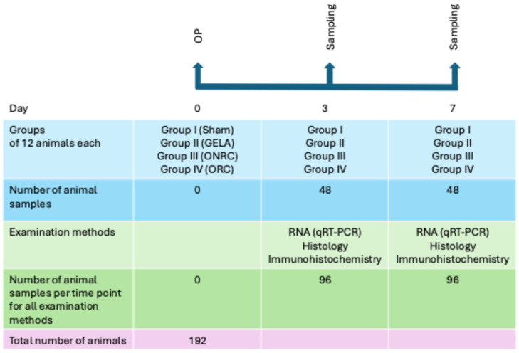 Figure 1
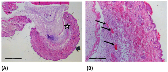 Figure 2
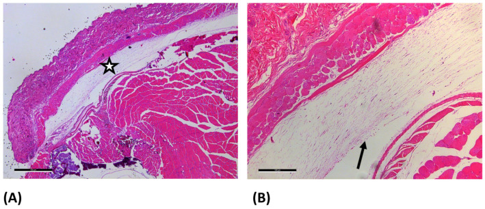 Figure 3
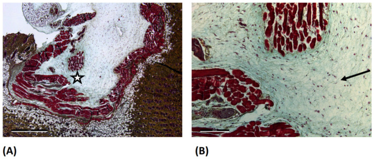 Figure 4
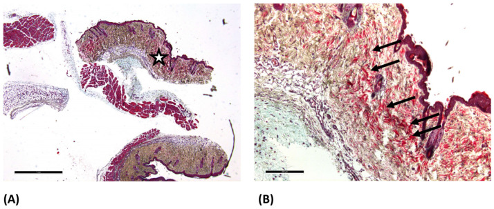 Figure 5
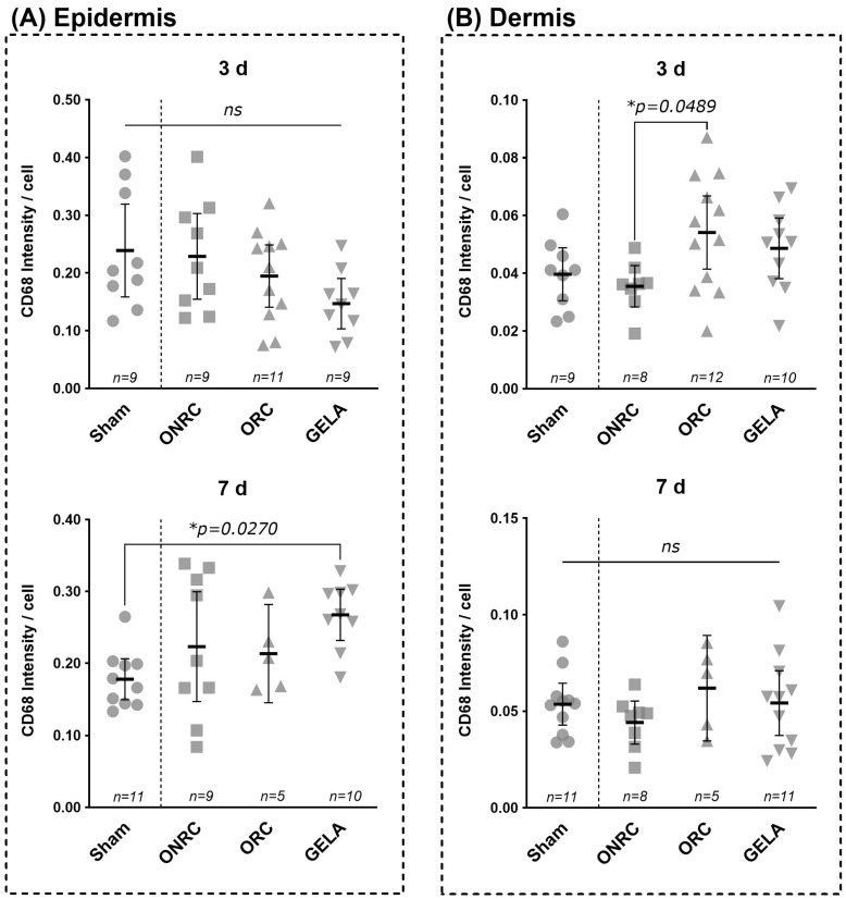 Figure 6
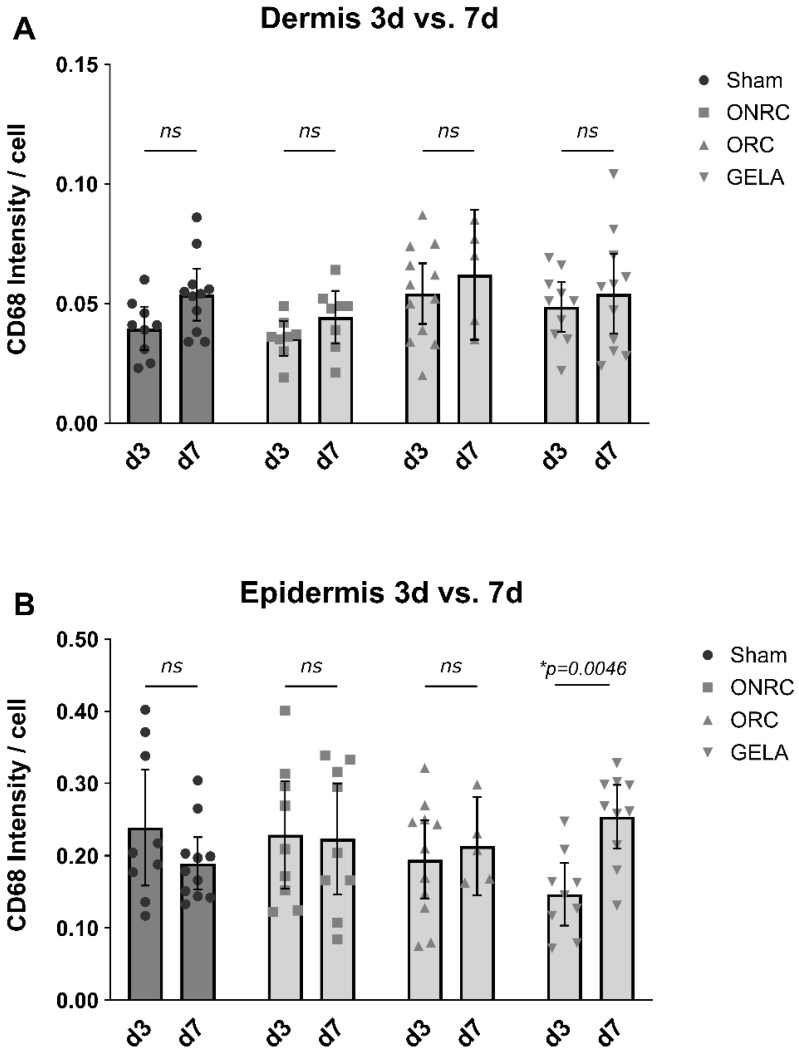 Figure 7
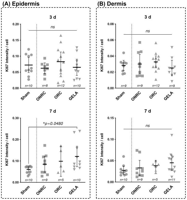 Figure 8
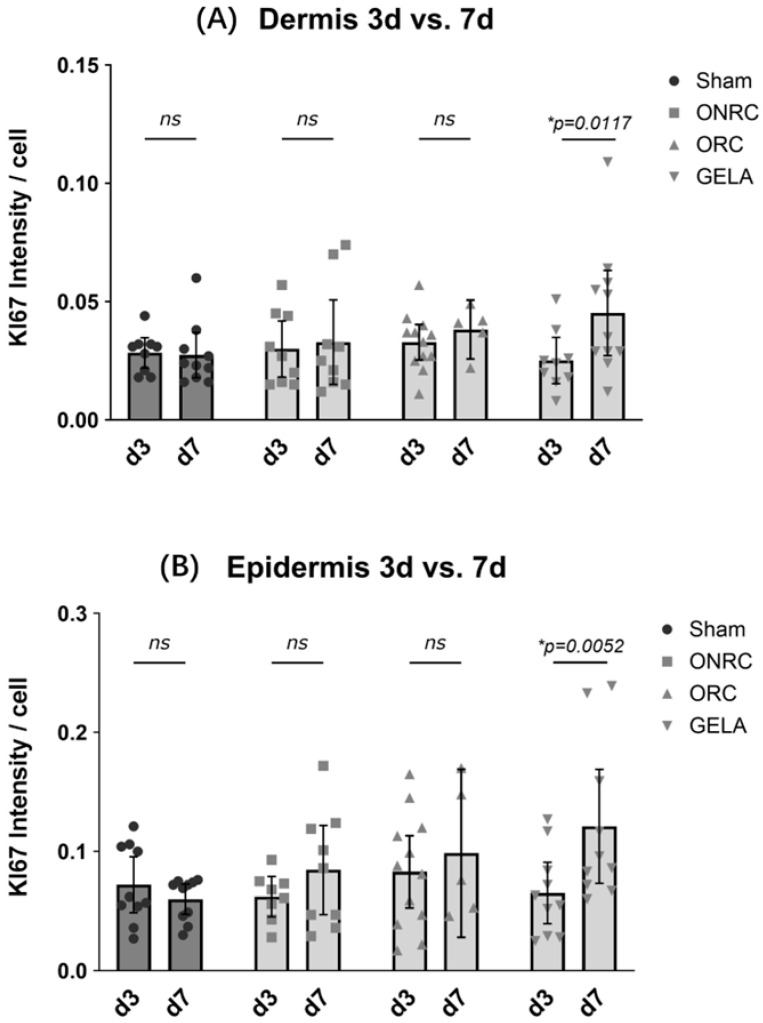 Figure 9
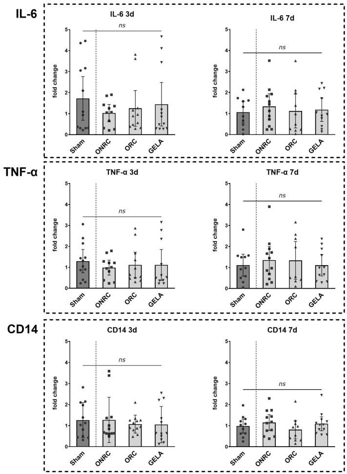 Figure 10
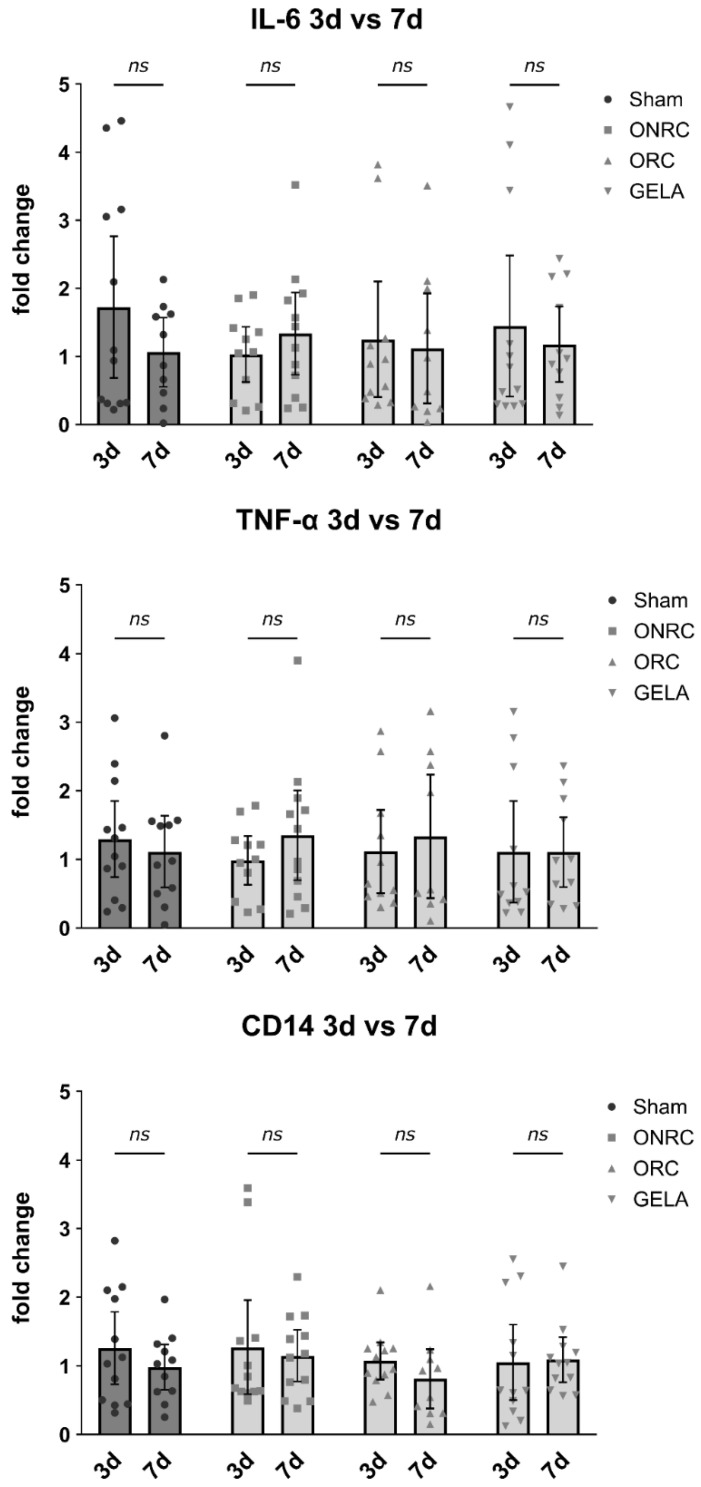 Figure 11
Peer Reviews
No public reviews on file for this paper yet. If you reviewed it on a platform where reviews are public (OpenReview, ICLR, NeurIPS, ICML), you can paste yours below so the community can read it here.
Videos
No videos yet. Explain this paper in a talk, walkthrough, or lecture? Add one.
Taxonomy
TopicsWound Healing and Treatments · Hemostasis and retained surgical items · Surgical Sutures and Adhesives