Circulating Extracellular Vesicles Downregulate NOS3 Expression in Endothelial Cells in Atrial Fibrillation
Nyozin Leimon, Anna Suzuki, Kohei Kawajiri, Giichi Nitta, Junji Yamaguchi, Satoshi Iwamiya, Satomi Hamada, Yasuhiro Shirai, Lai Wei, Masahiro Yamazoe, Kensuke Ihara, Tetsushi Furukawa, Tetsuo Sasano

TL;DR
Atrial fibrillation increases circulating extracellular vesicles, which may impair endothelial function by reducing NOS3 expression.
Contribution
This study identifies a novel mechanism linking atrial fibrillation to endothelial dysfunction via extracellular vesicles.
Findings
Patients with AF have higher plasma EV concentrations and reduced endothelial function compared to those with SR.
EVs from AF patients and rapidly paced cardiomyocytes reduce NOS3 mRNA expression in endothelial cells.
Irregular pacing of cardiomyocytes increases EV release compared to regular pacing or non-paced cells.
Abstract
Background: Atrial fibrillation (AF) is closely linked to endothelial dysfunction, yet its mechanisms remain unclear. Extracellular vesicles (EVs), including exosomes, are released by most cell types and mediate intercellular communication. We therefore investigated the role of EVs in endothelial dysfunction associated with AF. Methods: Vascular endothelial function in patients with sinus rhythm (SR), premature ventricular contractions (PVCs), or AF was assessed by peripheral arterial tonometry. Plasma-derived EVs were isolated from these three groups. Conditioned medium was collected from cultured cardiomyocytes (CMs), which were paced either regularly or irregularly at 1 Hz or 10 Hz or were non-paced, and EVs were subsequently isolated from the conditioned media. The isolated EVs were applied to endothelial cells (ECs), and mRNA levels of vasoactive genes were quantified. Results: The…
Genes, proteins, chemicals, diseases, species, mutations and cell lines named across the full text — each resolved to its canonical identifier and authoritative record.
Click any figure to enlarge with its caption.
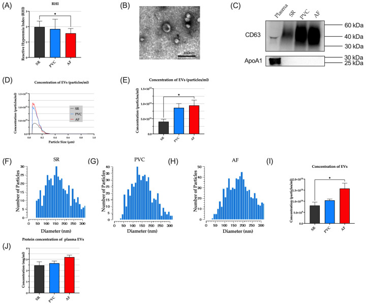 Figure 1
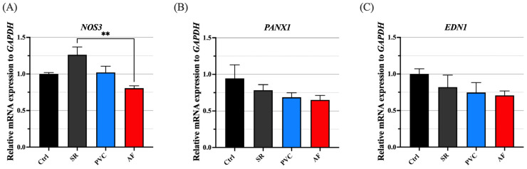 Figure 2
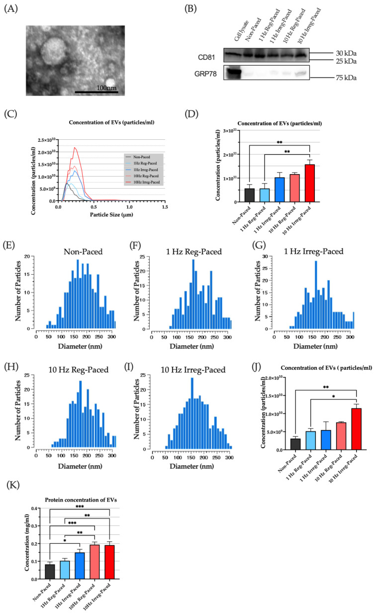 Figure 3
 Figure 4
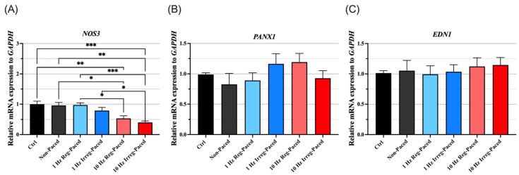 Figure 5
Peer Reviews
No public reviews on file for this paper yet. If you reviewed it on a platform where reviews are public (OpenReview, ICLR, NeurIPS, ICML), you can paste yours below so the community can read it here.
Videos
No videos yet. Explain this paper in a talk, walkthrough, or lecture? Add one.
Taxonomy
TopicsExtracellular vesicles in disease · Cardiovascular Disease and Adiposity · IL-33, ST2, and ILC Pathways